. 2000 Dec;131(8):1809-16.
doi: 10.1038/sj.bjp.0703758.

Selective phenylalkylamine block of I(Kr) over other K(+) currents in guinea-pig ventricular myocytes

Affiliations

Selective phenylalkylamine block of I(Kr) over other K(+) currents in guinea-pig ventricular myocytes

S E Jones et al. Br J Pharmacol. 2000 Dec.

Abstract

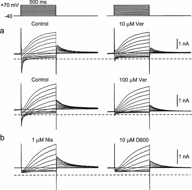
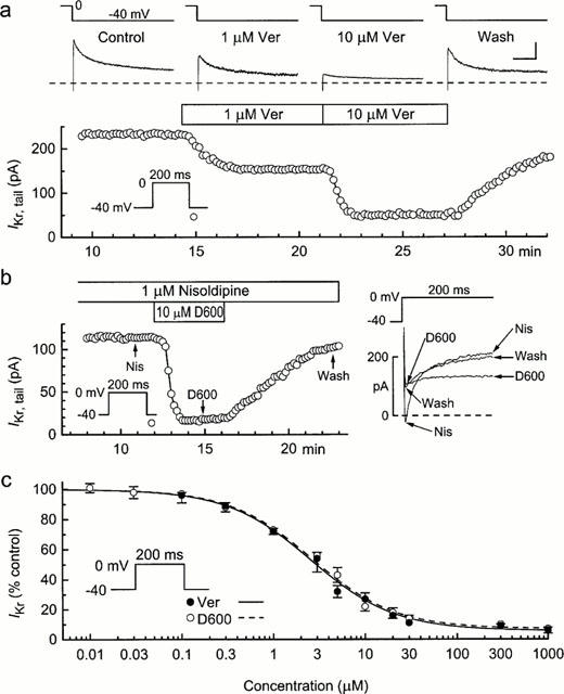
Previous studies on verapamil and D600 have established that the Ca(2+)-channel blockers also inhibit delayed-rectifier K(+) currents in cardiac tissues and myocytes. However, estimated IC(50) values range over two to three orders of concentration, and it is unclear whether this reflects a high selectivity by one or both of the phenylalkylamines for particular K(+) channels. The purpose of the present study was to determine the concentration-dependent actions of verapamil and D600 on three defined cardiac K(+) currents. Guinea-pig ventricular myocytes in the conventional whole-cell configuration were bathed with normal Tyrode's or K(+)-free solution, and pulsed from -80 mV for measurement of the effects of 0.01 microM to 3 mM verapamil and D600 on the inwardly-rectifying K(+) current (I:(Kl)) and the two delayed-rectifier K(+) currents, rapidly-activating I:(Kr) and slowly-activating I:(Ks). The phenylalkylamines inhibited both inward- and outward-directed I:(Kl). The IC(50) values for outward I:(Kl) were approximately 220 microM. Verapamil and D600 were approximately equipotent inhibitors of the delayed-rectifier K(+) currents. They inhibited I:(Kr) with IC(50) near 3 microM, and I:(Ks) with IC(50) > or =280 microM. These results are discussed in relation to previous findings on K(+) currents and to the clinical actions of the drugs.


Figures

Figure 1
Figure 1
Effects of verapamil and D600 on membrane currents in guinea-pig ventricular myocytes. The myocytes were bathed in normal Tyrode's solution, held at −80 mV, depolarized to prepulse −40 mV for 200 ms, and then depolarized to more positive potentials for 500 ms at 0.1 Hz; tail currents (IK,tail) were recorded on repolarizations to −40 mV. (a) Near-complete inhibition of inward ICa,L and partial inhibition of IK,tail after 10-min exposures of myocytes to 10 μM (top) and 100 μM (bottom) verapamil. (b) Effect of 10 μM D600 on membrane currents in a myocyte pretreated with 1 μM nisoldipine to suppress ICa,L. The dashed lines on the records here and in other figures indicate zero-current levels.
Figure 2
Figure 2
IK,tail–V relationships from myocytes treated with verapamil, D600, and E4031. The myocytes were bathed in normal Tyrode's solution, and tail currents on repolarizations to −40 mV after 500-ms pulses to potentials V were recorded before and 7–10 min after addition of drug. (a) Top: tail currents obtained from a myocyte that was sequentially exposed to 1, 5 and 20 μM verapamil, and then washed for 10 min with drug-free solution. The dotted line indicates steady current at −40 mV, and the calibration bars indicate 200 pA and 200 mS. Left hand plot: IK,tail–V relationships obtained from measurements of the IK,tail amplitudes. Right hand plot: verapamil-sensitive current obtained by subtracting drug data from control data. (b) Effects of 3 μM D600. Left: mean IK,tail–V relationships from five experiments. Right: D600-sensitive current. The latter data are significantly different than zero (P<0.01) at all potentials ⩾−10 mV (one-way ANOVA and Dunnetts Multiple Comparison test). (c) Effects of the selective IKr inhibitor E4031 (3 μM) for comparison with those of the phenylalkylamines. Left: mean IK,tail–V relationships from five experiments. Right: E4031-sensitive current. The latter data are significantly different than zero (P<0.01) at all potentials ⩾−10 mV.
Figure 3
Figure 3
Inhibition of IKr by verapamil and D600. Myocytes bathed in Tyrode's solution were held at −80 mV and depolarized for 200 ms from prepulse −40 mV to 0 mV at 0.1 Hz; IKr,tail was measured on the repolarizations to −40 mV. (a) Time course of changes in IKr,tail amplitude before, during and after exposure of a myocyte to 1 and 10 μM verapamil. (b) Effects of 10 μM D600 in a myocyte pretreated with 1 μM nisoldipine. Left: time course of changes in IKr,tail. Right: superimposition of currents elicited by the 200-ms pulses to 0 mV at the times marked on the time plot. Note the inhibition and recovery of outward current at 200 ms. (c) Dependence of IKr inhibition on drug concentration. The Hill equation fitting the verapamil data has an IC50 of 2.6±0.2 μM and a Hill coefficient of 0.95 (solid curve); the parameters describing the D600 data are 2.7±0.2 μM and 0.96 (dashed curve), respectively. Myocytes were exposed to one or two concentrations of a drug for 6–10 min, and the numbers of observations ranged from 4 to 20.
Figure 4
Figure 4
Phenylalkylamine-induced inhibition of IKs in myocytes bathed with normal Tyrode's solution. The myocytes were held at −80 mV and depolarized from prepulse −40 mV to more positive potentials V for 500 ms at 0.1 Hz for measurement of IK,tail on repolarizations to −40 mV. (a) The effect of 300 μM verapamil (n=5) on the IK-tail–V relationship. (b) The effect of 1000 μM on the IK,tail–V relationship. Also depicted are the measurements taken for calculation of the effect of drug on the amplitude of IKs. (c) Dependence of IKs inhibition on drug concentration. The Hill equation fitting the verapamil data has an IC50 of 1080±240 μM and a coefficient of 0.87. Myocytes were exposed to one or two concentrations of a drug for 6–10 min, and the numbers of observations are shown in parentheses.
Figure 5
Figure 5
Time course and reversibility of phenylalkylamine-induced inhibition of IKs in myocytes bathed with K+-, Ca2+-free Cd2+ solution. (a) Inhibition by 300 μM D600. The myocytes were held at −30 mV and depolarized to +50 mV for 500 ms at 0.1 Hz. Left: changes in the amplitude of IKs,tail. Right: example records from this experiment; the records on the 500-ms pulses are (top to bottom) control, wash, and D600. (b) Inhibition by 200 and 500 μM verapamil. The records on the 500-ms pulses are (top to bottom) wash, control, 200 and 500 μM verapamil.
Figure 6
Figure 6
Phenylalkylamine concentration-dependent inhibition of IKs in myocytes bathed in K+-, Ca2+-free Cd2+ solution. The myocytes were held at −30 mV and depolarized to more positive potentials for 2-s at 0.1 Hz. (a,b) Records and IK,tail–V relationships from representative experiments. The calibration bars beside the records indicate 200 pA and 200 ms. (c) Dependence of inhibition on drug concentration. The amplitude of IKs,tail elicited after 2-s depolarizations to +70 mV was measured before and 5 to 8 min after addition of drug. The Hill equation fitting the verapamil data has an IC50 of 280±26 μM and a coefficient of 0.96. Myocytes were exposed to one or two concentrations of a drug for 6–10 min, and the numbers of observations are shown in parentheses.
Figure 7
Figure 7
Inhibition of IK1 by phenylalkylamines. Myocytes were bathed in Tyrode's solution±0.2 mM Cd2+, and held at −80 mV. (a) Data from a myocyte treated with 300 μM verapamil for 9 min, Left: records obtained when the myocyte was pulsed from prepulse −40 mV to more positive and negative potentials. Right: end-of-pulse current amplitudes measured in this experiment. (b) Time courses of inhibition of outward IK1 in representative experiments. (c) Dependence of inhibition of outward IK1 at −40 mV on phenylalkylamine concentration. The Hill equation that describes the verapamil data has an IC50 of 220±14 μM and a coefficient of 0.96. The numbers of myocytes are shown in parentheses.

Similar articles

Cited by

References

    1. BARROS F., DELGADO L.M., DEL CAMINO D., DE LA PENA P. Characteristics and modulation by thyrotropin-releasing hormone of an inwardly rectifying K+ current in patch-perforated GH3 anterior pituitary cells. Pflügers Arch. 1992;422:31–39. - PubMed
    1. BAYER R., KALUSCHE D., KAUFMANN R., MANNHOLD R. Inotropic and electrophysiological actions of verapamil and D 600 in mammalian myocardium. III. Effects of the optical isomers on transmembrane action potentials. Naunyn Schmiedeberg's Arch. Pharmacol. 1975;290:81–97. - PubMed
    1. CHOUABE C., DRICI M.D., ROMEY G., BARHANIN J., LAZDUNSKI M. HERG and KvLQT1/IsK, the cardiac K+ channels involved in long QT syndromes, are targets for calcium channel blockers. Mol. Pharmacol. 1998;54:695–703. - PubMed
    1. CRANEFIELD P.F., ARONSON R.S., WIT A.L. Effect of verapamil on the normal action potential and on a calcium-dependent slow response of canine cardiac Purkinje fibers. Circ. Res. 1974;34:204–213. - PubMed
    1. DECOURSEY T.E. Mechanism of K+ channel block by verapamil and related compounds in rat alveolar epithelial cells. J. Gen. Physiol. 1995;106:745–779. - PMC - PubMed

LinkOut - more resources