. 2001 Jan 15;29(2):387-96.
doi: 10.1093/nar/29.2.387.

Characterization of Schizosaccharomyces pombe RNA triphosphatase

Affiliations

Characterization of Schizosaccharomyces pombe RNA triphosphatase

Y Pei et al. Nucleic Acids Res. .

Abstract

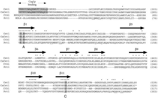
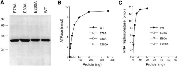
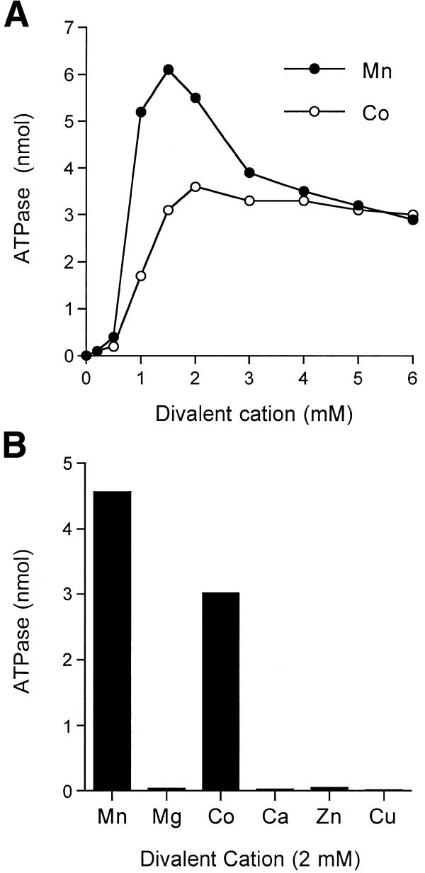
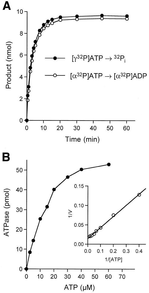
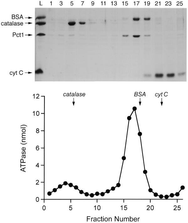
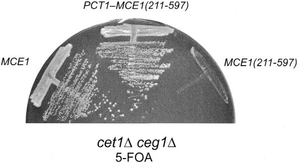
RNA triphosphatase catalyzes the first step in mRNA cap formation which entails the cleavage of the beta-gamma phosphoanhydride bond of triphosphate-terminated RNA to yield a diphosphate end that is then capped with GMP by RNA guanylyltransferase. Here we characterize a 303 amino acid RNA triphosphatase (Pct1p) encoded by the fission yeast SCHIZOSACCHAROMYCES: pombe. Pct1p hydrolyzes the gamma phosphate of triphosphate-terminated poly(A) in the presence of magnesium. Pct1p also hydrolyzes ATP to ADP and P(i) in the presence of manganese or cobalt (K(m) = 19 microM ATP; k(cat) = 67 s(-1)). Hydrolysis of 1 mM ATP is inhibited with increasing potency by inorganic phosphate (I(0.5) = 1 mM), pyrophosphate (I(0.5) = 0.4 mM) and tripolyphosphate (I(0.5) = 30 microM). Velocity sedimentation indicates that Pct1p is a homodimer. Pct1p is biochemically and structurally similar to the catalytic domain of Saccharomyces cerevisiae RNA triphosphatase Cet1p. Mechanistic conservation between Pct1p and Cet1p is underscored by a mutational analysis of the putative metal-binding site of Pct1p. Pct1p is functional in vivo in S.cerevisiae in lieu of Cet1p, provided that it is coexpressed with the S.pombe guanylyltransferase. Pct1p and other yeast RNA triphosphatases are completely unrelated, mechanistically and structurally, to the metazoan RNA triphosphatases, suggesting an abrupt evolutionary divergence of the capping apparatus during the transition from fungal to metazoan species.


Figures

Figure 1
Figure 1
Schizosaccharomyces pombe RNA triphosphatase is structurally related to the RNA triphosphatases of S.cerevisiae and C.albicans. The complete amino acid sequence of S.pombe Pct1p is aligned to the sequences of the C-terminal catalytic domains of S.cerevisiae Cet1p (residues 241–539) and C.albicans CaCet1p (residues 202–520) and to S.cerevisiae Cth1p (residues 1–317). Gaps in the alignment are indicated by dashes. The β strands that comprise the triphosphate tunnel of Cet1p are denoted above the sequence. The peptide domain of Cet1p that mediates its interaction with the guanylyltransferase Ceg1p is conserved in CaCet1p and is shaded. This segment is not conserved in either Cth1p or Pct1p. Positions of side-chain identity or structural similarity in all four fungal RNA triphosphatases are denoted by dots. Conserved motifs A (β1) and C (β11) that define the metal-dependent RNA triphosphatase family are indicated below the sequence. Residues comprising the homodimer interface of Cet1p that are conserved in Pct1p are underlined below the Pct1p sequence. Residues Glu78, Glu80 and Glu260 of Pct1p that were targeted for alanine substitution in the present study are also shaded.
Figure 2
Figure 2
RNA triphosphatase and ATPase activities of Pct1p. (A) Protein purification. Aliquots (4 µg) of the phosphocellulose fractions of wild-type Pct1p (WT) and mutants E78A, E80A and E260A were analyzed by electrophoresis in a 12% polyacrylamide gel containing 0.1% SDS. Polypeptides were visualized by staining with Coomassie blue dye. The positions and sizes (in kDa) of marker proteins are indicated to the left of the gel. (B) ATPase activity. Reaction mixtures (10 µl) containing 50 mM Tris–HCl pH 7.5, 5 mM DTT, 2 mM MnCl2, 1 mM [γ-32P]ATP and either wild-type (WT) or mutant proteins as specified were incubated for 15 min at 30°C. The reactions were quenched by adding 2.5 µl of 5 M formic acid. Aliquots of the mixtures were applied to a polyethyleneimine–cellulose thin-layer chromatography (TLC) plate, which was developed with 1 M formic acid, 0.5 M LiCl. 32Pi release was quantitated by scanning the chromatogram with a FUJIX phosphorimager and was plotted as a function of input protein. (C) RNA triphosphatase activity. Reaction mixtures (10 µl) containing 50 mM Tris–HCl pH 7.5, 5 mM DTT, 1 mM MgCl2, 20 pmol (of triphosphate termini) of [γ-32P]poly(A) and either WT or mutant proteins as specified were incubated for 15 min at 30°C. 32Pi release is plotted as a function of input protein.
Figure 3
Figure 3
Divalent cation requirement for ATP hydrolysis. (A) Dependence of ATPase activity on manganese and cobalt cation concentration. Reaction mixtures (10 µl) containing 50 mM Tris–HCl pH 7.5, 1 mM [γ-32P]ATP, 20 ng of Pct1p and MnCl2 or CoCl2 as specified were incubated for 15 min at 30°C. 32Pi release is plotted as a function of divalent cation concentration. (B) Divalent cation specificity. Reaction mixtures (10 µl) containing 50 mM Tris–HCl pH 7.5, 1 mM [γ-32P]ATP, 20 ng of Pct1p and 2 mM divalent cation as specified were incubated for 15 min at 30°C. Mg, Mn, Ca and Co were added as chloride salts; Cu and Zn were added as sulfates.
Figure 4
Figure 4
Kinetic analysis of ATP hydrolysis. (A) Reaction mixtures (150 µl) containing 50 mM Tris–HCl pH 7.5, 5 mM DTT, 2 mM MnCl2, 1 mM [α-32P]ATP or [γ-32P]ATP and 1.5 µg of Pct1p were incubated at 30°C. Aliquots (10 µl) were withdrawn at the times indicated and quenched immediately with formic acid. The reaction products were analyzed by polyethyleneimine–cellulose TLC. The extent of 32Pi or [α-32P]ADP formation (from 10 nmol of input ATP per sample) is plotted as a function of time. (B) Dependence of ATP hydrolysis on ATP concentration. Reaction mixtures (20 µl) containing 50 mM Tris–HCl pH 7.5, 5 mM DTT, 2 mM MnCl2, 40 pg of Pct1p and [γ-32P]ATP as specified were incubated for 15 min at 30°C. The extent of 32Pi release is plotted as a function of ATP concentration. The insert shows a double-reciprocal plot of the rate of 32Pi formation (s–1 = pmol 32Pi formed/900) versus [ATP].
Figure 5
Figure 5
Glycerol gradient sedimentation. Pct1p (40 µg) was mixed with catalase (40 µg), BSA (40 µg) and cytochrome c (40 µg) in 0.2 ml of buffer G (50 mM Tris–HCl pH 8.0, 100 mM NaCl, 1 mM EDTA, 2 mM DTT, 0.05% Triton X-100). The mixture was layered onto a 4.8 ml 15–30% glycerol gradient containing buffer G. The gradient was centrifuged in a Beckman SW50 rotor at 50 000 r.p.m. for 15 h at 4°C. Fractions (∼0.2 ml) were collected from the bottom of the tube. (Top) Aliquots (18 µl) of odd numbered fractions were analyzed by SDS–PAGE along with samples of the input protein mixture (L). Polypeptides were visualized by staining with Coomassie blue dye. The identities of the polypeptides are indicated at the left of the gel. (Bottom) ATPase activity profile. Reaction mixtures (20 µl) containing 50 mM Tris–HCl pH 7.5, 5 mM DTT, 2 mM MnCl2, 1 mM [γ-32P]ATP and 2 µl of the indicated glycerol gradient fractions were incubated for 15 min at 30°C. The peaks of the marker proteins are indicated by arrows.
Figure 6
Figure 6
Inhibition of ATP hydrolysis by phosphate, pyrophosphate and tripolyphosphate. Reaction mixtures (10 µl) containing 50 mM Tris–HCl pH 7.5, 5 mM DTT, 2 mM MnCl2, 1 mM [γ-32P]ATP, 11 ng of Pct1p and either sodium phosphate, sodium pyrophosphate or sodium tripolyphosphate as specified were incubated for 15 min at 30°C. The reactions were initiated by the addition of Pct1p. The extents of ATP hydrolysis were normalized to those of control reactions from which inhibitory phosphates were omitted (values of 2.5–3.5 nmol 32P released). The normalized activities (control value = 1.0) are plotted as a function of inhibitor concentration.
Figure 7
Figure 7
Genetic interaction between S.pombe RNA triphosphatase and S.pombe RNA guanylyltransferase. Yeast strain YBS20 (cet1Δ) was transformed with CEN TRP1 plasmids containing either CET1, PCT1, PCT1 + PCE1 or PCT1 + CEG1 and with a 2µ plasmid containing PCT1. Trp+ isolates were streaked on an agar plate containing 0.75 mg/ml 5-FOA. The plate was photographed after incubation for 3 days at 30°C.
Figure 8
Figure 8
Pct1p functions in vivo in S.cerevisiae when fused to the guanylyltransferase domain of mammalian capping enzyme. Yeast strain YBS50 (cet1Δ ceg1Δ) was transformed with CEN TRP1 plasmids containing either MCE1, MCE1(211–597), or the PCT1–MCE1(211–597) chimera. Trp+ isolates were streaked on an agar plate containing 0.75 mg/ml 5-FOA. The plate was photographed after incubation for 5 days at 30°C.

References

    1. Shuman S. (2000) Structure, mechanism and evolution of the mRNA capping apparatus. Prog. Nucleic Acid Res. Mol. Biol., 66, 1–40. - PubMed
    1. Takagi T., Moore,C.R., Diehn,F. and Buratowski,S. (1997) An RNA 5′-triphosphatase related to the protein tyrosine phosphatases. Cell, 89, 867–873. - PubMed
    1. Wen Y., Yue,Z. and Shatkin,A.J. (1998) Mammalian capping enzyme binds RNA and uses protein tyrosine phosphatase mechanism. Proc. Natl Acad. Sci. USA, 95, 12226–12231. - PMC - PubMed
    1. Ho C.K., Sriskanda,V., McCracken,S., Bentley,D., Schwer,B. and Shuman,S. (1998) The guanylyltransferase domain of mammalian mRNA capping enzyme binds to the phosphorylated carboxyl-terminal domain of RNA polymerase II. J. Biol. Chem., 273, 9577–9585. - PubMed
    1. Martins A. and Shuman,S. (2000) Mechanism of phosphoanhydride cleavage by baculovirus phosphatase. J. Biol. Chem., 275, 35070–35076. - PubMed

Publication types

MeSH terms