Comparative Study
. 2001 Jan 15;29(2):407-14.
doi: 10.1093/nar/29.2.407.

Enzymatic processing of DNA containing tandem dihydrouracil by endonucleases III and VIII

Affiliations
Comparative Study

Enzymatic processing of DNA containing tandem dihydrouracil by endonucleases III and VIII

R Venkhataraman et al. Nucleic Acids Res. .

Abstract

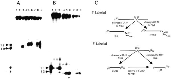
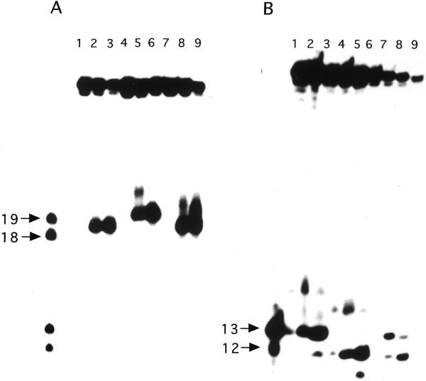
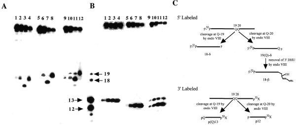
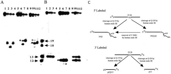
Endonuclease III from Escherichia coli, yeast (yNtg1p and yNtg2p) and human and E.coli endonuclease VIII have a wide substrate specificity, and recognize oxidation products of both thymine and cytosine. DNA containing single dihydrouracil (DHU) and tandem DHU lesions were used as substrates for these repair enzymes. It was found that yNtg1p prefers DHU/G and exhibits much weaker enzymatic activity towards DNA containing a DHU/A pair. However, yNtg2p, E. coli and human endonuclease III and E.coli endonuclease VIII activities were much less sensitive to the base opposite the lesion. Although these enzymes efficiently recognize single DHU lesions, they have limited capacity for completely removing this damaged base when DHU is present on duplex DNA as a tandem pair. Both E.coli endonuclease III and yeast yNtg1p are able to remove only one DHU in DNA containing tandem lesions, leaving behind a single DHU at either the 3'- or 5'-terminus of the cleaved fragment. On the other hand, yeast yNtg2p can remove DHU remaining on the 5'-terminus of the 3' cleaved fragment, but is unable to remove DHU remaining on the 3'-terminus of the cleaved 5' fragment. In contrast, both human endonuclease III and E.coli endonuclease VIII can remove DHU remaining on the 3'-terminus of a cleaved 5' fragment, but are unable to remove DHU remaining on the 5'-terminus of a cleaved 3' fragment. Tandem lesions are known to be generated by ionizing radiation and agents that generate reactive oxygen species. The fact that these repair glycosylases have only a limited ability to remove the DHU remaining at the terminus suggests that participation of other repair enzymes is required for the complete removal of tandem lesions before repair synthesis can be efficiently performed by DNA polymerase.


Figures

Figure 1
Figure 1
Substrate specificity of yNtg1p and yNtg2p on oligonucleotides containing a DHU lesion. (A) 100 fmol of 5′-32P-labeled duplex DHU-19/A (lanes 1–3), DHU-20/G (lanes 4–6) and DHU/AG (lanes 7–9) were incubated with 40 ng of yeast yNtg1p or yNtg2p at 37°C for 30 min in a standard reaction mix. Lane 1, control, DHU-19/A; lane 2, DHU-19/A + yNtg1p; lane 3, DHU-19/A + yNtg2p; lane 4, control, DHU-20/G; lane 5, DHU-20/G + yNtg1p; lane 6, DHU-20/G + yNtg2p; lane 7, control, DHU/AG; lane 8, DHU/AG + yNtg1p; lane 9, DHU/AG + yNtg2p. Arrows indicate the positions of 5′-32P-labeled 18mer and 19mer standards. (B) 100 fmol of 3′-32P-labeled duplex DHU-19/A (lanes 1–3), DHU-20/G (lanes 4–6) and DHU/AG (lanes 7–9) were incubated with 40 ng of yeast yNtg1p or yNtg2p at 37°C for 30 min in a standard reaction mix. Lane 1, control, DHU-19/A; lane 2, DHU-19/A + yNtg1p; lane 3, DHU-19/A + yNtg2p; lane 4, control, DHU-20/G; lane 5, DHU-20/G + yNtg1p; lane 6, DHU-20/G + yNtg2p; lane 7, control, DHU/AG; lane 8, DHU/AG + yNtg1p; lane 9, DHU/AG + yNtg2p. Arrows indicate the positions of 3′-32P-labeled 12mer and 13mer standards. (C) A scheme describing the ability of yNtg2p to further remove the DHU lesion remaining on the 5′-terminus.
Figure 2
Figure 2
Substrate specificity of yNtg1p and yNtg2p on oligonucleotides containing a DHU lesion. (A) 100 fmol of 5′-32P-labeled duplex DHU-19/G (lanes 1–3), DHU-20/A (lanes 4–6) and DHU/GA (lanes 7–9) were incubated with 40 ng of yeast yNtg1p or yNtg2p at 37°C for 30 min in a standard reaction mix. Lane 1, control, DHU-19/G; lane 2, DHU-19/G + yNtg1p; lane 3, DHU-19/G + yNtg2p; lane 4, control, DHU-20/A; lane 5, DHU-20/A + yNtg1p; lane 6, DHU-20/A + yNtg2p; lane 7, control, DHU/GA; lane 8, DHU/GA + yNtg1p; lane 9, DHU/GA + yNtg2p. Arrows indicate the positions of 5′-32P-labeled 18mer and 19mer standards. (B) 100 fmol of 3′-32P-labeled duplex DHU-19/G (lanes 1–3), DHU-20/A (lanes 4–6) and DHU/GA (lanes 7–9) were incubated with 40 ng of yeast yNtg1p or yNtg2p at 37°C for 30 min in a standard reaction mix. Lane 1, control, DHU-19/G; lane 2, DHU-19/G + yNtg1p; lane 3, DHU-19/G + yNtg2p; lane 4, control, DHU-20/A; lane 5, DHU-20/A + yNtg1p; lane 6, DHU-20/A + yNtg2p; lane 7, control, DHU/GA; lane 8, DHU/GA + yNtg1p; lane 9, DHU/GA + yNtg2p. Arrows indicate the positions of 3′-32P-labeled 12mer and 13mer standards.
Figure 3
Figure 3
Substrate specificity of eNTH, hNTH and eNEI on oligonucleotides containing a DHU lesion. (A) 100 fmol of 5′-32P-labeled duplex DHU-19/A (lanes 1–4), DHU-20/G (lanes 5–8) and DHU/AG (lanes 9–12) were incubated with 40 ng of eNTH, hNTH or eNEI at 37°C for 30 min in a standard reaction mix. Lane 1, control, DHU-19/A; lane 2, DHU-19/A + eNTH; lane 3, DHU-19/A + eNEI; lane 4, DHU-19/A + hNTH; lane 5, control, DHU-20/G; lane 6, DHU-20/G + eNTH; lane 7, DHU-20/G + eNEI; lane 8, DHU-20/G + hNTH lane 9, control, DHU/AG; lane 10, DHU/AG + eNTH; lane 11, DHU/AG + eNEI; lane 12, DHU/AG + hNTH. (B) 100 fmol of 3′-32P-labeled duplex DHU-19/A (lanes 1–4), DHU-20/G (lanes 5–8) and DHU/AG (lanes 9–12) were incubated with 40 ng of eNTH, hNTH or eNEI at 37°C for 30 min in a standard reaction mix. Lane 1, control, DHU-19/A; lane 2, DHU-19/A + eNTH; lane 3, DHU-19/A + eNEI; lane 4, DHU-19/A + hNTH; lane 5, control, DHU-20/G; lane 6, DHU-20/G + eNTH; lane 7, DHU-20/G + eNEI; lane 8, DHU-20/G + hNTH; lane 9, control, DHU/AG; lane 10, DHU/AG + eNTH; lane 11, DHU/AG + eNEI; lane 12, DHU/AG + hNTH. (C) A scheme describing the ability of eNEI to further process the DHU remaining at the 3′-terminus, generating a β-elimination product, 18-β. Reactions described in (A) and (B) were analyzed in the same sequencing gel, separated with the four DNA standards as indicated by arrows. Arrows indicate the positions of 5′-32P-labeled 18mer and 19mer standards and 3′-32P-labeled 12mer and 13mer standards.
Figure 4
Figure 4
Substrate specificity of eNTH, hNTH and eNEI on oligonucleotides containing a DHU lesion. (A) 100 fmol of 5′-32P-labeled duplex DHU-19/G (lanes 1–4), DHU-20/A (lanes 5–8) and DHU/GA (lanes 9–12) were incubated with 40 ng of eNTH, hNTH or eNEI at 37°C for 30 min in a standard reaction mix. Lane 1, control, DHU-19/G; lane 2, DHU-19/G + eNTH; lane 3, DHU-19/G + eNEI; lane 4, DHU-19/G + hNTH; lane 5, control, DHU-20/A; lane 6, DHU-20/A + eNTH; lane 7, DHU-20/A + eNEI; lane 8, DHU-20/A + hNTH; lane 9, control, DHU/GA; lane 10, DHU/GA + eNTH; lane 11, DHU/GA + eNEI; lane 12, DHU/GA + hNTH. (B) 100 fmol of 3′-32P-labeled duplex DHU-19/G (lanes 1–4), DHU-20/A (lanes 5–8) and DHU/GA (lanes 9–12) were incubated with 40 ng of eNTH, hNTH or eNEI at 37°C for 30 min in a standard reaction mix. Lane 1, control, DHU-19/G; lane 2, DHU-19/G + eNTH; lane 3, DHU-19/G + eNEI;. lane 4, DHU-19/G + hNTH; lane 5, control, DHU-20/A; lane 6, DHU-20/A + eNTH; lane 7, DHU-20/A + eNEI; lane 8, DHU-20/A + hNTH; lane 9, control, DHU/GA; lane 10, DHU/GA + eNTH; lane 11, DHU/GA + eNEI; lane 12, DHU/GA + hNTH. (C) A scheme describing the ability of hNTH to further process the DHU remaining at the 3′-terminus, generating a β-elimination product, 18-β. Reactions described in (A) and (B) were analyzed in the same sequencing gel, separated with the four DNA standards as indicated by arrows. Arrows indicate the positions of 5′-32P-labeled 18mer and 19mer standards and 3′-32P-labeled 12mer and 13mer standards.

Similar articles

Cited by

References

    1. Huttermann J., Kuhnlein,W. and Teoule,R. (1978) Effects of Ionizing Radiation on DNA. Physical, Chemical, and Biological Aspects. Springer-Verlag, Berlin. - PubMed
    1. Von Sonntag C. (1987) The Chemical Basis of Radiation Biology. Taylor and Francis, London, UK.
    1. Ward J.F. (1981) Some biochemical consequences of the spatial distribution of ionizing radiation-produced free radicals. Radiat. Res., 86, 185–195. - PubMed
    1. Ward J.F. (1988) DNA damage produced by ionizing radiation in mammalian cells: identities, mechanisms of formation, and repairability. Prog. Nucleic Acids Res. Mol. Biol., 35, 95–125. - PubMed
    1. Ward J.F., Limoli,C.L., Calabrp-Jones,P. and Evans,J.W. (1988) In Nygaard,O.F., Simic,M. and Cerutti,P. (eds), Anticarcinogenesis and Radiation Protection. Plenum, New York, NY.

Publication types