. 2001 Jan 15;29(2):455-63.
doi: 10.1093/nar/29.2.455.

TectoRNA: modular assembly units for the construction of RNA nano-objects

Affiliations

TectoRNA: modular assembly units for the construction of RNA nano-objects

L Jaeger et al. Nucleic Acids Res. .

Abstract

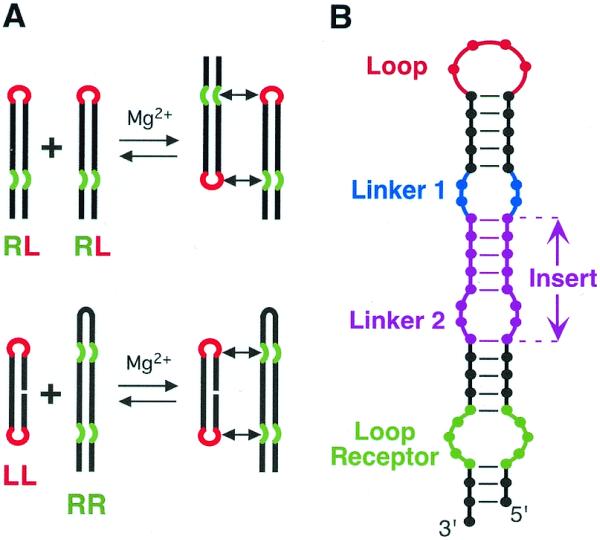
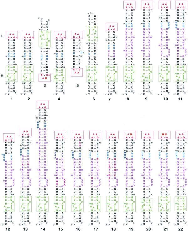
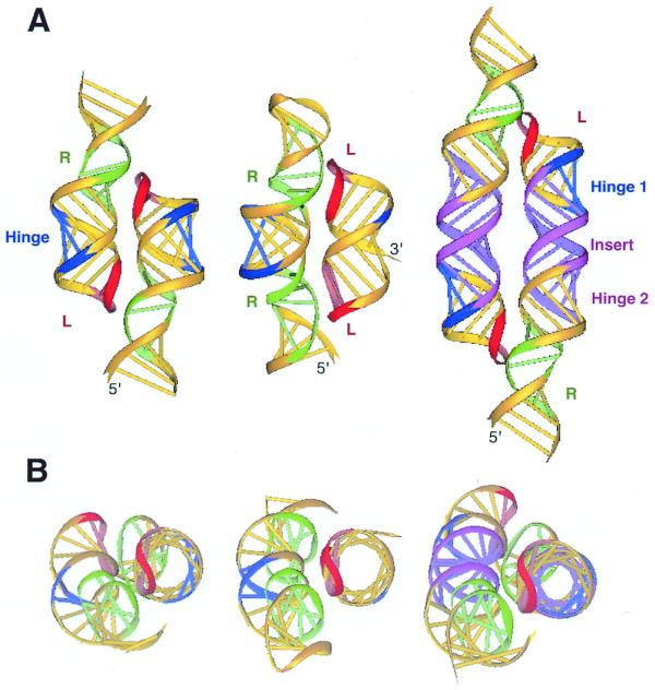
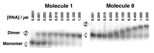
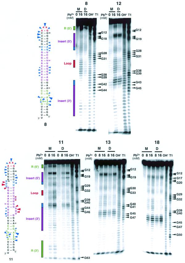
Structural information on complex biological RNA molecules can be exploited to design tectoRNAs or artificial modular RNA units that can self-assemble through tertiary interactions thereby forming nanoscale RNA objects. The selective interactions of hairpin tetraloops with their receptors can be used to mediate tectoRNA assembly. Here we report on the modulation of the specificity and the strength of tectoRNA assembly (in the nanomolar to micromolar range) by variation of the length of the RNA subunits, the nature of their interacting motifs and the degree of flexibility of linker regions incorporated into the molecules. The association is also dependent on the concentration of magnesium. Monitoring of tectoRNA assembly by lead(II) cleavage protection indicates that some degree of structural flexibility is required for optimal binding. With tectoRNAs one can compare the binding affinities of different tertiary motifs and quantify the strength of individual interactions. Furthermore, in analogy to the synthons used in organic chemistry to synthesize more complex organic compounds, tectoRNAs form the basic assembly units for constructing complex RNA structures on the nanometer scale. Thus, tectoRNA provides a means for constructing molecular scaffoldings that organize functional modules in three-dimensional space for a wide range of applications.


Figures

Figure 1
Figure 1
TectoRNAs employing two loop–receptor motifs and their modes of assembly. (A) Two modes of assembly used in this study. (B) Schematic of RNA assembly unit showing elements varied for this study. The tetraloop (L) is shown in red, the tetraloop receptor (R) in green, the linker (or hinge) in blue and the insert [comprising a helix and a second linker (or hinge)] in magenta (refer also to the 2D and 3D models in Figs 2 and 3).
Figure 2
Figure 2
Secondary structures of all molecules reported (see Table 1). All molecules in the second row are derived from molecule 11. The bases that differ from molecule 11 are shown in bold. A red box is drawn around the tetraloop and a green box around the tetraloop receptor. The color code is the same as Figure 1. Molecules 3 and 4 as well as 5 and 6 are drawn to show their mode of interaction. All molecules were synthesized as described in the Materials and Methods.
Figure 3
Figure 3
3D models of representative tectoRNA units. (A) Dimers formed by molecules 1 (left), 5 interacting with 6 (center), and 8 (right). (B) The respective top views are shown. The color code is the same as in Figures 1 and 2. L, tetraloop; R, tetraloop receptor.
Figure 4
Figure 4
Autoradiograms of native gels used to characterize the dimerization of tectoRNAs. Experiments were carried out in the presence of 15 mM Mg(OAc)2, as described in the Materials and Methods. The behavior of molecule 1 (left) shows two distinct bands, one for the monomer and one for the dimer. By contrast, molecule 8 (right) is typical of molecules showing fast exchange kinetics. The mobility of the dimer band of 8 varies continuously with concentration, in the concentration range where significant populations of each form exist in solution, indicating a dynamic equilibrium between monomer and dimer forms. Because of their different size, molecules 1 and 8 have different migration mobility on native polyacrylamide gels.
Figure 5
Figure 5
Lead(II)-induced cleavage patterns for molecules 8, 11, 12, 13 and 18 in their monomeric and dimeric states. Monomer lanes are indicated by M and dimer lanes by D. To maintain the RNA in the monomer state, RNA concentrations were set to 1 nM. To achieve dimerization, the RNA concentration was adjusted to 5 µM for molecules 8 and 12, and 1 µM for 11, 13 and 18, well above the Kd in all cases. Lanes labeled OH and T1 correspond to alkaline treatment and digestion with RNase T1, respectively. Lead(II)-induced cleavage was performed as described in the Materials and Methods. Phosphates that are cleaved in the monomeric RNA but are mostly protected in the dimeric RNA are indicated with blue arrows on the secondary structures for 8 and 11. Sites that are cleaved in both monomeric and dimeric forms are shown with red arrows. The size of the arrows is roughly proportional to the extent of cleavage in the monomer.

Similar articles

Cited by

References

    1. Seeman N.C. (1998) DNA nanotechnology: novel DNA constructions. Annu. Rev. Biophys. Biomol. Struct., 27, 225–248. - PubMed
    1. Seeman N.C. (1999) DNA engineering and its application to nanotechnology. Trends Biotechnol., 17, 437–443. - PubMed
    1. Zhang Y. and Seeman,N.C. (1994) The construction of a DNA octahedron. J. Am. Chem. Soc., 116, 1661–1669.
    1. Chen J.H. and Seeman,N.C. (1991) Synthesis from DNA of a molecule with the connectivity of a cube. Nature, 350, 631–633. - PubMed
    1. Winfree E., Liu,F., Wenzler,L.A. and Seeman,N.C. (1998) Design and self-assembly of two-dimensional DNA crystals. Nature, 394, 539–544. - PubMed

Publication types