. 2001 Jan 15;29(2):488-98.
doi: 10.1093/nar/29.2.488.

Cis-acting requirements in flanking DNA for the programmed elimination of mse2.9: a common mechanism for deletion of internal eliminated sequences from the developing macronucleus of Tetrahymena thermophila

Affiliations

Cis-acting requirements in flanking DNA for the programmed elimination of mse2.9: a common mechanism for deletion of internal eliminated sequences from the developing macronucleus of Tetrahymena thermophila

J S Fillingham et al. Nucleic Acids Res. .

Abstract

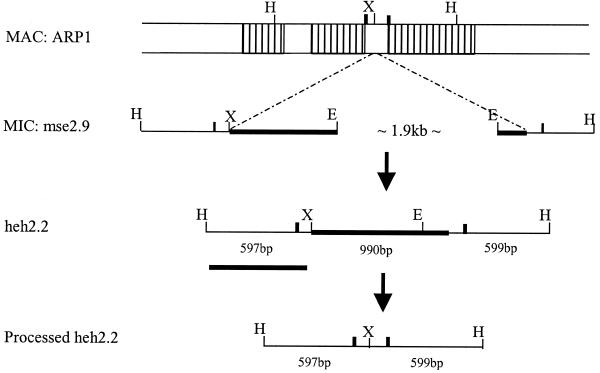
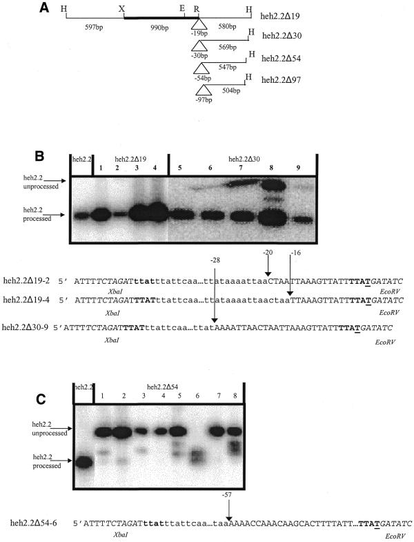
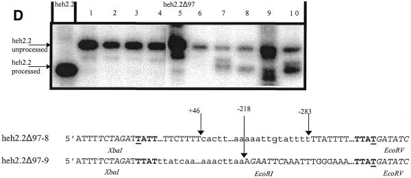
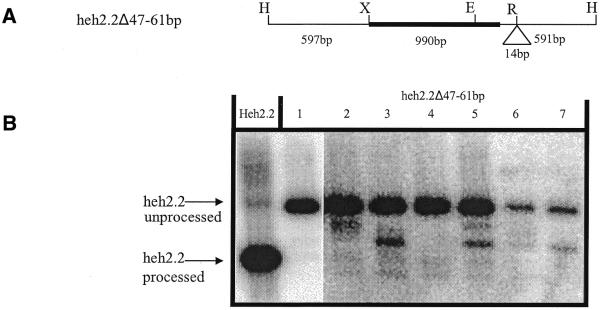
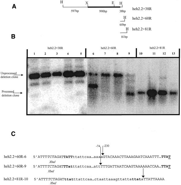
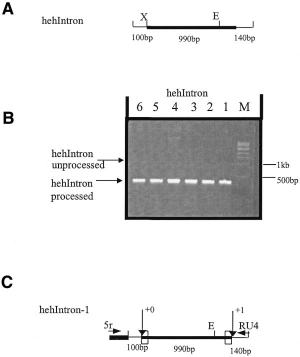
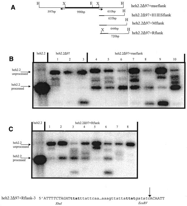
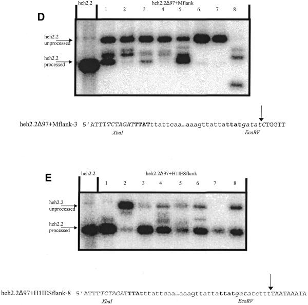
During macronuclear development in the ciliated protozoan Tetrahymena thermophila, extensive DNA deletions occur, eliminating thousands of internal eliminated sequences (IESs). Using an rDNA-based transformation assay we have analyzed the role during DNA deletion of DNA flanking mse2.9, an IES within the second intron of a gene encoding an as yet incompletely characterized protein. We establish that a cis-acting sequence for mse2.9 deletion acts at a distance to specify deletion boundaries. A complex sequence element necessary for efficient and accurate mse2.9 deletion is located in the region 47-81 bp from the right side of mse2.9. The ability of a variety of IES flanking sequences to rescue a processing deficient mse2.9 construct indicates that some cis-acting signal is shared among different IESs. In addition, the short intronic sequence that flanks mse2.9 is able to direct efficient and accurate processing. Despite no obvious sequence similarity between mse2.9 and other IESs, we suggest that a common mechanism is used to delete different families of IESs in Tetrahymena.


Figures

Figure 1
Figure 1
Organization of T.thermophila DNA at the mse2.9 locus and construction of heh2.2. MAC, macronuclear DNA; MIC, micronuclear DNA; ARP1, acidic repetitive protein. The area inside the dotted lines indicates micronuclear-limited DNA. Outside the lines macro- and micronuclear sequence are identical. Hatched and clear boxes denote ARP1 exons and introns, respectively. Construction of heh2.2 has been described previously (12). The solid line beneath unprocessed heh2.2 represents the probe used for Southern analysis. Intron boundaries that flank mse2.9 are indicated by thick vertical lines. H, HindIII; X, XbaI; E, EcoRI.
Figure 2
Figure 2
Analysis of a series of small deletions at the right flank of heh2.2. (A) The thick horizontal line represents the micronuclear-limited sequence of heh2.2. The triangle (Δ) indicates the site of the deletions. (B) Southern blot analysis of total genomic DNA from cells transformed with heh2.2, heh2.2Δ19 (lanes 1–4) and heh2.2Δ30 (lanes 5–9) constructs. The amount of DNA (∼0.5 µg) digested in each experiment in this study ensures that only heh2.2 constructs cloned onto the high-copy rDNA-based vector are seen. DNA was digested with HindIII and probed with the HindIII–XbaI macronuclear-retained fragment of heh2.2 (Fig. 1). Sequence of several transformant deletion junctions is shown below the Southern blot. Macronuclear-destined sequence is in capital letters and deleted sequence is in lowercase. The TTAT direct repeats that flank mse2.9/heh2.2 are in bold text. Arrows emphasize shifted right (and one left) boundaries. The numbering corresponds to the distance from the underlined nucleotide within the respective direct repeat TTAT (left side of heh2.2) and TTAT (right side of heh2.2). Restriction sites are italicized. (C) Southern blot and sequence analysis of heh2.2Δ54 transformants (as in B). (D) Southern blot and sequence analysis of heh2.2Δ97 transformants (as in B). H, HindIII; X, XbaI; E, EcoRI; R, EcoRV.
Figure 2
Figure 2
Analysis of a series of small deletions at the right flank of heh2.2. (A) The thick horizontal line represents the micronuclear-limited sequence of heh2.2. The triangle (Δ) indicates the site of the deletions. (B) Southern blot analysis of total genomic DNA from cells transformed with heh2.2, heh2.2Δ19 (lanes 1–4) and heh2.2Δ30 (lanes 5–9) constructs. The amount of DNA (∼0.5 µg) digested in each experiment in this study ensures that only heh2.2 constructs cloned onto the high-copy rDNA-based vector are seen. DNA was digested with HindIII and probed with the HindIII–XbaI macronuclear-retained fragment of heh2.2 (Fig. 1). Sequence of several transformant deletion junctions is shown below the Southern blot. Macronuclear-destined sequence is in capital letters and deleted sequence is in lowercase. The TTAT direct repeats that flank mse2.9/heh2.2 are in bold text. Arrows emphasize shifted right (and one left) boundaries. The numbering corresponds to the distance from the underlined nucleotide within the respective direct repeat TTAT (left side of heh2.2) and TTAT (right side of heh2.2). Restriction sites are italicized. (C) Southern blot and sequence analysis of heh2.2Δ54 transformants (as in B). (D) Southern blot and sequence analysis of heh2.2Δ97 transformants (as in B). H, HindIII; X, XbaI; E, EcoRI; R, EcoRV.
Figure 3
Figure 3
Deletion of a 14 bp A-rich sequence that is situated in DNA flanking the right side of micronuclear sequence inhibits heh2.2 processing. (A) An EcoRV site replaces the 14 bp at a distance of 47–60 bp from the right boundary of mse2.9. (B) Southern blot analysis of whole-cell DNA purified from heh2.2Δ47–60 bp transformants. DNA was digested and probed as in Figure 2. H, HindIII; X, XbaI; E, EcoRI; R, EcoRV.
Figure 4
Figure 4
Deletion analysis reveals that mse2.9 requires 81 bp of sequence on the right side for efficient and accurate processing. (A) The thick horizontal line represents micronuclear-limited sequence of heh2.2. (B) Southern blot analysis of genomic DNA purified from transformants of heh2.2+38R (lanes 1–5), heh2.2+60R (lanes 6–9) and heh2.2+81R (lanes 10–13) clones. Whole-cell DNA was digested and probed as in Figure 2. Longer exposure for some lanes was performed to increase visualization of DNA fragments in low abundance. (C) Sequence analysis of several deletion clone transformants as in Figure 2.
Figure 5
Figure 5
Intronic flanking sequences are sufficient for processing of heh2.2. (A) The thick horizontal line represents micronuclear-limited sequence of heh2.2. (B) Agarose gel analysis (1.2%) of PCR amplification of whole-cell DNA from hehIntron transformants. Primers 5r and RU4 were used to ensure only transformant junctions were amplified. Correct processing yields a product of ∼400 bp. (C) Arrows designate deletion junctions indicating that processing is accurate. The boxes represent the TTAT direct repeats that border mse2.9. The numbering corresponds to the distance from the underlined nucleotide within the respective direct repeat TTAT (left side of heh2.2) and TTAT (right side of heh2.2). Primers 5r (complementary to rDNA sequence) and RU4 (complementary to mse2.9 flanking sequence) are shown. X, XbaI; E, EcoRI.
Figure 6
Figure 6
Partial rescue of the heh2.2Δ97 processing defect. (A) Insertion of flanking sequence from the left flank of mse2.9, and right flanking sequence from the M and R element, and the H1 IES (thick horizontal rule). The arrows at the left and right flanks of construct heh2.2Δ97+mseflanks emphasize that they are in an inverted repeat configuration. (B) Southern blot analysis of whole-cell DNA from heh2.2Δ97+mseflanks transformants, as well as heh2.2Δ97 transformants (from a separate experiment than Fig. 2C), and a heh2.2 control for fully processed DNA. DNA was digested and probed as in Figure 2. (C) Southern blot analysis of heh2.2Δ97+Rflank (as in B). Sequence from a transformant deletion junction is shown under the Southern blot, as described in Figure 2. (D) Southern blot and sequence analysis of heh2.2Δ97+Mflank (as in C). (E) Southern blot and sequence analysis of heh2.2Δ97+H1IESflank (as in C). H, HindIII; X, XbaI; E, EcoRI; R, EcoRV.
Figure 6
Figure 6
Partial rescue of the heh2.2Δ97 processing defect. (A) Insertion of flanking sequence from the left flank of mse2.9, and right flanking sequence from the M and R element, and the H1 IES (thick horizontal rule). The arrows at the left and right flanks of construct heh2.2Δ97+mseflanks emphasize that they are in an inverted repeat configuration. (B) Southern blot analysis of whole-cell DNA from heh2.2Δ97+mseflanks transformants, as well as heh2.2Δ97 transformants (from a separate experiment than Fig. 2C), and a heh2.2 control for fully processed DNA. DNA was digested and probed as in Figure 2. (C) Southern blot analysis of heh2.2Δ97+Rflank (as in B). Sequence from a transformant deletion junction is shown under the Southern blot, as described in Figure 2. (D) Southern blot and sequence analysis of heh2.2Δ97+Mflank (as in C). (E) Southern blot and sequence analysis of heh2.2Δ97+H1IESflank (as in C). H, HindIII; X, XbaI; E, EcoRI; R, EcoRV.

Similar articles

Cited by

References

    1. Borst P. and Greaves,D.R. (1987) Programmed gene rearrangements altering gene expression. Science, 235, 658–667. - PubMed
    1. Lewis S.M. (1994) The mechanism of V(D)J joining: lessons from molecular, immunological, and comparative analyses. Adv. Immunol., 56, 27–150. - PubMed
    1. Rudenko G., Cross,M. and Borst,P. (1998) Changing the end: antigenic variation orchestrated at the telomeres of African trypanosomes. Trends Microbiol., 3, 113–117. - PubMed
    1. Prescott D.M. (1994) The DNA of ciliated protozoa. Microbiol. Rev., 58, 233–267. - PMC - PubMed
    1. Klobutcher L.A. and Herrick,G. (1997) Developmental genome reorganization in ciliated protozoa: the transposon link. Prog. Nucleic Acids Res. Mol. Biol., 56, 1–62. - PubMed

Publication types

MeSH terms