. 2001 Jan 15;29(2):527-35.
doi: 10.1093/nar/29.2.527.

Highly conserved features of DNA binding between two divergent members of the Myb family of transcription factors

Affiliations

Highly conserved features of DNA binding between two divergent members of the Myb family of transcription factors

B Pinson et al. Nucleic Acids Res. .

Abstract

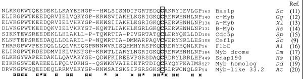
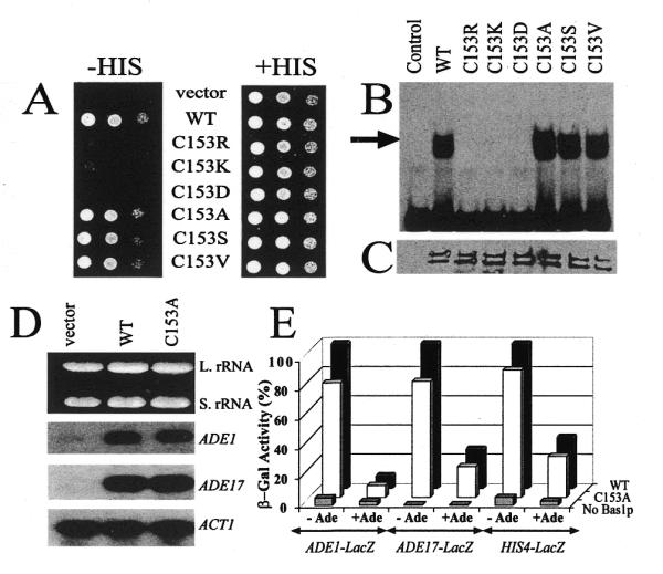
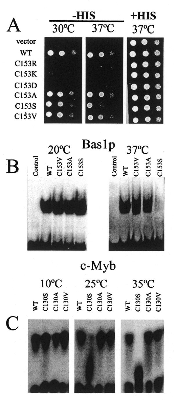
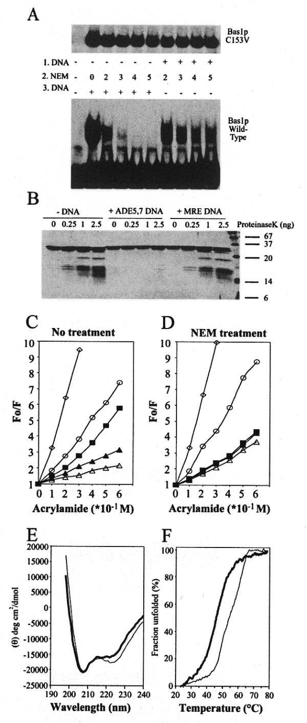
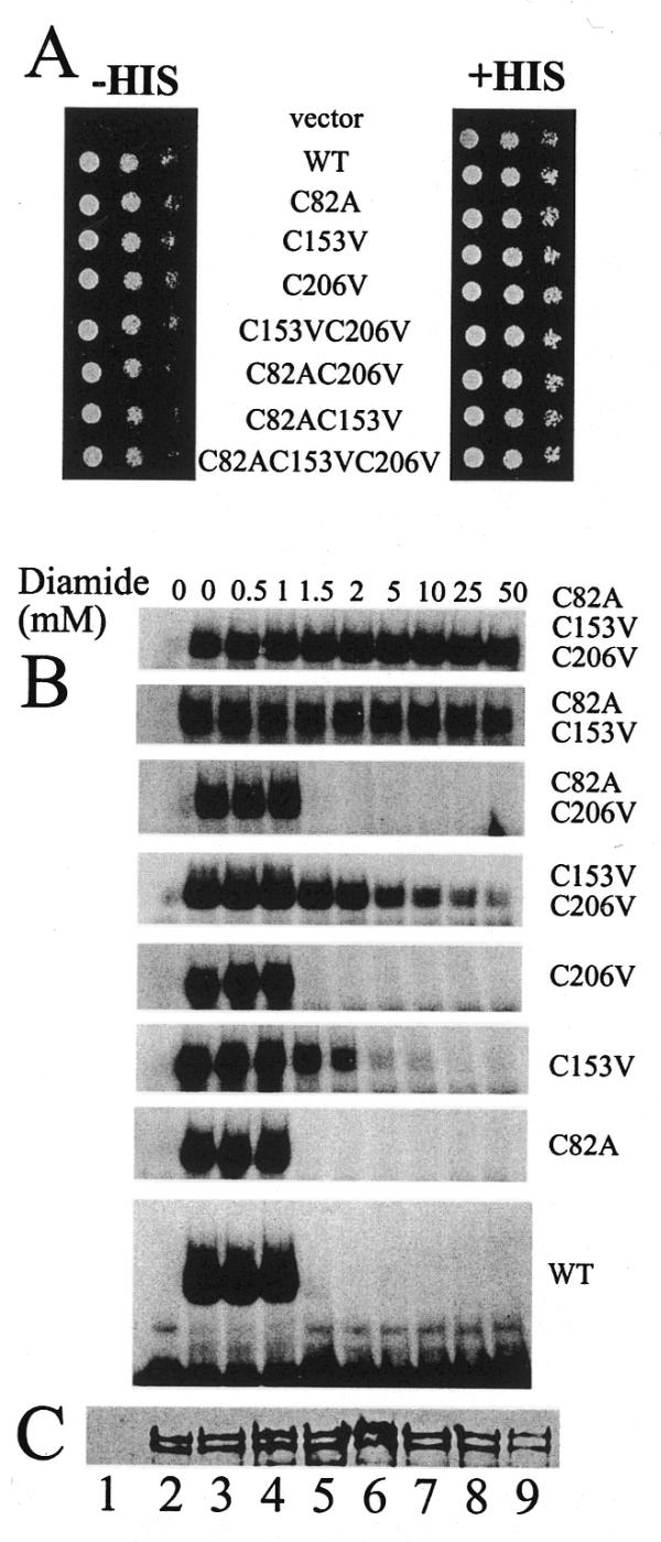
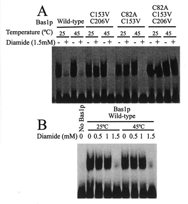
Bas1p, a divergent yeast member of the Myb family of transcription factors, shares with the proteins of this family a highly conserved cysteine residue proposed to play a role in redox regulation. Substitutions of this residue in Bas1p (C153) allowed us to establish that, despite its very high conservation, it is not strictly required for Bas1p function: its substitution with a small hydrophobic residue led to a fully functional protein in vitro and in vivo. C153 was accessible to an alkylating agent in the free protein but was protected by prior exposure to DNA. The reactivity of cysteines in the first and third repeats was much lower than in the second repeat, suggesting a more accessible conformation of repeat 2. Proteolysis protection, fluorescence quenching and circular dichroism experiments further indicated that DNA binding induces structural changes making Bas1p less accessible to modifying agents. Altogether, our results strongly suggest that the second repeat of the DNA-binding domain of Bas1p behaves similarly to its Myb counterpart, i.e. a DNA-induced conformational change in the second repeat leads to formation of a full helix-turn-helix-related motif with the cysteine packed in the hydrophobic core of the repeat.


Figures

Figure 1
Figure 1
Optimal sequence alignment of the second repeat of the DNA-binding domain of Bas1p and repeats in other various Myb family members. Letters on the right refer to the species from which the sequences used for alignment were extracted. Sc, Saccharomyces cerevisiae; Gg, Gallus gallus; Xl, Xenopus laevis; Sp, Schizosaccharomyces pombe; Al, Aspergillus nidulans; Dm, Drosophila melanogaster; Hs, Homo sapiens; Dd, Dictyostelium discoideum; At, Arabidopsis thaliana. The identical and the highly conserved residues in all sequences aligned are marked by the symbols * and ¤, respectively. The conserved cysteine residue (C153 in Bas1p) is boxed. Numbers on the right refer to the position in the protein sequence.
Figure 2
Figure 2
In vivo and in vitro effect of substitutions of the C153 residue of Bas1p. (A) In vivo effect of mutations in the C153 codon of BAS1. Y329 cells were transformed with either the B836 control plasmid (vector), the wild-type BAS1 gene (WT) or the various C153 substitution mutants. Transformants in exponential growth phase were resuspended in water. Three drops of cellular suspension corresponding to serial dilutions of the cells (104, 103 and 102 cells) were spotted on SC medium lacking uracil and supplemented (right) or not (left) with histidine. Cells were grown for 36 h at 30°C. (B) In vitro DNA-binding activity of Bas1p mutants. Purified GST–HA (control lane) or GST–HA–Bas1p wild-type (WT) or C153 mutants (0.5 µg each protein) were incubated for 15 min at room temperature with 100 fmol radiolabelled ADE5,7 promoter probe and were then separated by 4% non-denaturing gel electrophoresis. The gel was dried on paper and radioactivity was revealed by autoradiography. GST–HA–Bas1p/DNA complexes are indicated by the black arrow. (C) Western blot analysis of wild-type Bas1p and C153 mutants. Each purified GST–HA–Bas1p (4 µg each protein) was subjected to 12.5% SDS–PAGE and electroblotting on PVDF membrane. The blot was incubated with anti-HA antibodies (0.5 µg/ml) followed by horseradish peroxidase-conjugated IgG (1:2500) as secondary antibodies and, finally, luminescent substrate before exposure to film. The GST–HA protein (25 kDa) in the control lane is much smaller than the GST–HA–Bas1p fusion (115 kDa) and therefore does not appear in the figure. (D) Effect of C153A substitution in Bas1p on ADE1 and ADE17 transcription. Strain Y329 was transformed with plasmids B836 (vector), P79 (WT, wild-type BAS1) or P926 (C153A mutant). Transformants were grown in SD-CASA to an OD600 of 0.5. Total RNA was extracted, separated by electrophoresis, transferred and hybridised with radiolabelled probes. Equal loading of each lane was monitored by ethidium bromide staining of the gel showing the large (L. rRNA) and small (S. rRNA) rRNAs and an ACT1-specific probe hybridisation. Each hybridisation was done independently and assembled for the figure. (E) In vivo effect of C153A substitution in Bas1p on expression of lacZ reporter fusions. Y329 cells were co-transformed with a plasmid containing the lacZ fusion and with a centromeric plasmid harboring the BAS1 wild-type or C153A mutant gene. –Ade and +Ade correspond to cells grown in the absence or presence of adenine, respectively. The control lane (No Bas1p) corresponds to a transformation with the B836 plasmid not containing the BAS1 gene. For each lacZ fusion β-Gal units are expressed as per cent expression measured in the absence of adenine with the wild-type (WT) BAS1 gene.
Figure 3
Figure 3
Temperature sensitivity of Cys→Ser replacement in the second repeat of the c-Myb and Bas1p DNA-binding domains. (A) In vivo effect of mutations in the C153 codon of BAS1 at 37°C. Y329 cells were transformed with either the B836 control plasmid (vector), the wild-type BAS1 gene or the various C153 substitution mutants and treated as in Figure 2A, but cells were grown at 37°C. The panel –His 30°C is a copy of the panel from Figure 2A added to facilitate a comparison of growth between the two temperatures tested. (B) In vitro DNA-binding activity of Bas1p mutants at different temperatures. Two microlitres (0.5 µg protein) of purified GST–HA (control lane) or GST–HA–Bas1p wild-type or C153 mutants were treated and analysed by EMSA as in Figure 2B at 20 and 37°C. (C) In vitro DNA binding activity of c-Myb mutants at different temperatures. Purified Myb R2R3 proteins (10 fmol both wild-type and mutants as indicated) were incubated with 10 fmol MRE probe at the indicated temperature for 10 min. The complex formed was separated by native electrophoresis using a thermostatic equipment set-up as described in Materials and Methods.
Figure 4
Figure 4
A DNA-induced conformational change in Bas1p. (A) Sensitivity of wild-type Bas1p and the C153A mutant to alkylation by NEM. (Lower) Two microlitres (0.5 µg protein) of purified wild-type GST–HA–Bas1p were incubated for 15 min at room temperature with increasing concentrations of NEM either before (1.DNA, left of pannel) or after (3.DNA, right) incubation with 100 fmol radiolabelled ADE5,7 promoter probe. Samples were then analysed by EMSA as in Figure 2B. (Upper) As above but carried out with the GST–HA–Bas1p C153V mutant. (B) Partial digestion of Bas1p, either free or complexed to DNA, with proteinase K. Bas1p[1–272] (0.25 µg) free (–DNA), in complex with ADE5,7 DNA (+ADE5,7 DNA) or in complex with unrelated DNA (+MRE DNA) was treated with increasing amounts of proteinase K. Proteins were then separated by 17.5% SDS–PAGE and revealed by silver staining. (C and D) Fluorescence quenching by acrylamide of free and DNA-bound Bas1p. The fluorescence emission of 2 µM Bas1p was quenched by successive addition of acrylamide to Bas1p treated (D) or not (C) with 5 mM NEM. In each condition fluorescence was measured at the wavelength corresponding to the emission maximum for Bas1p and the results are presented as the ratio between the unquenched fluorescence (Fo) and the fluorescence measured after acrylamide addition (F). Results correspond to the average of between six and nine independent measurements. Inter-assay variation was found to be <15 %. Closed square, Free Bas1p; open triangle, Bas1p bound to ADE5,7 specific DNA; closed triangle, Bas1p bound to unrelated DNA; open circle, Bas1p denatured for 60 min in 6 M guanidinium chloride; open diamond, 10 µM free tryptophan solution used as a reference. (E) CD spectrum of Bas1p[1–272] either alone (thick line) or in complex with specific DNA (ADE5,7 DNA, thin line). The protein concentration was 0.15 mg/ml in phosphate buffer and equimolar concentrations of DNA were added. The α-helical contents of Bas1p[1–272] alone and in complex with specific DNA were calculated as described in Materials and Methods and found to be 40 and 45%, respectively. (F) Thermal denaturation curves of Bas1p[1–272] either alone (thick line) or in complex with specific DNA (thin line). The apparent fraction of folded protein, obtained by monitoring the CD value at 222 nm, is shown as a function of temperature.
Figure 5
Figure 5
Comparison of the sensitivity to oxidation of the three cysteine residues in Bas1p. (A) In vivo effect of mutations of the three Cys residues in Bas1p. Y329 cells were transformed with either the B836 control plasmid (vector), the wild-type BAS1 gene or the various Cys substitution mutants and treated as in Figure 2A. (B) In vitro sensitivity to diamide of the different cysteine mutants of Bas1p. Purified wild-type GST–HA–Bas1p or cysteine mutants were incubated for 15 min at room temperature with increasing concentrations of diamide. Samples were then incubated with radiolabelled ADE5,7 promoter probe and analysed by EMSA as in Figure 2B. (C) Western blot analysis of purified wild-type and mutant GST–HA–Bas1p. Purified wild-type GST–HA–Bas1p and mutants fusions (10 µl) were separated by 12.5% SDS–PAGE and electrotransferred to PVDF membrane. GST–HA–Bas1p proteins were revealed by western blot analysis as in Figure 2C. Lane 1, GST–HA control; lanes 2–9, wild-type Bas1p and the C82A C153V C206V, C153V C206V, C82A C206V, C82A C153V, C206V, C153V and C82A Bas1p mutants, respectively.
Figure 6
Figure 6
Effect of a partial denaturation on accessibility of the cysteine residues in Bas1p. (A) Effect of increased temperature on oxidation of C82 and C206 in Bas1p. Purified wild-type and mutant GST–HA–Bas1p proteins were incubated with or without 1.5 mM diamide at the indicated temperature (either 25 or 45°C) and immediately allowed to slowly return to room temperature. Samples were then incubated for 15 min with 100 fmol radiolabelled ADE5,7 promoter probe and analysed by EMSA as in Figure 2B. (B) Increased temperature has no major effect on diamide reactivity. Purified wild-type GST–HA–Bas1p was incubated with increasing concentrations of diamide at either 25 or 45°C and immediately allowed to slowly return to room temperature. Samples were then treated as in (A).

Similar articles

Cited by

References

    1. Ness S.A. (1996) The Myb oncoprotein: regulating a regulator. Biochim. Biophys. Acta, 1288, 123–139. - PubMed
    1. Oh I.H. and Reddy,E.P. (1999) The myb gene family in cell growth, differentiation and apoptosis. Oncogene, 18, 3017–3033. - PubMed
    1. Frampton J., Gibson,T.J., Ness,S.A., Doderlein,G. and Graf,T. (1991) Proposed structure for the DNA-binding domain of the Myb oncoprotein based on model building and mutational analysis. Protein Eng., 4, 891–901. - PubMed
    1. Gabrielsen O.S., Sentenac,A. and Fromageot,P. (1991) Specific DNA binding by c-Myb: evidence for a double helix-turn-helix-related motif. Science, 253, 1140–1143. - PubMed
    1. Ogata K., Morikawa,S., Nakamura,H., Sekikawa,A., Inoue,T., Kanai,H., Sarai,A., Ishii,S. and Nishimura,Y. (1994) Solution structure of a specific DNA complex of the Myb DNA-binding domain with cooperative recognition helices. Cell, 79, 639–648. - PubMed

Publication types

MeSH terms