Review
. 2001 Jan;233(1):9-17.
doi: 10.1097/00000658-200101000-00003.

Muscle cachexia: current concepts of intracellular mechanisms and molecular regulation

Affiliations
Review

Muscle cachexia: current concepts of intracellular mechanisms and molecular regulation

P O Hasselgren et al. Ann Surg. 2001 Jan.

Abstract

Objective: To review present knowledge of intracellular mechanisms and molecular regulation of muscle cachexia.

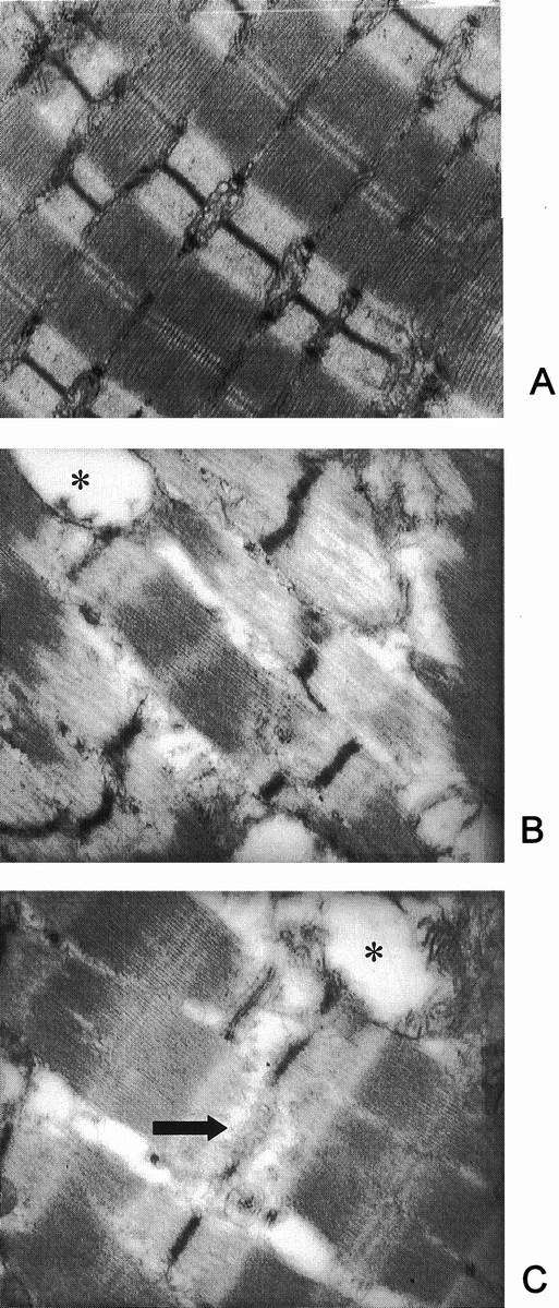
Summary background data: Muscle cachexia, mainly reflecting degradation of myofibrillar proteins, is an important clinical feature in patients with severe injury, sepsis, and cancer. The catabolic response in skeletal muscle may result in muscle wasting and weakness, delaying or preventing ambulation and rehabilitation in these patients and increasing the risk for pulmonary complications.

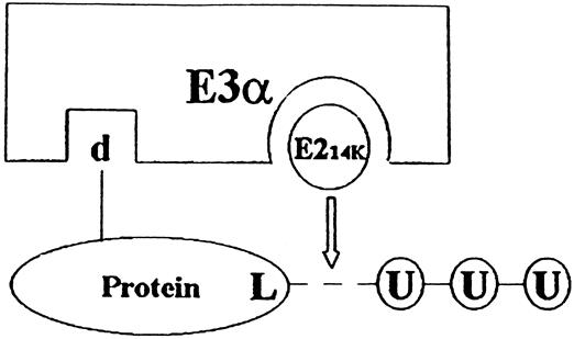
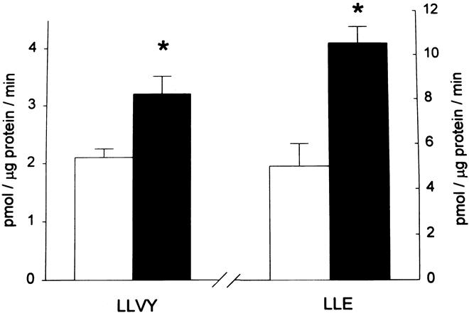
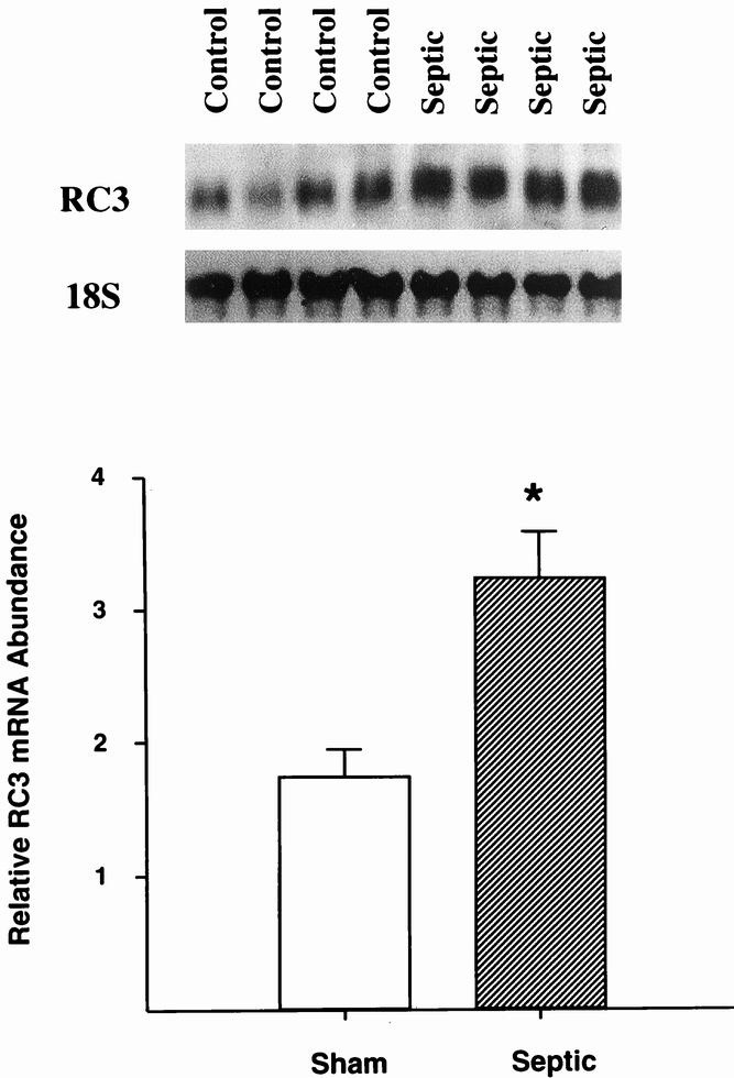
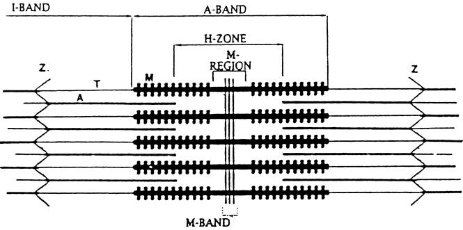
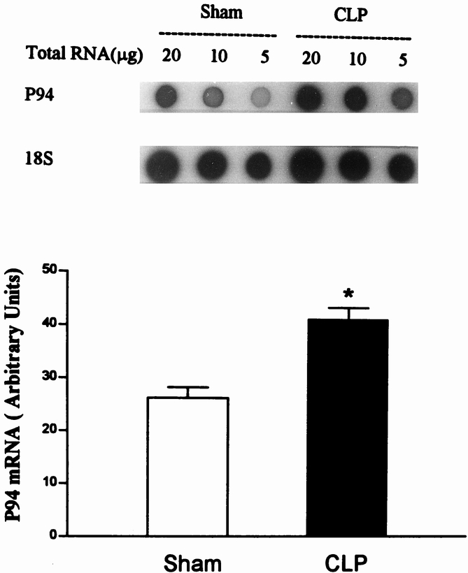
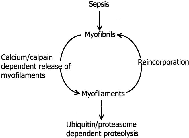
Results: Muscle cachexia, induced by severe injury, sepsis, and cancer, is associated with increased gene expression and activity of the calcium/calpain- and ubiquitin/proteasome-proteolytic pathways. Calcium/calpain-regulated release of myofilaments from the sarcomere is an early, and perhaps rate-limiting, component of the catabolic response in muscle. Released myofilaments are ubiquitinated in the N-end rule pathway, regulated by the ubiquitin-conjugating enzyme E2(14k) and the ubiquitin ligase E3 alpha, and degraded by the 26S proteasome.

Conclusions: An understanding of the mechanisms regulating muscle protein breakdown is important for the development of therapeutic strategies aimed at treating or preventing muscle cachexia in patients with severe injury, sepsis, cancer, and perhaps other catabolic conditions as well.


Figures

None
Figure 1. Simplified scheme of the ubiquitin-proteasome proteolytic pathway. In this pathway, ubiquitinated proteins are recognized and degraded by the 26S proteasome. The steps involved in the breakdown of proteins by this mechanism include 1) activation of ubiquitin by the ubiquitin activating enzyme E1; 2) transfer of ubqiutin to the ubiquitin conjugating enzyme E2; 3) interaction between the substrate protein and the ubiquitin ligase E3; 4) interaction between E2 and E3 resulting in 5) multiubiquitination of the substrate protein; 6) degradation of the ubiquitinated protein by the 26S proteasome; and 7) deubiquitination resulting in the release and reuse of ubiquitin in the pathway. Energy is required for at least two steps in the pathway: activation of ubiquitin by E1 (step 1) and the proteolytic activity in the 26S proteasome (step 6). (From Hershko A. Lessons from the discovery of the ubiquitin system. Trends Biochem Sci 1996; 21:445–449, with permission.)
None
Figure 2. Simplified scheme of the interaction between protein substrate, E214k, and E3α in the N-end rule pathway. Proteins with a destabilizing N-end (d) are ligated to ubiquitin ligase E3α. Specific interaction between E3α and the ubiquitin conjugating enzyme E214k (ubiquitin carrier protein) allows for the transfer and conjugation of ubiquitin to a lysine residue (L) of the substrate protein. This is followed by the conjugation of additional ubiquitin molecules, resulting in a multiubiquitinated protein that is subsequently recognized and degraded by the 26S proteasome.
None
Figure 3. Sepsis results in increased activity of the 20S proteasome in skeletal muscle. 20S proteasomes were isolated from muscles of sham-operated (open bars) or septic rats (filled bars), and the chymotrypsinlike and peptidyl-glutamyl peptidase activities were determined by using the fluorogenic substrates LLVY and LLE, respectively. * P < .05. (Hobler SC, Williams AB, Fischer D, et al. Activity and expression of the 20S proteasome are increased in skeletal muscle during sepsis. Am J Physiol 1999; 277:R434–R440, with permission.)
None
Figure 4. Sepsis results in increased gene expression of the 20S α subunit RC3 in skeletal muscle. Northern blots from four sham-operated control rats and four septic rats (sepsis induced by cecal ligation and puncture) are shown in the upper panel, and quantitation of the blots is shown in the lower panel. * P < .05. (Data from Hobler SC, Williams AB, Fischer D, et al. Activity and expression of the 20S proteasome are increased in skeletal muscle during sepsis. Am J Physiol 1999; 277:R434–R440, with permission.)
None
Figure 5. Total (upper panel) and myofibrillar (lower panel) protein breakdown in incubated muscles from septic rats is inhibited by the specific proteasome blocker lactacystin (100 μM). * P < .05. (Hobler SC, Tiao G, Fischer JE, et al. The sepsis-induced increase in muscle proteolysis is blocked by specific proteasome inhibitors. Am J Physiol 1998; 274:R30–R37, with permission.)
None
Figure 6. Simplified scheme of the muscle sarcomere. The sarcomere extends from one Z-disk to the next. Myosin (M) is anchored to the Z-disk by titin (T) and actin (A) by nebulin. Z, Z-band.
None
Figure 7. Electron microscopy of muscles from sham-operated (A) and septic (B, C) rats. Note disintegration or complete loss (arrow in C) of Z-bands in muscles from septic rats. Swollen mitochondria (*) were frequently seen in septic muscles. (From Williams AB, DeCourten-Myers GM, Fischer JE, et al. Sepsis stimulates release of myofilaments in skeletal muscle by a calcium-dependent mechanism. FASEB J 1999; 13:1435–1443, with permission.)
None
Figure 8. Sepsis results in increased gene expression of the muscle-specific calpain p94. Dot blots of RNA extracted from control and septic muscles are shown in the upper panel and quantitation of the blots is shown in the lower panel. (From Williams AB, DeCourten-Myers GM, Fischer JE, et al. Sepsis stimulates release of myofilaments in skeletal muscle by a calcium-dependent mechanism. FASEB J 1999; 13:1435–1443, with permission.)
None
Figure 9. Model of sepsis-induced muscle cachexia. In this model, sepsis results in calcium/calpain-dependent release of myofilaments from the sarcomere. The myofilaments are ubiquitinated in the N-end rule pathway and degraded by the 26S proteasome or reincorporated into the myofibrils. This model offers two levels of possible therapeutic intervention: inhibition of the calcium/calpain-dependent release of myofilaments (e.g., with dantrolene) or inhibition of the ubiquitin-proteasome pathway (e.g.,with proteasome inhibitor).

References

    1. Rosenblatt S, Clowes GHA, George BC,et al. Exchange of amino acids by muscle and liver in sepsis. Comparative studies in vivo and in vitro. Arch Surg 1983; 118: 167–175. - PubMed
    1. Windmueller HG, Spaeth AE. Respiratory fuels and nitrogen metabolism in vivo in small intestine of fed rats. J Biol Chem 1980; 255: 107–112. - PubMed
    1. Newsholme EA, Parry-Billings M. Properties of glutamine release from muscle and its importance for the immune system. J Parenteral Enteral Nutr 1990; 14: 63S–67S. - PubMed
    1. Reid WD, MacGowan NA. Respiratory muscle injury in animal models and humans. Mol Cell Biochem 1998; 179: 63–80. - PubMed
    1. Andreyev HJN, Norman AR, Oates J, Cunningham D. Why do patients with weight loss have a worse outcome when undergoing chemotherapy for gastrointestinal malignancies? Eur J Cancer 1998; 34: 503–509. - PubMed

Publication types