. 2001 Jan;158(1):63-73.
doi: 10.1016/s0002-9440(10)63945-4.

Abeta-induced inflammatory processes in microglia cells of APP23 transgenic mice

Affiliations

Abeta-induced inflammatory processes in microglia cells of APP23 transgenic mice

K D Bornemann et al. Am J Pathol. 2001 Jan.

Abstract

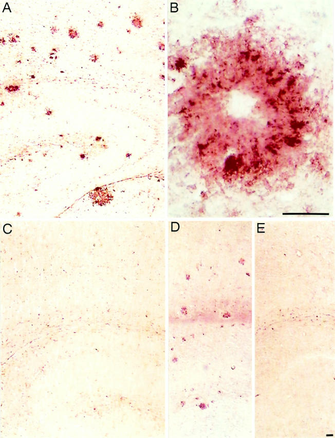
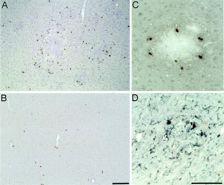
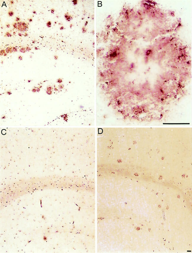
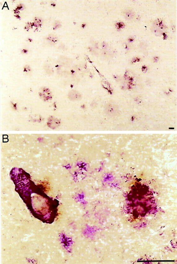
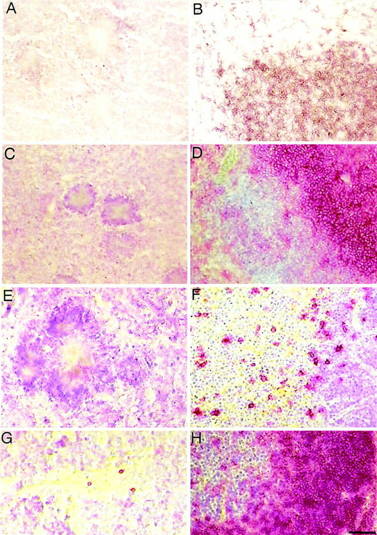
A microglial response is part of the inflammatory processes in Alzheimer's disease (AD). We have used APP23 transgenic mice overexpressing human amyloid precursor protein with the Swedish mutation to characterize this microglia response to amyloid deposits in aged mice. Analyses with MAC-1 and F4/80 antibodies as well as in vivo labeling with bromodeoxyuridine demonstrate that microglia in the plaque vicinity are in an activated state and that proliferation contributes to their accumulation at the plaque periphery. The amyloid-induced microglia activation may be mediated by scavenger receptor A, which is generally elevated, whereas the increased immunostaining of the receptor for advanced glycation end products is more restricted. Although components of the phagocytic machinery such as macrosialin and Fc receptors are increased in activated microglia, efficient clearance of amyloid is missing seemingly because of the lack of amyloid-bound autoantibodies. Similarly, although up-regulation of major histocompatibility complex class II (IA) points toward an intact antigen-presenting function of microglia, lack of T and B lymphocytes does not indicate a cell-mediated immune response in the brains of APP23 mice. The similar characteristics of microglia in the APP23 mice and in AD render the mouse model suitable to study the role of inflammatory processes during AD pathogenesis.


Figures

Figure 1.
Figure 1.
Amyloid plaque-associated microglia are activated. A: MAC-1-immunostained microglia cells associated with compact amyloid deposits in the neocortex and hippocampus of a 23-month-old APP23 transgenic mouse. Deposits of various sizes are visualized. B: Higher magnification of a single amyloid plaque showing several clusters of MAC-1-positive microglial cell bodies that frame the amyloid. Microglial processes cover nearly the entire amyloid, thereby revealing the morphology of this dense-core plaque. The center of the plaque (core) is not always stained. C: No MAC-1 immunostaining is obtained in equivalent brain regions of an age-matched control mouse under the labeling conditions used. D: F4/80 staining of sections containing neocortex and hippocampus of a 23-month-old APP23 mouse. The staining shows microglia cells closely associated with compact amyloid plaques. E: Lack of F4/80 reactivity in a corresponding brain section from a control mouse. Scale bar, 50 μm.
Figure 2.
Figure 2.
Activated microglia near amyloid plaques proliferate. A: BrdU-positive cells in the neocortex of a 12.5-month-old APP23 mouse are predominantly associated with amyloid plaques. B: BrdU labeling of a wild-type mouse of the same age shows only a few positive cells in the neocortex. C: Higher magnification shows BrdU labeled cells around an amyloid plaque. D: Double-labeling with BrdU (black nuclear staining) and the microglia marker MAC-1 (blue staining of plasma membrane processes) identifies the amyloid-associated BrdU-labeled cells as microglia. Scale bar, 50 μm.
Figure 3.
Figure 3.
Scavenger receptor A may contribute to microglia activation. A: Immunostaining of a single neocortical amyloid plaque with the 2F8 antibody directed to SRA shows strongest reactivity at the outer edge of the deposit, where microglial processes cover the amyloid. The weaker staining throughout the plaque may be related to a lower density of microglial processes. The contours of cell bodies surrounding the plaques are also visualized. B: Microglial 2F8 reactivity at a blood vessel containing vascular amyloid. C: RAGE immunoreactivity of neocortical plaque-associated cells, revealing a strong, but very limited staining (the contours of the plaque are not well visible at this magnification). RAGE reactivity was only detected at a minority of the amyloid plaques. D: RAGE staining at the vascular endothelium shows the contours of the vessel. Scale bar, 50 μm.
Figure 4.
Figure 4.
Activated microglia are capable to phagocytose. A: FA-11 (macrosialin) immunostaining of a section through the neocortex and hippocampus of a 23-month-old APP23 transgenic mouse shows strong macrosialin-positive amyloid plaque-associated microglia cells. B: A higher magnification of a single amyloid plaque visualizes macrosialin-positive microglia cell bodies that are surrounding the amyloid. Weaker labeling is found at the amyloid, thereby revealing the contours of the plaque. C: Microglia in the neocortex and hippocampus of a control mouse show a basal macrosialin staining in agreement with published data. D: 2.4G2 staining of a section through the neocortex and hippocampus demonstrates an up-regulation of Fc receptors FcγRII and FcγRIII on activated microglia cells at amyloid plaques. Scale bar, 50 μm.
Figure 5.
Figure 5.
A subpopulation of activated microglia is MHC class II-positive. A: IA (MHC class II protein)-positive microglia are closely associated with amyloid plaques as well as capillary endothelium, as shown in a neocortex section of a 23-month-old APP23 transgenic mouse. B: Double-labeling of amyloid (purple reaction product) and IA (brown reaction product) reveals MHC class II protein reactivity within and around the compact amyloid deposits. Although MHC class II staining is also visible at a blood vessel and an amyloid plaque, it was not detected at diffuse, nonfibrillary amyloid as present in the center of the picture. Scale bar, 50 μm.
Figure 6.
Figure 6.
No detectable lymphocyte recruitment during inflammatory processes in the brains of APP23 mice. A: The CD3ε staining of a neocortex section with three compact amyloid plaques does not provide any evidence of T lymphocytes in aged APP23 mice. B: As a positive control the specific reactivity of CD3ε antibodies with T cells is shown in a staining of the splenic white pulp of a 6-week-old control mouse. C: CD4 staining did not identify T helper cells in the neocortex of APP23 transgenic animals. D: The reactivity of the CD4 antibodies was shown by labeling CD4-positive T helper cells (in red color) that are located at high density in a splenic white pulp section. Surrounding cells, mostly B cells, were counterstained with Mayer’s hemalaun (blue color). E: CD45R (B220) staining of a neocortex section showing a single compact amyloid plaque does not provide evidence for B lymphocytes. F: A control section stained with CD45R reveals B cell staining (red reaction product), located primarily in the medulla of the thymus of a 6-week-old control mouse. G: CD45R staining of a mouse brain section near a mechanical frontal cortex lesion that is infiltrated by three individual B lymphocytes demonstrates single cell detection. H: Control section showing part of a murine splenic white pulp packed with CD45R-positive B lymphocytes. Scale bar, 50 μm.

References

    1. Stewart WF, Kawas C, Corrada M, Meter EJ: Risk of Alzheimer’s disease and duration of NSAID use. Neurology 1997, 48:626-632 - PubMed
    1. McGeer PL, Schulzer M, McGeer EG: Arthritis and anti-inflammatory agents as possible protective factors for Alzheimer’s disease: a review of 17 epidemiologic studies. Neurology 1996, 47:425-432 - PubMed
    1. Mackenzie IR, Hao C, Munoz DG: Role of microglia in senile plaque formation. Neurobiol Aging 1995, 16:797-804 - PubMed
    1. McGeer PL, McGeer EG: The inflammatory response system of brain: implications for therapy of Alzheimer and other neurodegenerative diseases. Brain Res Rev 1995, 21:195-218 - PubMed
    1. Eikelenboom P, Zhan SS, Van Gool WA, Allsop D: Inflammatory mechanisms in Alzheimer’s disease. Trends Pharmacol Sci 1994, 15:447-450 - PubMed

Publication types

MeSH terms