. 2001 Jan;158(1):147-52.
doi: 10.1016/S0002-9440(10)63952-1.

Leukocyte-mediated endothelial cell injury and death in the diabetic retina

Affiliations

Leukocyte-mediated endothelial cell injury and death in the diabetic retina

A M Joussen et al. Am J Pathol. 2001 Jan.

Abstract

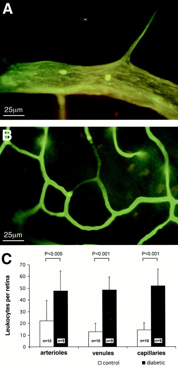
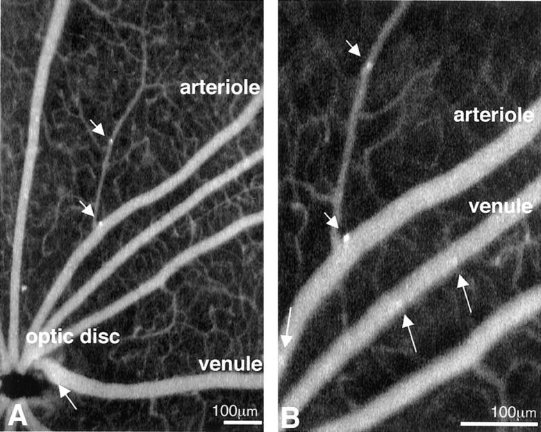
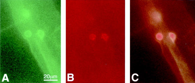
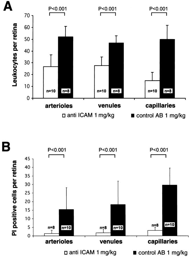
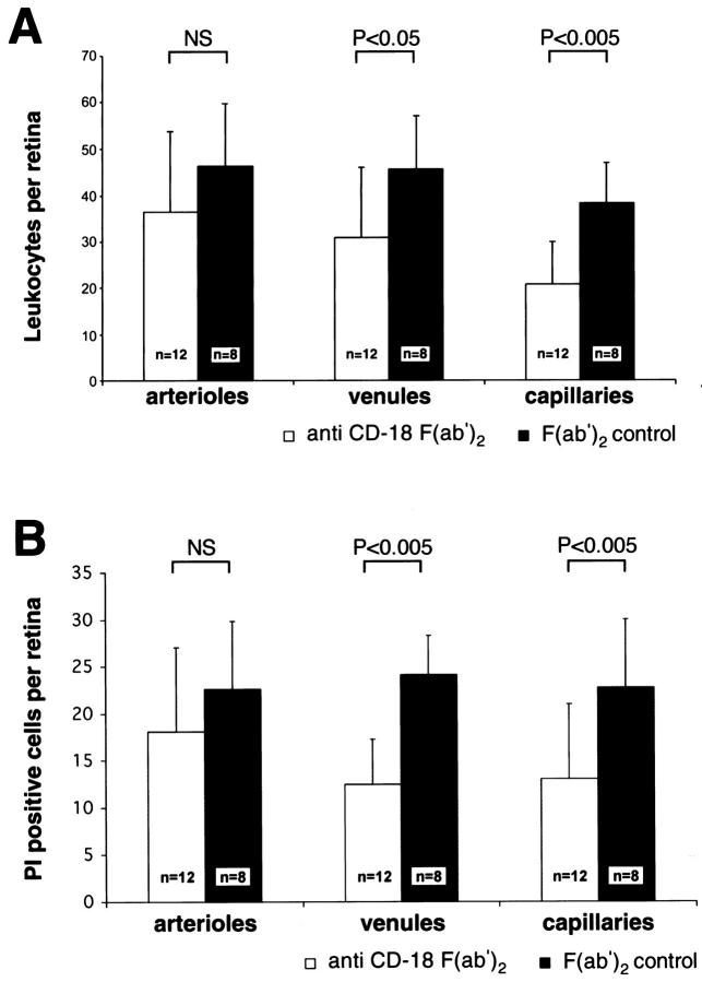
Endothelial cell death is a hallmark of diabetic retinopathy. Its occurrence is required for the formation of acellular (devitalized) capillaries, lesions that produce irreversible retinal ischemia through their inability to support blood flow. The mechanisms underlying diabetic retinal endothelial cell injury and death remain largely unknown. The current study demonstrates that adherent leukocytes are temporally and spatially associated with retinal endothelial cell injury and death within 1 week of streptozotocin-induced experimental diabetes in rats. Moreover, the antibody-based neutralization of intercellular adhesion molecule-1 and CD18 is shown to prevent both leukocyte adhesion and retinal endothelial cell injury and death. These data highlight the central and causal role of adherent leukocytes in the pathogenesis of diabetic retinopathy. They also underscore the potential utility of anti-intercellular adhesion molecule1- and anti-CD18-based therapies in the treatment of diabetic retinopathy, a newly recognized inflammatory disease.


Figures

Figure 1.
Figure 1.
Lectin staining highlights adherent leukocytes within the retinal vasculature. A: In this example, an adherent leukocyte is visible in the arteriole of a diabetic rat. B: Impedance of flow is occasionally seen downstream from adherent leukocytes in capillaries. C: The collated graphical data show a marked increase in the number of adherent leukocytes in the diabetic retinae.
Figure 2.
Figure 2.
In vivo leukocyte fluorography confirms the adhesion of leukocytes to retinal arterioles. A: A CFDASE fundus image of a diabetic rat (40° field setting) shows that leukocytes adhere not only to venules (arrow), but also to arterioles (arrowheads). B: The same fundus image in the 20° field setting, taken 30 seconds later, confirmed the static nature of the leukocyte. While many leukocytes flow through the retinal vessels (arrows), two leukocytes adhere to the walls of retinal arterioles (arrowheads). The direction of the flow of blood was used to distinguish arterioles from venules.
Figure 3.
Figure 3.
Anti-CD18 surface staining of a static leukocyte within a vessel. Green fluorescence from the FITC-linked Concanavalin A (A) and red fluorescence from the anti-CD18 antibody (B) identify the leukocytes as being CD18-positive when the images are superimposed (C).
Figure 4.
Figure 4.
Endothelial cell injury and death identified with propidium iodide (PI). A: PI-positive cells show sharply demarcated borders in a venule located in the mid-periphery of the diabetic retina. B: Capillaries, like larger vessels, exhibit clusters of PI-positive cells. Original magnification, ×400. C: The collated data show markedly increased numbers of PI-positive endothelial cells in the diabetic retinae.
Figure 5.
Figure 5.
ICAM-1 blockade prevents leukocyte adhesion and endothelial cell injury in the diabetic retina. A: After treatment with the anti-ICAM-1 antibody, diabetic retinal leukocyte adhesion was markedly decreased in all retinal vessels. B: Analysis with PI revealed a concomitant decrease in retinal endothelial cell injury and death.
Figure 6.
Figure 6.
CD18 blockade prevents leukocyte adhesion and endothelial cell injury and death in diabetic retinal venules and capillaries. A: After treatment with anti-CD-18 F(ab′)2, diabetic retinal leukocyte adhesion was markedly decreased in the retinal venules and capillaries. B: Analysis with PI revealed a concomitant decrease in retinal endothelial cell injury and death; however, no change in arteriolar endothelial cell injury and death was observed.

References

    1. Adamis AP, Shima DT, Tolentino M, Gragoudas ES, Ferrara N, Folkman J, D’Amore PA, Miller JW: Inhibition of VEGF prevents retinal ischemia-associated iris neovascularization in a primate. Arch Ophthalmol 1996, 114:66-71 - PubMed
    1. Aiello LP, Pierce EA, Foley ED, Takagi H, Chen H, Riddle L, Ferrara N, King G, Smith LEH: Suppression of retinal neovascularization in vivo by inhibition of vascular endothelial growth factor (VEGF) using soluble VEGF-receptor chimeric proteins. Proc Natl Acad Sci USA 1995, 92:10457-10461 - PMC - PubMed
    1. Mizutani M, Kern TS, Lorenzi M: Accelerated death of retinal microvascular cells in human and experimental diabetic retinopathy. J Clin Invest 1996, 97:2883-2890 - PMC - PubMed
    1. Schröder S, Palinski W, Schmid-Schönbein GW: Activated monocytes and granulocytes, capillary nonperfusion, and neovascularization in diabetic retinopathy. Am J Pathol 1991, 139:81-100 - PMC - PubMed
    1. Hammes HP, Martin S, Federlin K, Geisen K, Brownlee M: Aminoguanidine treatment inhibits the development of experimental diabetic retinopathy. Proc Natl Acad Sci USA 1991, 88:11555-11558 - PMC - PubMed

Publication types

MeSH terms

Substances