

Endothelial nitric oxide synthase gene-deficient mice demonstrate marked retardation in postnatal bone formation, reduced bone volume, and defects in osteoblast maturation and activity
- PMID: 11141498
- PMCID: PMC1850250
- DOI: 10.1016/S0002-9440(10)63963-6
Endothelial nitric oxide synthase gene-deficient mice demonstrate marked retardation in postnatal bone formation, reduced bone volume, and defects in osteoblast maturation and activity
Abstract
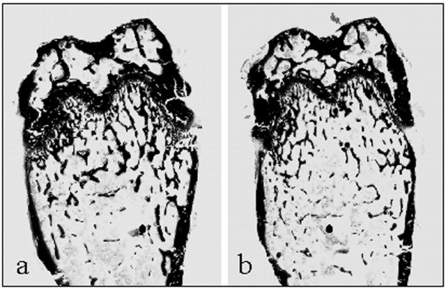
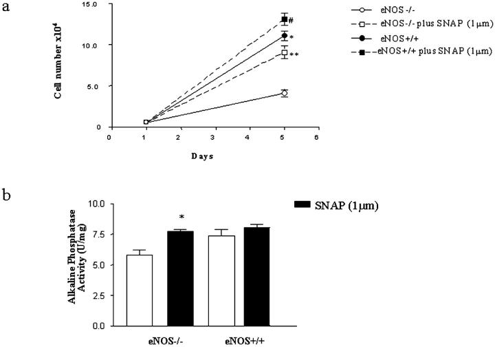
Nitric oxide (NO) has been implicated in the local regulation of bone metabolism. However, the contribution made by specific NO synthase (NOS) enzymes is unclear. Here we show that endothelial NOS gene knockout mice (eNOS-/-) have marked abnormalities in bone formation. Histomorphometric analysis of eNOS-/- femurs showed bone volume and bone formation rate was reduced by up to 45% (P: < 0.01) and 52% (P: < 0.01), respectively. These abnormalities were prevalent in young (6 to 9 weeks old) adults but by 12 to 18 weeks bone phenotype was restored toward wild-type. Dual energy X-ray absorptiometry analysis confirmed the age-related bone abnormalities revealing significant reductions in femoral (P: < 0.05) and spinal bone mineral densities (P: < 0.01) at 8 weeks that were normalized at 12 weeks. Reduction in bone formation and volume was not related to increased osteoclast numbers or activity but rather to dysfunctional osteoblasts. Osteoblast numbers and mineralizing activity were reduced in eNOS-/- mice. In vitro, osteoblasts from calvarial explants showed retarded proliferation and differentiation (alkaline phosphatase activity and mineral deposition) that could be restored by exogenous administration of a NO donor. These cells were also unresponsive to 17ss-estradiol and had an attenuated chemotactic response to transforming growth factor-beta. In conclusion, eNOS is involved in the postnatal regulation of bone mass and lack of eNOS gene results in reduced bone formation and volume and this is related to impaired osteoblast function.
Figures


References
-
- Bab A, Einhorn T: Polypeptide factors regulating osteogenesis and bone marrow repair. J Cell Biochem 1994, 55:358-365 - PubMed
-
- Parfitt A: Osteonal and hemi-osteonal remodeling: the spatial and temporal framework for signal traffic in adult human bone. J Cell Biochem 1994, 55:273-286 - PubMed
-
- Russel RGG, Croucher P, Oyajobi B, Rahman S, Rogers M, Scott A: Bone biology and pathophysiological mechanisms of bone disease. Hukkanen MVJ Polak JM Hughes SPF eds. Nitric Oxide in Bone and Joint Disease. 1998, :pp 21-38 Cambridge University Press, Cambridge
-
- Moncada S, Higgs A: The L-arginine-nitric oxide pathway. N Engl J Med 1993, 329:2002-2012 - PubMed
Publication types
MeSH terms
Substances
Grants and funding
LinkOut - more resources
Full Text Sources
Molecular Biology Databases
