. 2001 Jan;158(1):323-32.
doi: 10.1016/S0002-9440(10)63972-7.

Hyalinosis and Ym1/Ym2 gene expression in the stomach and respiratory tract of 129S4/SvJae and wild-type and CYP1A2-null B6, 129 mice

Affiliations

Hyalinosis and Ym1/Ym2 gene expression in the stomach and respiratory tract of 129S4/SvJae and wild-type and CYP1A2-null B6, 129 mice

J M Ward et al. Am J Pathol. 2001 Jan.

Abstract

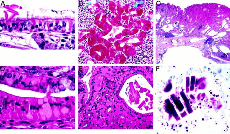
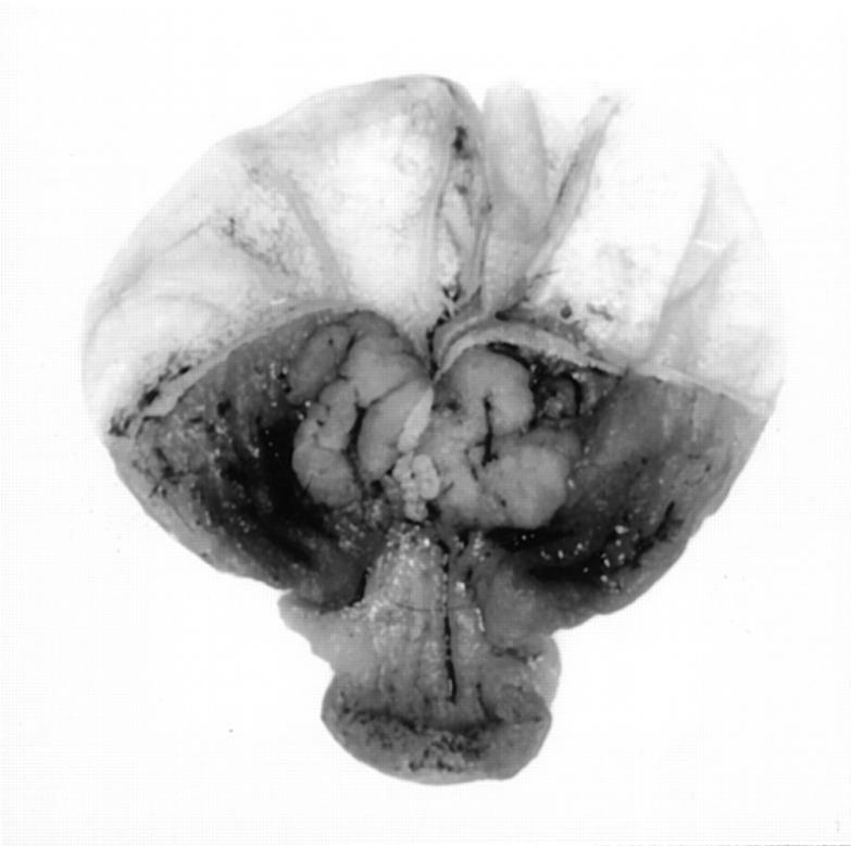
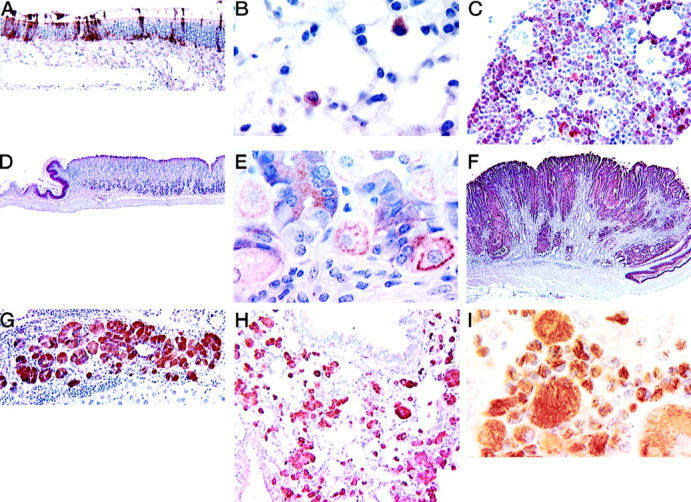
The C57BL/6, 129, and B6,129 mouse strains or stocks have been commonly used to generate targeted mutant mice. The pathology of these mice is not well characterized. In studies of these aging mice, we found high incidences of hyalinosis (eosinophilic cytoplasmic change) in the glandular stomach, respiratory tract, bile duct, and gall bladder of B6,129 CYP1A2-null and wild-type mice as well as in both sexes of the background 129S4/SvJae strain. The gastric lesions of the glandular stomach were found in 95.7% of female CYP1A2-null mice as well as in 45.7% of female 129S4/SvJae animals. The eosinophilic protein isolated from characteristic hyaline gastric lesions was identified as Ym2, a member of the chitinase family. Immunohistochemistry, using rabbit polyclonal antibodies to oligopeptides derived from the Ym1 sequence, detected focal to diffuse reactivity within both normal and abnormal nasal olfactory and respiratory epithelium, pulmonary alveolar macrophages, bone marrow myeloid cells, and the squamous epithelium of the forestomach and epithelium of the glandular stomach. Alveolar macrophages in acidophilic pneumonia, a major cause of death of aging 129 mice, and in mice with the me mutation also were highly immunoreactive. The possible cause of this protein excess in gastric and other lesions and its possible functions are discussed.


Figures

Figure 1.
Figure 1.
Histological appearance of hyalinosis in various tissues. A: Respiratory portion of nasal cavity showing hyaline intracellular inclusions and an extracellular crystal. H&E. B: Epithelium of hyaline bile duct epithelium stains deep red with Masson’s trinchrome. C: Portion of a large gastric lesion showing hyperplastic epithelium and cystic epithelium extending into the muscle wall with foci of lymphocytic inflammation. Note the eosinophilic epithelial areas within the hyperplastic lesion. H&E. D: Higher magnification of a gastric lesion showing eosinophilic epithelial cells. H&E. E: Gastric lesion showing large extracellular crystals within an area of hyalinosis. H&E. F: Dominici’s stain showing metachromatic extracellular crystals in a gastric lesion.
Figure 2.
Figure 2.
Gross appearance of two plaques in the glandular stomach at the limiting ridge of a 19-month-old female CYP1A2-null mutant (B6,129).
Figure 3.
Figure 3.
Ultrastructure of hyalin in the chief cells of a CYP1A2-null mouse. Note the dilated rough endoplasmic reticulum filled with fine granular material and few secretory granules. Uranyl acetate, lead citrate; original magnification, ×6,000.
Figure 4.
Figure 4.
Protein analyses of the stomach. Tissue homogenates (20 μg/lane) from normal wild-type glandular stomach (GS+/+), from a gastric lesion in a CYP1A2-null mouse (L−/−), and from a normal part of the same glandular stomach adjacent to the lesion (GS−/−) were run on 10% SDS-polyacrylamide gel and stained for protein.
Figure 5.
Figure 5.
Western blotting analyses of Ym2/1 proteins. Representative results are shown. A: Tissue homogenates from a normal part of the CYP1A2-null glandular stomach adjacent to a lesion (GS−/−), from a gastric lesion in a CYP1A2-null mouse (L−/−), from a normal wild-type glandular stomach (GS +/+), from a normal limiting ridge (LR+/+), and from a normal forestomach (FS +/+). Twenty μg of total protein was used per each lane, except for the lesion homogenate, L(−/−), in which 0.1 μg was used. B: Tissue homogenates from a normal part of a glandular stomach in a CYP1A2-null mouse adjacent to a gastric lesion (GS−/−), from a lesion in a CYP1A2-null mouse (L−/−), and from various normal organs of wild-type (+/+) mice as indicated were subjected to Western blotting. Twenty μg was used per each lane, except for the plaque-like lesion homogenate, L, in which 0.1 μg was used.
Figure 6.
Figure 6.
Northern blotting analyses of Ym2/1 mRNAs. Representative results are shown. A: Total RNAs from whole normal stomach (S +/+; glandular stomach + limiting ridge + forestomach), from a gastric lesion in a CYP1A2-null mouse (L−/−), and from a normal part of the glandular stomach adjacent to the lesion (GS−/−). Ten μg of total RNA/lane was used for the normal stomach samples, and 3 μg/lane for the lesion, L. B: Total RNAs from various organs as indicated were subjected to Northern blotting. Ten and 5 μg total RNA/lane were used for lung, spleen, and stomach samples, respectively.
Figure 7.
Figure 7.
Expression of Ym1/2 in tissues of mice as determined by ABC immunohistochemistry using a rabbit polyclonal antibody prepared against oligopeptides derived from the Ym1 protein sequence. All figures from formalin-fixed tissues unless otherwise noted and with hematoxylin counterstain. A: Olfactory region of a nasal cavity from 2-month-old wild-type B6,129 mouse. Note the many immunoreactive epithelial cells. B: Immunoreactive alveolar macrophages in a normal lung of a 4-month-old wild-type B6,129 mouse. C: Bone marrow from wild-type B6,129 mouse showing reactive myeloid cells. D: Normal stomach of a wild-type (CYP1A2+/+) mouse showing immunoreactivity in the squamous epithelium of the limiting ridge but no reactive staining in the glandular stomach. E: Higher magnification of the normal glandular stomach shown in D. Immunoreactivity is seen within chief and parietal cells. F: A gastric lesion from a CYP1A2-null animal showing abundant multifocal immunoreactivity. G: Hyalinosis in bile duct hyperplasia and cholangitis; the epithelium is strongly immunoreactive in a CYP1A2 null mouse. H: Macrophage pneumonia in a 129S4/SvJae mouse. The pulmonary macrophages are strongly immunoreactive. I: Macrophages in the lungs of a motheaten mouse. The intracytoplasmic needle-like crystals are reactive whereas the large extracellular crystals are not immunoreactive. Bouin’s fixation.

Similar articles

Cited by

References

    1. Guengerich FP, Humphreys WG, Yun CH, Hammons GJ, Kadlubar FF, Seto Y, Okazaki O, Martin MV: Mechanisms of cytochrome P450 1A2-mediated formation of N-hydroxy arylamines and heterocyclic amines and their reaction with guanyl residues. Princess Takamatsu Symp 1995, 23:78-84 - PubMed
    1. Nelson DR, Koymans L, Kamataki T, Stegeman JJ, Feyereisen R, Waxman DJ, Waterman MR, Gotoh O, Coon MJ, Estabrook RW, Gunsalus IC, Nebert DW: P450 superfamily: update on new sequences, gene mapping, accession numbers and nomenclature. Pharmacogenetics 1996, 6:1-42 - PubMed
    1. Windmill KF, McKinnon RA, Zhu X, Gaedigk A, Grant DM, McManus ME: The role of xenobiotic metabolizing enzymes in arylamine toxicity and carcinogenesis: functional and localization studies. Mutat Res 1997, 376:153-160 - PubMed
    1. Eaton DL, Gallagher EP, Bammler TK, Kunze KL: Role of cytochrome P4501A2 in chemical carcinogenesis: implications for human variability in expression and enzyme activity. Pharmacogenetics 1995, 5:259-274 - PubMed
    1. Kawajiri K, Hayashi S-I: The CYP1 family. Ioannides C eds. Cytochromes P450: Metabolic and Toxicological Aspects. 1996, :pp 77-97 CRC Press, London

Publication types

MeSH terms