. 2001 Jan-Feb;8(1):34-48.
doi: 10.1136/jamia.2001.0080034.

Integration of multidisciplinary sensory data: a pilot model of the human brain project approach

Affiliations

Integration of multidisciplinary sensory data: a pilot model of the human brain project approach

P L Miller et al. J Am Med Inform Assoc. 2001 Jan-Feb.

Abstract

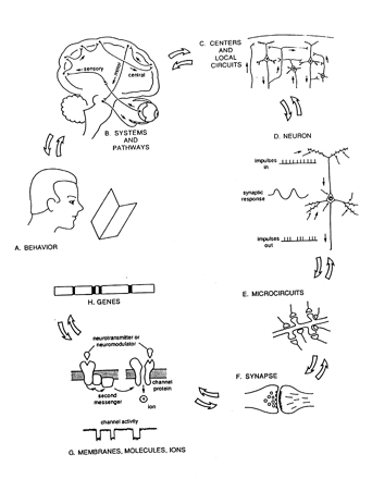
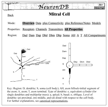
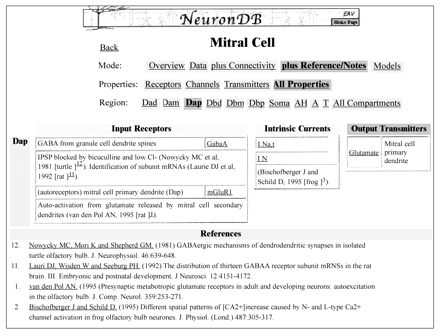
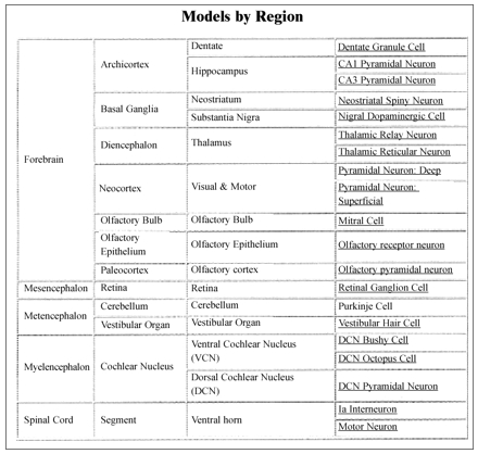
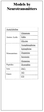
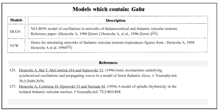
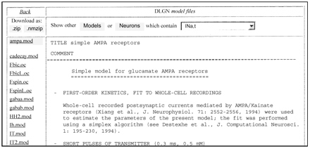
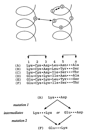
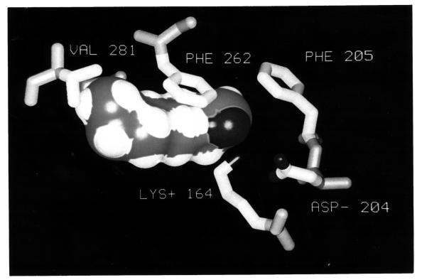
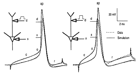
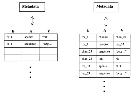
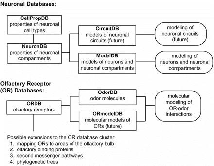
The paper provides an overview of neuroinformatics research at Yale University being performed as part of the national Human Brain Project. This research is exploring the integration of multidisciplinary sensory data, using the olfactory system as a model domain. The neuroinformatics activities fall into three main areas: 1) building databases and related tools that support experimental olfactory research at Yale and can also serve as resources for the field as a whole, 2) using computer models (molecular models and neuronal models) to help understand data being collected experimentally and to help guide further laboratory experiments, 3) performing basic neuroinformatics research to develop new informatics technologies, including a flexible data model (EAV/CR, entity-attribute-value with classes and relationships) designed to facilitate the integration of diverse heterogeneous data within a single unifying framework.


Figures

Figure 1
Figure 1
The different levels of organization of the nervous system. (From Shepherd. Used with permission.)
Figure 2
Figure 2
A NeuronDB screen designed to provide access to information about the mitral cell. The screen shows a sketch of an actual mitral cell (left) and a canonical representation of that cell (right). The canonical version provides a simplified representation of the cell's compartments and is designed to facilitate the organized storage, access, and comparison of cell properties.
Figure 3
Figure 3
This screen shows the data contained in NeuronDB about the properties (input receptors and their transmitters, intrinsic currents or channels, and output transmitters) of the proximal apical dendrite (Dap) compartment of the mitral cell. These data, which can be viewed at various levels of detail, include an annotated set of references to experimental articles about each property in each cell compartment.
Figure 4
Figure 4
Two screens that provide access to data in ModelDB, by region (left) or by neurotransmitter (right).
Figure 4
Figure 4
Two screens that provide access to data in ModelDB, by region (left) or by neurotransmitter (right).
Figure 5
Figure 5
ModelDB screen listing all models that contain the neurotransmitter Gaba.
Figure 6
Figure 6
ModelDB screen displaying information that describes a specific model.
Figure 7
Figure 7
Correlated mutation analysis theory and application. The theory is that when a mutation occurs in a structurally important residue (mutation 1), the intermediate has structural instability. Compensatory mutations are then selected (mutation 2), and the structural interaction is restored. Top, Several residues are shown in their structural context—in this example, two nearby alpha-helices. Middle, For these residues, six sequences (A–F) are shown as a multiple alignment. Positions 1 and 3 show correlated substitutions (connected by arrows), as do positions 5 and n. Bottom, For positions 1 and 3, the most parsimonious evolutionary pathways between sequences A and F. Correlated mutation analysis detects pairs of residue positions that show correlated substitutions without intermediates.
Figure 8
Figure 8
Details from the I7 olfactory receptor molecular model, showing octanal (rendered as a surface) in the predicted odor-binding pocket (rendered as sticks). White indicates carbon atoms, dark gray indicates oxygen, and the arrow indicates nitrogen. Medium gray indicates the carbon atoms of octanal. This model highlights a critical interaction between lysine 164 and the carbonyl (double-bonded oxygen) atom of octanal.
Figure 9
Figure 9
The results of two experiments (A and B), showing how neuronal modeling of the olfactory bulb mitral cell can help us understand experimental results. The dashed lines show recordings of membrane potential made at two locations (d and s). The solid lines show the predictions of our neuronal model. The goal of this work is to produce a model that matches the data through the rising phase of the spikes. In experiment A (left), location d is stimulated with a low current, whereas in experiment B (right), location d is stimulated with a high current. Intuitively, one would expect that the resulting action potential would start at site d and move toward site s, which is seen in experiment B with the high-current stimulus. In experiment A (with the low-current stimulus), however, the spike somehow manages to start at site s and propagate back out to site d (where the actual stimulation occurred). The neuronal model is able to explain this shift in the location of spike initiation in terms of the inhomogeneity of threshold and of voltage gradients along the dendrite. (From Shen et al. Used with permission.)
Figure 10
Figure 10
Simplified examples of data stored in EAV format (left) and EAV/CR format (right).
Figure 11
Figure 11
An outline of the current SenseLab project, with potential future extensions.

Comment in

References

    1. Shepherd GM. Neurobiology. New York: Oxford University Press, 1994.
    1. Buck L, Axel R. A novel multigene family may encode odorant receptors: a molecular basis for odor recognition. Cell. 1991;65:175–87. - PubMed
    1. Mori K, Yoshihara Y. Molecular recognition and olfactory processing in the mammalian olfactory system. Prog Neurobiol. 1995;45:585–619. - PubMed
    1. Hildebrand JG, Shepherd GM. Molecular mechanisms of olfactory discrimination: converging evidence for common principles across phyla. Annu Rev Neurosci. 1997;20:595–631. - PubMed
    1. Shepherd GM, Greer CA. Olfactory bulb. In: Shepherd GM (ed). The Synaptic Organization of the Brain. New York: Oxford University Press, 1998.

Publication types