Broca's region subserves imagery of motion: a combined cytoarchitectonic and fMRI study
- PMID: 11144756
- PMCID: PMC6872088
- DOI: 10.1002/1097-0193(200012)11:4<273::aid-hbm40>3.0.co;2-0
Broca's region subserves imagery of motion: a combined cytoarchitectonic and fMRI study
Abstract
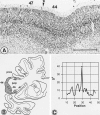
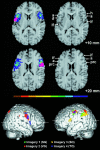
Broca's region in the dominant cerebral hemisphere is known to mediate the production of language but also contributes to comprehension. Here, we report the differential participation of Broca's region in imagery of motion in humans. Healthy volunteers were studied with functional magnetic resonance imaging (fMRI) while they imagined movement trajectories following different instructions. Imagery of right-hand finger movements induced a cortical activation pattern including dorsal and ventral portions of the premotor cortex, frontal medial wall areas, and cortical areas lining the intraparietal sulcus in both cerebral hemispheres. Imagery of movement observation and of a moving target specifically activated the opercular portion of the inferior frontal cortex. A left-hemispheric dominance was found for egocentric movements and a right-hemispheric dominance for movement characteristics in space. To precisely localize these inferior frontal activations, the fMRI data were coregistered with cytoarchitectonic maps of Broca's areas 44 and 45 in a common reference space. It was found that the activation areas in the opercular portion of the inferior frontal cortex were localized to area 44 of Broca's region. These activations of area 44 can be interpreted to possibly demonstrate the location of the human analogue to the so-called mirror neurones found in inferior frontal cortex of nonhuman primates. We suggest that area 44 mediates higher-order forelimb movement control resembling the neuronal mechanisms subserving speech.
Figures


References
-
- Amunts K, Schleicher A, Schormann T, Bürgel U, Mohlberg, H. , Uylings, H.B.M. , Zilles, K (1997): The cytoarchitecture of Broca's regions and its variability. Neuroimage 5: S353.
-
- Amunts K, Schleicher A, Bürgel U, Mohlberg H, Uylings HBM, Zilles K (1999): Broca's region re‐visited: Cytoarchitecture and intersubject variability. J Comp Neurol 412: 319–341. - PubMed
-
- Berthoz A (1996): The role of inhibition in the hierarchical gating of executed and imagined movements. Brain Res Cogn Brain Res 3: 101–113. - PubMed
-
- Binkofski F, Dohle C, Posse S, Stephan KM, Hefter H, Seitz RJ, Freund H‐J (1998): Human anterior intraparietal area subserves prehension. A combined lesion and functional MRI activation study. Neurology 50: 1253–1259. - PubMed
-
- Binkofski F, Buccino G, Posse S, Seitz RJ, Rizzolatti G, Freund H‐J (1999): A fronto‐parietal circuit for object manipulation in man. Evidence from an fMRI‐study. Eur J Neurosci 11: 3276–3286. - PubMed
Publication types
MeSH terms
LinkOut - more resources
Full Text Sources

