. 2000 Apr;5(2):121-31.
doi: 10.1379/1466-1268(2000)005<0121:iohspb>2.0.co;2.

Induction of heat shock protein 70 by herbimycin A and cyclopentenone prostaglandins in smooth muscle cells

Affiliations

Induction of heat shock protein 70 by herbimycin A and cyclopentenone prostaglandins in smooth muscle cells

L Hamel et al. Cell Stress Chaperones. 2000 Apr.

Abstract

This study characterizes Hsp70 induction in human smooth muscle cells (SMC) by herbimycin A and cyclopentenone prostaglandins. The magnitude of Hsp70 induction by cyclopentenone prostaglandins was 8- to 10-fold higher than induction by herbimycin A. Hsp70 induction by delta12PGJ2 was first observed at 10 microM, rose to 4000-5000 ng/mL within one log unit and a maximum response was not observed; concentrations of delta12PGJ2 higher than 30 microM were toxic to the cells. A maximum response with herbimycin A (500 ng/mL) was reached at 0.05 microM and maintained to 1 microM without toxicity. Both, delta12PGJ2 and herbimycin A, were inhibited by dithiothreitol (DTT, 100 microM) at lower concentrations and became less sensitive to inhibition at higher concentrations. Hsp70 induction after incubation of SMC with delta12PGJ2 followed by addition of herbimycin A was significantly higher than Hsp70 induction after incubation with herbimycin A followed by addition of delta12PGJ2. When cells were incubated with [3H]-PGJ2, followed by protein denaturation, substantial radioactivity remained protein-bound suggesting that the prostaglandin must be covalently bound. Covalent binding was largely insensitive to DTT. Maximal Hsp70 induction was observed after 5 minutes of exposure of the cells to herbimycin A followed by a 20 hour recovery period in agent-free medium. Cells required 3-4 hours of exposure to delta12PGJ2 followed by a 20 hour recovery period in order to see high Hsp70 induction. Binding of the heat shock factor (HSF) to the heat shock element (HSE) in the presence of herbimycin A or delta12PGJ2, and the effects of DTT, mirrored the results of Hsp70 induction. The results suggest that probable differences between the 2 agents are at the level of the signal transduction prior to HSF activation.


Figures

Fig. 1.
Fig. 1.
Hsp70 induction by herbimycin A and the cyclopentenone prostaglandin Δ12PGJ2. Determination of Hsp70 induction by herbimycin A and Δ12PGJ2 by Western Analysis (inset). Human primary SMC were preincubated with various concentrations of herbimycin A or Δ12PGJ2 for 4 hours. Cells were then allowed to recover in drug-free medium for 20 hours. Western analysis was carried as specified in materials and methods using mouse monoclonal anti-hsp70 as a primary antibody and goat anti-mouse immunoglobulin G conjugated to horseradish peroxidase as secondary antibody. Cell lysates from cells treated with prostaglandin were diluted 5-fold. C, control cells; V, DMSO vehicle 0.5%; S, Hsp70 standard (10 ng). Determination of Hsp70 induction by herbimycin A and Δ12PGJ2 by ELISA. Human primary SMC were preincubated with various concentrations of herbimycin A (n = 4) or Δ12PGJ2 (n = 4), for 4 hours in the presence or absence of DTT (100 μM). After preincubation, cells were allowed to recover in drug-free medium for 20 hours. Cell lysates were prepared and Hsp70 levels were determined by ELISA as specified in materials and methods. Error bars are standard error of the mean; they are shown only for the dose response when DTT is absent for clarity. Error bars in the presence of DTT were similar.
Fig. 2.
Fig. 2.
Cytotoxicity. Human primary SMC were incubated with various concentrations of herbimycin A or Δ12-PGJ2 for 4 hours. Drugs were removed and cytotoxicity, measured by MTS (materials and methods), was determined. Triplicate runs for each concentration were carried out in each of 3 independent experiments. Percent toxicity is the average of the results for the 3 experiments; error bars represent the standard error of the mean.
Fig. 3.
Fig. 3.
Protein synthesis in SMC after treatment with herbimycin A, prostaglandin, and DTT. Human primary SMC were incubated for 4 hours with various concentrations (μM) of herbimycin A (HA), Δ12-PGJ2 (PG), DTT, and combinations of herbimycin A (1 μM) or Δ12-PGJ2 (30 μM) with DTT (100 μM). Cells were then washed twice with PBS and further incubated for 2 hours in medium containing 35S methionine and cysteine. After incubation with the radiolabeled amino acids, cells were washed with PBS (3 ×), cell lysates prepared and SDS-PAGE carried out. The level of radioactivity on each lane was quantitated with a phosphorImager after 3-day exposure to a phosphoscreen. The radioactivity level obtained with untreated cells was set to 100% and other readings were normalized to this value. U, untreated; D, DMSO vehicle (0.5%); CH, cycloheximide (50 mg/mL) positive control.
Fig. 4.
Fig. 4.
Effect of order of drug addition on Hsp70 induction. Human primary SMC were incubated for 48 hours in serum free adapting medium and then incubated with Δ12PGJ2 (25 μM, 4 hours) followed by addition of herbimycin A (1 μM, last hour) (PG then HA) or with herbimycin A (1 μM, 1 hour) followed by addition of Δ12PGJ2 (25 μM, 4 hours) (HA then PG). Control assays included herbimycin A alone (1 μM, 1 hour) and Δ12PGJ2 alone (25 μM, 4 hours). Times of exposure to the 2 agents were determined based on the results shown in Figure 5. After drug exposure, cells were allowed to recover for 20 hours in serum-containing medium. Hsp70 protein levels were determined by ELISA. Results were normalized to the hsp level when cells were incubated with prostaglandin only. Error bars represent standard error of the mean from 5 independent experiments. * P < 0.001 vs PG then HA
Fig. 5.
Fig. 5.
Time of drug exposure required for Hsp70 induction. Human primary SMC were incubated for the indicated times with herbimycin A (1 μM, n = 5) or Δ12PGJ2 (20 μM, n = 4). Cells were then allowed to recover in drug-free medium for an additional 20 hours. Levels of Hsp70 in cell lysates were determined by ELISA. Data were normalized to maximal level of Hsp70 observed for each drug in each experiment respectively. Error bars represent standard error of the mean
Fig. 6.
Fig. 6.
Kinetics of binding of HSF to [32P]-HSE after cell exposure to herbimycin A or Δ12PGJ2. (a) Rat SMC were heat shocked 1 hour at 43°C (heat shock) or incubated with herbimycin A (1 μM) or Δ12PGJ2 (20 μM) for the times indicated. At the end of each time point, a cell lysate was prepared and a binding experiment with [32P]-HSE carried out. After electrophoresis, results were quantitated with a PhosphorImager (STORM 840). A representative gel shift assay is shown. HSE, 50-fold excess HSE over [32P]-HSE. NFκB, 100-fold excess of NFκB DNA probe. Serum, preimmune rabbit serum. anti-HSF1 ab, antibody to HSF1. (b) Densitometry analysis of the [HSE-HSF] signals from 3 independent experiments. Data were normalized to the most intense signal in each treatment. Error bars represent the standard error of the mean
Fig. 7.
Fig. 7.
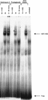
Time of cell exposure to drugs required for HSF activation. Rat SMC were heat shocked for 1 hour at 43°C (heat shock) or incubated with herbimycin A (1 μM) or Δ12PGJ2 (20 μM) for 5 minutes and allowed to recover for 3 hours without drug (5’ + 3 h recov.) or 3 hours without recovery (3h). After treatment, cell lysates were prepared and a gel shift assay was carried out as in the experiment illustrated in Figure 6.
Fig. 8.
Fig. 8.
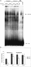
Effect of DTT on HSF binding to [32P]-HSE. (a) Rat SMC were incubated with herbimycin A (0.1 or 1 μM) or prostaglandin Δ12PGJ2 (20 or 30 μM) in the presence (+) or absence (-) of DTT (100 μM), for 3 hours. Cell lysates were then prepared and incubated with [32P]-HSE followed by gel electrophoresis. Binding was quantitated with a PhosphorImager (STORM 840). A representative gel shift assay is shown. Heat Shock, heat shocked cells (1 hour at 43°C). HSE, 50-fold excess HSE over [32P]-HSE. Free, unbound γ-[32P]HSE probe (b) Densitometry analysis of the signals from 3 independent experiments similar to that shown in (a). Data were normalized to the most intense HSF-HSE band. Error bars are the standard error of the mean. ** P < 0.05 vs (-) DTT. * P < 0.05 vs (-) DTT
None
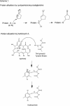
Scheme 1

Similar articles

Cited by

References

    1. Amici C, Sistonen L, Santoro MG, Morimoto RI. Antiproliferative prostaglandins activate heat shock transcription factor. Proc Natl Acad Sci U S A. 1992;89:6227–6231. - PMC - PubMed
    1. Benjamin IJ, McMillan DR. Stress (heat shock) proteins: molecular chaperones in cardiovascular biology and disease. Circ Res. 1998;83:117–132. - PubMed
    1. Conde AG, Lau SS, Dillmann WH, Mestril R. Induction of heat shock proteins by tyrosine kinase inhibitors in rat cardiomyocytes and myogenic cells confers protection against simulated ischemia. J Mol Cell Cardiol. 1997;26:1927–1938. - PubMed
    1. Feige U, Polla BS. Hsp70—a multi-gene, multi-structure, muti-function family with potential clinical applications. Experientia. 1994;50:979–986. - PubMed
    1. Fernandes M, O'Brien T, and Lis JT 1994 Structure and Regulation of Heat Shock Gene Promoters. In: The Biology of Heat Shock Proteins and Molecular Chaperones, eds Morimoto RI, Tissieres A, Georgopoulos C. Cold Spring Harbor Laboratory Press, Cold Spring Harbor, NY , 375–393.

MeSH terms

LinkOut - more resources