Review
. 2001 Jan;14(1):15-37.
doi: 10.1128/CMR.14.1.15-37.2001.

Diagnosis of noncultivatable gastroenteritis viruses, the human caliciviruses

Affiliations
Review

Diagnosis of noncultivatable gastroenteritis viruses, the human caliciviruses

R L Atmar et al. Clin Microbiol Rev. 2001 Jan.

Abstract

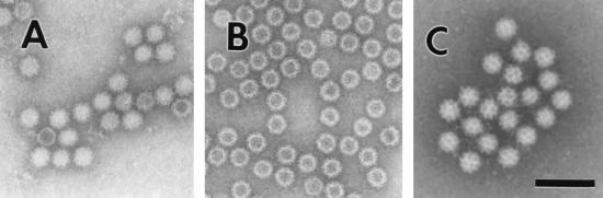
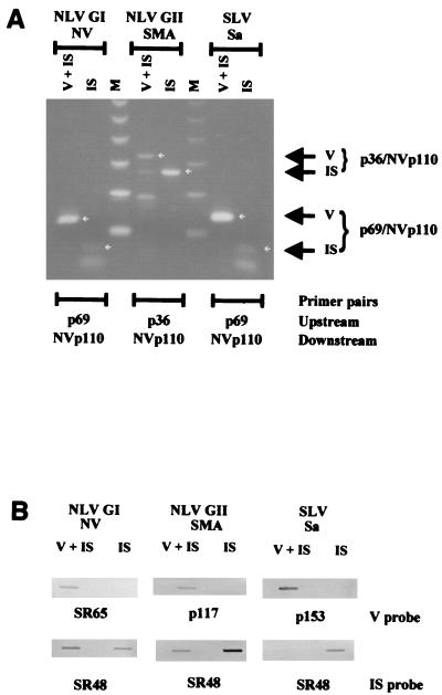
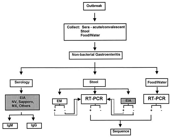
Gastroenteritis is one of the most common illnesses of humans, and many different viruses have been causally associated with this disease. Of those enteric viruses that have been established as etiologic agents of gastroenteritis, only the human caliciviruses cannot be cultivated in vitro. The cloning of Norwalk virus and subsequently of other human caliciviruses has led to the development of several new diagnostic assays. Antigen detection enzyme immunoassays (EIAs) using polyclonal hyperimmune animal sera and antibody detection EIAs using recombinant virus-like particles have supplanted the use of human-derived reagents, but the use of these assays has been restricted to research laboratories. Reverse transcription-PCR assays for the detection of human caliciviruses are more widely available, and these assays have been used to identify virus in clinical specimens as well as in food, water, and other environmental samples. The application of these newer assays has significantly increased the recognition of the importance of human caliciviruses as causes of sporadic and outbreak-associated gastroenteritis.


Figures

FIG. 1
FIG. 1
Electron micrographs of (A) NV, (B) baculovirus-expressed NVL particles, and (C) Sapporo virus. Bar, 100 nm.
FIG. 2
FIG. 2
Schematic of the genomic organization of viruses from the four different genera of the Caliciviridae. Strains [GenBank numbers] for which the entire genomic sequence is available are presented for each of the genera: NV [M87661] , , Manchester virus [X86560] , , rabbit hemorrhagic disease virus [M67473] , and feline calicivirus [M86379] . A nonstructural polyprotein (aa 1719 to 1789) is encoded by the 5′ end of the genome. The major capsid protein (shaded area) is in frame for the SLVs and lagoviruses, while it is in the +1 frame for NLVs and −1 frame for the vesiviruses. A basic protein is encoded at the 3′ end of the genome for all four genera. SLVs have another ORF (+1) overlapping the capsid ORF that is not seen in the other genera.
FIG. 3
FIG. 3
Unrooted phylogram generated using the entire capsid amino acid sequence of nine genogroup II (GII) strains and one genogroup I (NV) strain and the PAUP program in the Genetics Computer Group suite of programs . Strains within different genogroup II genetic clusters are indicated, with the genogroup I strain NV serving as an outgroup. GenBank accession numbers used for this analysis are as follows: NV, M87661; HV, U07611; SMA, U75682; Melksham virus, X81879; Oth-25, L23830; MX, U22498; TV, U02030; Camberwell virus, U46500; Bristol virus, X76716; and LV, X86557.
FIG. 4
FIG. 4
(A) Schematic representation of the calicivirus genome and the regions amplified by common primer pairs. Modified from reference with permission from Technomic Publishing Co, Inc., copyright 2000. See Table 6 for further details about primer sequences. hel, helicase; pol, polymerase. (B) Schematic representation of the NLV genome from which an internal standard control for genogroup I NLVs was made. The relative locations of selected primers and probes are noted for virus and internal standard (Int. Std.) RNA. The internal standard control RNA yields amplicons that are 123 bp shorter (347 bp) than those from NV genomic RNA (470 bp). A portion of the genomic sequence targeted by virus-specific probes (e.g., SR65 and p116) is not present in the internal standard control, allowing differentiation of virus-specific and internal standard amplicons by nucleic acid hybridization .
FIG. 5
FIG. 5
Representation of RT-PCR detection of NLVs and SLVs by agarose gel electrophoresis (A) and confirmation by slot blot hybridization (B). Virus (V)- and internal standard control (IS)-specific RNAs are visible after amplification of NLVs (NV and SMA) and an SLV (Sapporo virus [Sa]) with primers NVp110 and p36 or p69. Virus-specific amplicons are detected by probes SR65 (NV), p117 (SMA), and p153 (Sapporo virus), while the internal standard control is detected by probe SR48 (3,159).
FIG. 6
FIG. 6
Schematic of evaluation strategies for the identification of infection caused by HuCVs. Once an outbreak is identified, clinical samples (serum or stool) and potential vehicles of transmission (e.g., water and food) are collected. Stool samples can be evaluated by any of three methods: electron microscopy (EM), RT-PCR, or EIA. If EIA or EM is positive, calicivirus infection is confirmed. Samples negative by EIA or EM and those for which further characterization is desired can be evaluated by RT-PCR. Virus-specific amplicons from stool or food and water samples may be sequenced and compared to determine the similarity of the strains. Serologic studies may also be performed using a number of different VLP antigens. IgM tests are performed on single sera, and IgG tests are performed on paired sera. The shaded boxes show those tests that are currently only available in selected research laboratories. Reprinted from reference , with permission from Technomic Publishing Co, Inc., copyright 2000.

Similar articles

Cited by

References

    1. Adler J L, Zickl R. Winter vomiting disease. J Infect Dis. 1969;119:668–673. - PubMed
    1. Anderson T F, Stanley W M. A study by means of the electron microscope of the reaction between tobacco mosaic virus and its antiserum. J Biol Chem. 1941;139:339–344.
    1. Ando T, Monroe S S, Gentsch J R, Jin Q, Lewis D C, Glass R I. Detection and differentiation of antigenically distinct small round-structured viruses (Norwalk-like viruses) by reverse transcription-PCR and Southern hybridization. J Clin Microbiol. 1995;33:64–71. - PMC - PubMed
    1. Ando T, Monroe S S, Noel J S, Glass R I. A one-tube method of reverse transcription-PCR to efficiently amplify a 3-kilobase region from the RNA polymerase gene to the poly(A) tail of small round-structured viruses (Norwalk-like viruses) J Clin Microbiol. 1997;35:570–577. - PMC - PubMed
    1. Ando T, Mulders M N, Lewis D C, Estes M K, Monroe S S, Glass R I. Comparison of the polymerase region of small round structured virus strains previously characterized in three serotypes by solid-phase immune electron microscopy. Arch Virol. 1994;135:217–226. - PubMed

Publication types

MeSH terms