Review
. 2001 Jan;14(1):59-97.
doi: 10.1128/CMR.14.1.59-97.2001.

Emergence of diverse Helicobacter species in the pathogenesis of gastric and enterohepatic diseases

Affiliations
Review

Emergence of diverse Helicobacter species in the pathogenesis of gastric and enterohepatic diseases

J V Solnick et al. Clin Microbiol Rev. 2001 Jan.

Abstract

Since Helicobacter pylori was first cultivated from human gastric biopsy specimens in 1982, it has become apparent that many related species can often be found colonizing the mucosal surfaces of humans and other animals. These other Helicobacter species can be broadly grouped according to whether they colonize the gastric or enterohepatic niche. Gastric Helicobacter species are widely distributed in mammalian hosts and are often nearly universally prevalent. In many cases they cause an inflammatory response resembling that seen with H. pylori in humans. Although usually not pathogenic in their natural host, these organisms serve as models of human disease. Enterohepatic Helicobacter species are an equally diverse group of organisms that have been identified in the intestinal tract and the liver of humans, other mammals, and birds. In many cases they have been linked with inflammation or malignant transformation in immunocompetent hosts and with more severe clinical disease in immunocompromised humans and animals. The purpose of this review is to describe these other Helicobacter species, characterize their role in the pathogenesis of gastrointestinal and enterohepatic disease, and discuss their implications for our understanding of H. pylori infection in humans.


Figures
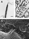
FIG. 1
FIG. 1
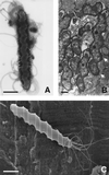
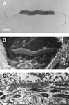
(A and B) Transmission electron micrographs of Helicobacter sp. flexispira (Lockard type 1) taken from a pure culture (A) and from an intestinal crypt in a mouse (B). Bipolar flagella and periplasmic fibers are apparent. (C) Scanning electron micrograph of Helicobacter sp. flexispira shows the organisms among intestinal microvilli in a mouse. Bars, 1 μm. Reprinted from reference with permission from the publisher.
FIG. 2
FIG. 2
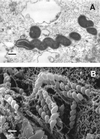
(A) Negatively stained preparation of H. felis (Lockard type 2) isolated from the gastric mucosa of an adult cat shows the multiple bipolar flagella. Bar, 1 μm. (C) Although faintly seen in Panel A, the characteristic periplasmic fibers are better visualized on a scanning electron micrograph. Bar, 1 μm. (B) Transmission electron micrograph of H. felis in the gastric mucosa of a germ-free mouse shows the characteristic spiral morphology. Pairs of periplasmic fibers can be seen en face (arrows). Bar, 0.5 μm. Photos courtesy of Adrian Lee, Jani O'Rourke, and Lucinda Thompson.
FIG. 3
FIG. 3
Transmission (A) and scanning (B) electron micrographs of bacteria in the gastric mucosa of a healthy pet cat. The helical morphology without periplasmic fibers (Lockard type 3) is characteristic of “H. heilmannii” and H. bizzozeronii. Bars, 0.5 μm. (A) Reprinted from reference with permission from the American Society for Microbiology. (B) Photo courtesy of Robert Munn.
FIG. 4
FIG. 4
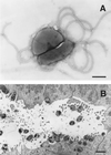
(A) Negatively stained preparation of H. mustelae shows the bipolar and lateral flagella (bar, 0.5 μm). (B) Thin section of a ferret antral gastric pit shows the intimate association of the bacterium with the epithelial surface, including the apparent formation of adhesion pedestals, Bar, 1 μm. Reprinted from reference with permission from the publisher.
FIG. 5
FIG. 5
(A) Scanning electron micrograph of H. pylori shows the gently curved morphology, multiple bipolar flagella, and absence of periplasmic fibers. Bar, 1 μm. Photo courtesy of Adrian Lee, Jani O'Rourke, and Lucinda Thompson. (B) Transmission electron micrograph of human gastric epithelium with large numbers of H. pylori intimately attached to the surface. Bar, 1 μm. Reprinted from reference with permission from the publisher.
FIG. 6
FIG. 6
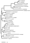
Phylogenetic tree for 19 validated Helicobacter species and 9 additional provisional species, based on 16S rRNA sequence. The scale bar represents a 1% difference in nucleotide sequence as determined by measuring the lengths of horizontal lines connecting any two species. The tree was constructed from the differences matrix (Table 3) using TREEVIEW (314a).
FIG. 7
FIG. 7
(A and B) Negatively stained preparation (A) and transmission electron micrograph (B) of H. hepaticus show the absence of periplasmic fibers and the single bipolar flagella. (C) Transmission electron micrograph of a liver section in a mouse reveals H. hepaticus in a bile canaliculus. Panel C is reprinted from reference with permission from the American Society for Microbiology. Bars, 0.5 μm.

Similar articles

Cited by

References

    1. Adam W S, Calhoun M L, Smith E M, Stinson A W. Microscopic anatomy of the dog. Springfield, Il: Charles C Thomas; 1970.
    1. Akin O Y, Tsou V M, Werner A L. Gastrospirillum hominis-associated chronic active gastritis. Pediatr Pathol Lab Med. 1995;15:429–435. - PubMed
    1. Akopyants N, Bukanov N O, Westblom T U, Kresovich S, Berg D E. DNA diversity among clinical isolates of Helicobacter pylori detected by PCR-based RAPD fingerprinting. Nucleic Acids Res. 1992;20:5137–5142. - PMC - PubMed
    1. Alder J D, Ewing P J, Mitten M J, Oleksijew A, Tanaka S K. Relevance of the ferret model of Helicobacter-induced gastritis to evaluation of antibacterial therapies. Am J Gastroenterol. 1996;91:2347–2354. - PubMed
    1. al-Himyary A J, Zabaneh R I, Zabaneh S S, Barnett S. Gastrospirillum hominis in acute gastric erosion. South Med J. 1994;87:1147–1150. - PubMed

Publication types

MeSH terms

Substances

LinkOut - more resources