. 2000 Dec;12(12):2395-2408.
doi: 10.1105/tpc.12.12.2395.

Cloning and characterization of IAR1, a gene required for auxin conjugate sensitivity in Arabidopsis

Affiliations

Cloning and characterization of IAR1, a gene required for auxin conjugate sensitivity in Arabidopsis

J Lasswell et al. Plant Cell. 2000 Dec.

Abstract

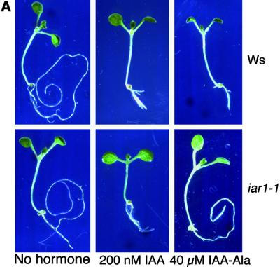
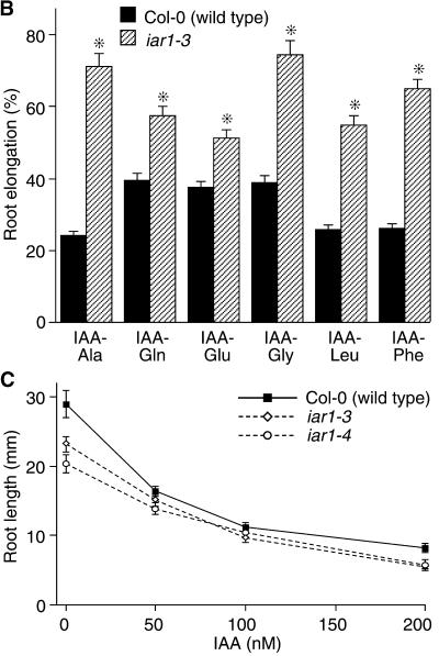
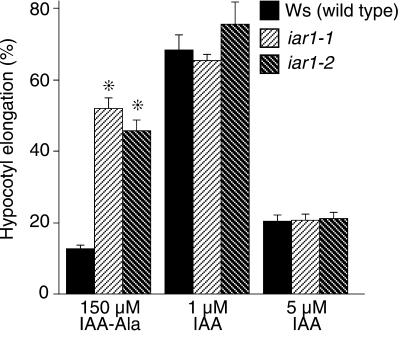
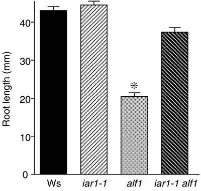
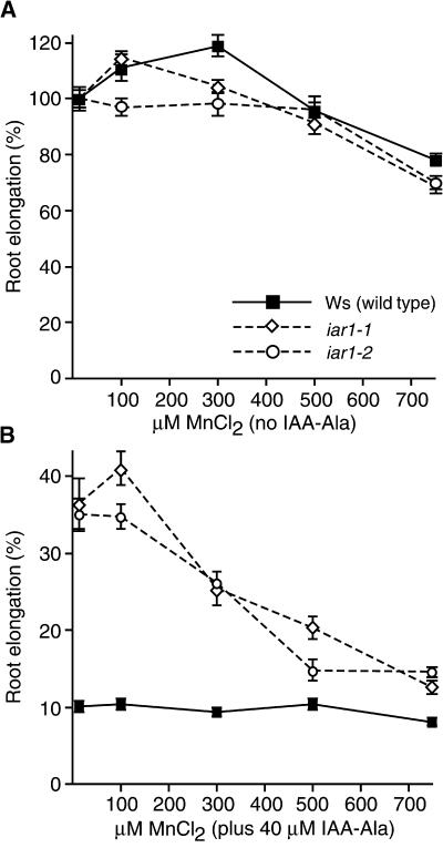
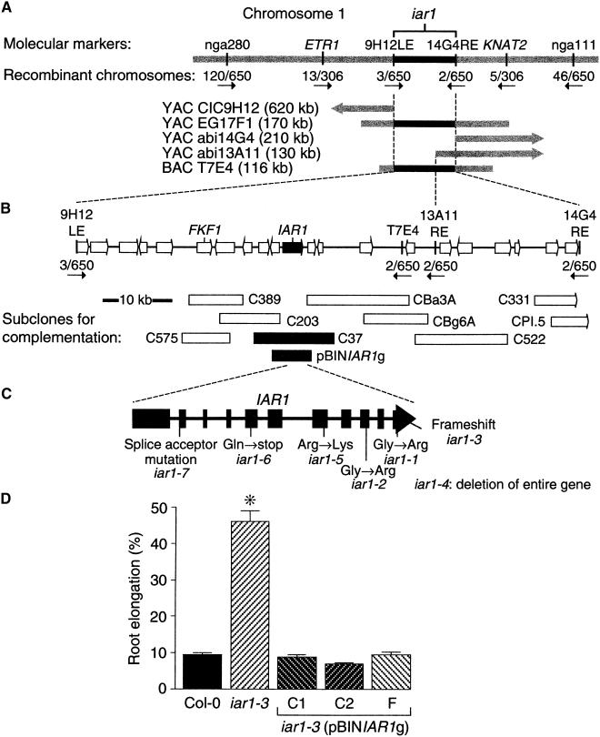
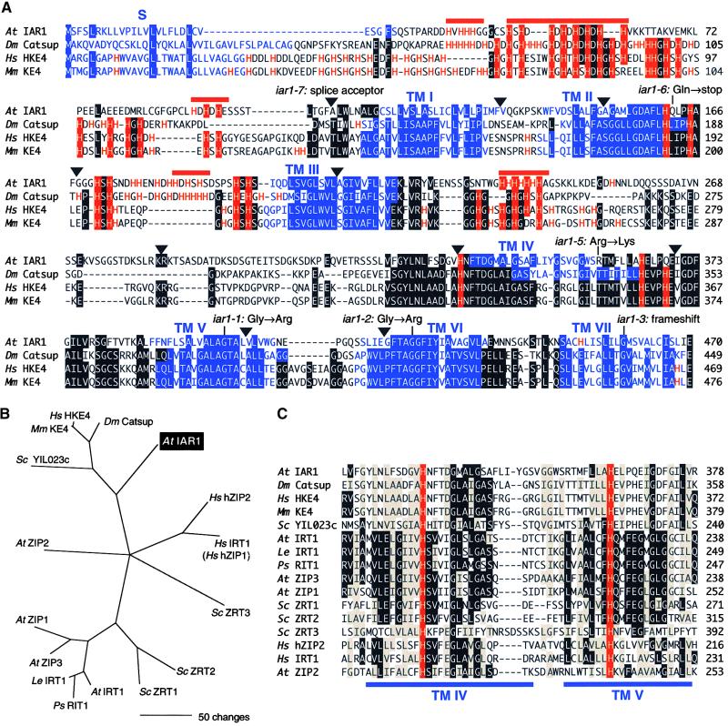
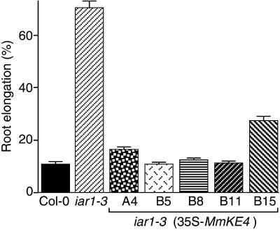
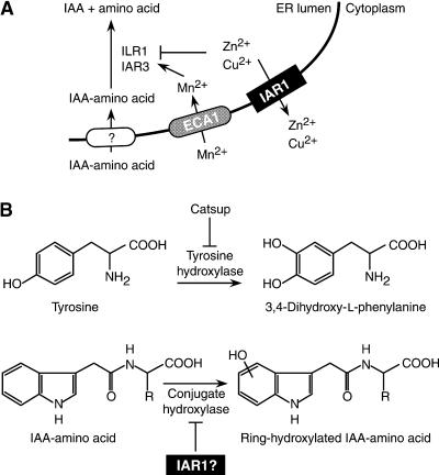
Most indole-3-acetic acid (IAA) in higher plants is conjugated to amino acids, sugars, or peptides, and these conjugates are implicated in regulating the concentration of the free hormone. We identified iar1 as an Arabidopsis mutant that is resistant to the inhibitory effects of several IAA-amino acid conjugates but remains sensitive to free IAA. iar1 partially suppresses phenotypes of a mutant that overproduces IAA, suggesting that IAR1 participates in auxin metabolism or response. We used positional information to clone IAR1, which encodes a novel protein with seven predicted transmembrane domains and several His-rich regions. IAR1 has homologs in other multicellular organisms, including Drosophila, nematodes, and mammals; in addition, the mouse homolog KE4 can functionally substitute for IAR1 in vivo. IAR1 also structurally resembles and has detectable sequence similarity to a family of metal transporters. We discuss several possible roles for IAR1 in auxin homeostasis.


Figures

Figure 1.
Figure 1.
iar1 Roots Have Decreased Sensitivity to IAA–Amino Acid Conjugates and Normal Sensitivity to IAA. (A) Eight-day-old iar1-1 mutant and Wassilewskija (Ws) wild-type seedlings were removed from the agar and photographed after growth on medium containing no hormone, 200 nM IAA, or 40 μM IAA-Ala. (B) Wild-type Columbia (Col-0) and iar1 mutant seed were plated on medium containing 20 μM of the indicated IAA–amino acid conjugates. After 8 days, the primary root of each seedling was measured and normalized against growth on unsupplemented medium. Error bars indicate standard errors of the means (formula image), and asterisks indicate significant differences from the wild type (Student's t test, P < 0.0001). (C) Wild-type (Col-0) and iar1 mutant seed were plated on medium containing the indicated concentrations of IAA. After 8 days, the primary root of each seedling was measured. Error bars indicate standard errors of the means (formula image).
Figure 1.
Figure 1.
iar1 Roots Have Decreased Sensitivity to IAA–Amino Acid Conjugates and Normal Sensitivity to IAA. (A) Eight-day-old iar1-1 mutant and Wassilewskija (Ws) wild-type seedlings were removed from the agar and photographed after growth on medium containing no hormone, 200 nM IAA, or 40 μM IAA-Ala. (B) Wild-type Columbia (Col-0) and iar1 mutant seed were plated on medium containing 20 μM of the indicated IAA–amino acid conjugates. After 8 days, the primary root of each seedling was measured and normalized against growth on unsupplemented medium. Error bars indicate standard errors of the means (formula image), and asterisks indicate significant differences from the wild type (Student's t test, P < 0.0001). (C) Wild-type (Col-0) and iar1 mutant seed were plated on medium containing the indicated concentrations of IAA. After 8 days, the primary root of each seedling was measured. Error bars indicate standard errors of the means (formula image).
Figure 2.
Figure 2.
iar1 Hypocotyls Have Decreased Sensitivity to IAA-Ala. Wild-type and iar1 seed were plated on medium containing the indicated concentrations of IAA or IAA-Ala. After 8 days in yellow-filtered light at 22°C, hypocotyls were measured and normalized against growth on unsupplemented medium. Error bars indicate standard errors of the means (formula image for Ws and iar1-1, formula image for iar1-2), and asterisks indicate significant differences from the wild type (Student's t test, P < 0.0001).
Figure 3.
Figure 3.
iar1 Suppresses the alf1 Root Elongation Defect. Seed of Ws (wild type), iar1-1 and alf1 single mutants, and iar1-1 alf1 double mutants were plated on unsupplemented medium. After 10 days under yellow-filtered light at 22°C, the primary root of each seedling was measured. Homozygous alf1 mutants were identified among the progeny of an alf1/ALF1 parent based on the epinastic cotyledon phenotype. Error bars indicate standard errors of the means (n ⩾ 18), and the asterisk indicates a significant difference from the wild type (Student's t test, P < 0.0001).
Figure 4.
Figure 4.
Manganese Suppresses iar1 IAA-Ala Resistance. Wild-type (Ws) and iar1 mutant seed were plated on medium containing 14 to 750 μM manganese either with or without 40 μM IAA-Ala. After 8 days under yellow-filtered light at 22°C, the primary root of each seedling was measured and normalized to growth on unsupplemented medium (which contained 14 μM manganese). Error bars indicate standard errors of the means (formula image). (A) Without IAA-Ala. (B) With 40 μM IAA-Ala.
Figure 5.
Figure 5.
Positional Cloning of IAR1. (A) Recombination mapping with PCR-based markers nga111 and nga280 (Bell and Ecker, 1994) localized iar1 to the bottom of chromosome 1. This position was refined to between ETR1 (Chang et al., 1993) and KNAT2 (Lincoln et al., 1994). Additional markers from nearby YAC clones included 9H12LE and 14G4RE (Nelson et al., 2000) and a marker made from the right end of YAC abi13A11 (see Methods). These three markers hybridized to BAC T7E4 (Choi et al., 1995). (B) A complementation library was constructed (see Methods) from BAC T7E4, and subclones that were tested for iar1 complementation are indicated by rectangles below the predicted genes (arrows) in the region. Subclones shown in black rescued the iar1 IAA-Ala resistance, and those shown in white did not. (C) Positions of the seven iar1 mutations are shown below a scheme of the IAR1 coding region. Exons are indicated by closed rectangles, and introns are represented by lines. (D) A genomic construct containing the predicted IAR1 open reading frame controlled by its own promoter rescued the iar1 mutant phenotype. Seed from the wild type (Col-0), the iar1-3 mutant, and three iar1-3 transgenic lines homozygous for the pBINIAR1g construct, shown in (B), were plated on medium containing 40 μM IAA-Ala. After 8 days of growth under yellow-filtered light at 22°C, the primary root of each seedling was measured and standardized against growth on unsupplemented medium. Error bars indicate standard errors of the means (n ⩾ 17), and the asterisk indicates a significant difference from the wild type (Student's t test, P < 0.0001).
Figure 6.
Figure 6.
Alignment of IAR1 and Similar Proteins from Other Organisms. (A) IAR1, Drosophila (Dm) Catsup, human (Hs) HKE4 (Janatipour et al., 1992; Ando et al., 1996), and mouse (Mm) KE4 (Abe et al., 1988; St.-Jaques et al., 1990) predicted proteins were aligned with the Megalign program (DNAStar, Madison, WI) by using the Clustal method (Higgins and Sharp, 1989) with PAM250 residue weights. Residues conserved in at least three proteins are shaded. Triangles indicate the positions of introns in the IAR1 coding sequence. Positions of iar1 mutations are shown above the IAR1 sequence, except for iar1-4, which is a deletion of the entire IAR1 gene. The His residues are highlighted in red, and IAR1 potential metal-binding sequences of the type (HX)3-6 are indicated by red bars above the sequence. Regions predicted by the SMART program (Schultz et al., 2000) to be transmembrane domains (TM I to VII) are indicated in blue. (B) Phylogenetic tree of IAR1 and its relatives. The tree reconstructs the evolutionary relationship between IAR1 family members from (A) and ZIP proteins, including Arabidopsis (At; Korshunova et al., 1999) and tomato (Le) IRT1 (GenBank accession number AF136579); pea (Ps) RIT1 (GenBank accession number AF065444); Arabidopsis ZIP1, ZIP2, and ZIP3 (Grotz et al., 1998); yeast (Sc) ZRT1 (Zhao and Eide, 1996a), ZRT2 (Zhao and Eide, 1996b), ZRT3 (MacDiarmid et al., 2000), and YIL023c (GenBank accession number P40544); and human (Hs) IRT1 (hZIP1) and hZIP2 (Lioumi et al., 1999; Gaither and Eide, 2000). Sequences (from predicted transmembrane domain IV to the end of the proteins) were aligned as described in (A), and the phylogenetic tree was generated by using PAUP 4.0b3a (Swofford, 2000). The bootstrap method was performed for 100 replicates with a maximum parsimony optimality criterion. All characters were weighted equally. Starting trees for the heuristic search were obtained by random stepwise addition, and tree-bisection-reconnection was the branch-swapping algorithm. (C) Alignment of predicted transmembrane domains IV and V from the ZIP family members described in (B) with the similar region from IAR1 family members. Identical residues and conservative changes seen in at least seven proteins are shown in black and gray boxes, respectively. Transmembrane domains in the ZIP family described by MacDiarmid et al. (2000) are indicated by blue bars below the alignment. The His residues conserved with those in Arabidopsis IRT1 that are required for zinc transport (Rogers et al., 2000) are indicated in red.
Figure 7.
Figure 7.
IAR1 Encodes a Potential Transmembrane Protein. Hydropathy was calculated for IAR1, Drosophila (Dm) Catsup, mouse (Mm) KE4 (Abe et al., 1988; St.-Jaques et al., 1990), and Arabidopsis (At) ZIP1 (Grotz et al., 1998) with a window size of 13 and a linear weight variation model (Kyte and Doolittle, 1982). Positive and negative values indicate hydrophobic and hydrophilic regions, respectively. Predicted signal sequences are indicated by S, and the transmembrane domains predicted by the SMART program (Schultz et al., 2000) are numbered.
Figure 8.
Figure 8.
The Mouse KE4 Gene Can Functionally Substitute for IAR1. Wild-type (Col-0), iar1-3, and homozygous T3 progeny of five independent lines of iar1-3 plants transformed with 35S-MmKE4, a construct containing the mouse KE4 cDNA (Abe et al., 1988; St.-Jaques et al. 1990) under the control of the cauliflower mosaic virus 35S promoter, were grown for 8 days under yellow-filtered light at 22°C on medium containing 40 μM IAA-Ala. The primary root of each seedling was measured and standardized against growth on unsupplemented medium. Error bars indicate standard errors of the means (n ⩾ 20).
Figure 9.
Figure 9.
Possible Models for IAR1 Function. (A) IAR1 may transport inhibitory metals out of the ER. This model is suggested by the weak similarity of IAR1 to ZIP family metal transporters, the suppression of the iar1 phenotype by the conjugate hydrolase cofactor Mn2+, and the inhibition of conjugate hydrolases by Cu2+ or Zn2+. (B) IAR1 may inhibit an auxin conjugate hydroxylase. This model is suggested by the similarity of IAR1 to the Drosophila tyrosine hydroxylase inhibitor Catsup (Stathakis et al., 1999) and is consistent with the presence of ring-hydroxylated IAA conjugates in plants. Flat-tipped arrows indicate inhibitory interactions. See text for details.

Similar articles

Cited by

References

    1. Abe, K., Wei, J.-F., Wei, F.-S., Hsu, Y.-C., Uehara, H., Artzt, K., and Bennett, D. (1988). Searching for coding sequences in the mammalian genome: The H-2K region of the mouse MHC is replete with genes expressed in embryos. EMBO J. 7 3441–3449. - PMC - PubMed
    1. Ando, A., Kikuti, Y.Y., Shigenari, A., Kawata, H., Okamoto, N., Shiina, T., Chen, L., Ikemura, T., Abe, K., Kimura, M., and Inoko, H. (1996). cDNA cloning of the human homologues of the mouse Ke4 and Ke6 genes at the centromeric end of the human MHC region. Genomics 35 600–602. - PubMed
    1. Ausubel, F., Brent, R., Kingston, R.E., Moore, D.D., Seidman, J.G., Smith, J.A., and Struhl, K. (1995). Short Protocols in Molecular Biology. (New York: John Wiley and Sons).
    1. Bandurski, R.S., Cohen, J.D., Slovin, J.P., and Reinecke, D.M. (1995). Auxin biosynthesis and metabolism. In Plant Hormones, P.J. Davies, ed (Dordrecht, The Netherlands: Kluwer Academic Publishers), pp. 39–65.
    1. Bartel, B. (1997). Auxin biosynthesis. Annu. Rev. Plant Physiol. Plant Mol. Biol. 48 51–66. - PubMed

Publication types

LinkOut - more resources