. 2000 Dec;12(12):2455-2472.
doi: 10.1105/tpc.12.12.2455.

The molybdenum cofactor biosynthetic protein Cnx1 complements molybdate-repairable mutants, transfers molybdenum to the metal binding pterin, and is associated with the cytoskeleton

Affiliations

The molybdenum cofactor biosynthetic protein Cnx1 complements molybdate-repairable mutants, transfers molybdenum to the metal binding pterin, and is associated with the cytoskeleton

G Schwarz et al. Plant Cell. 2000 Dec.

Abstract

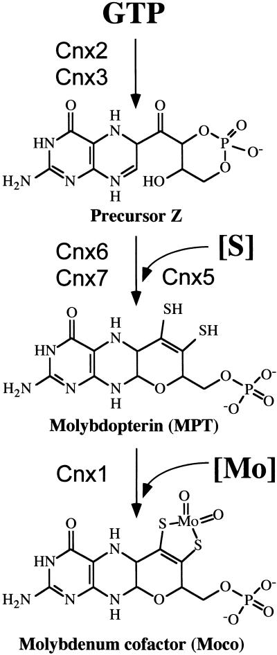
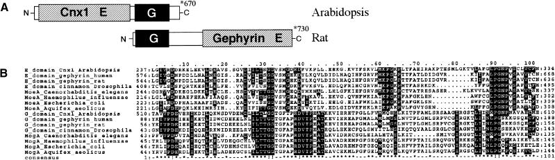
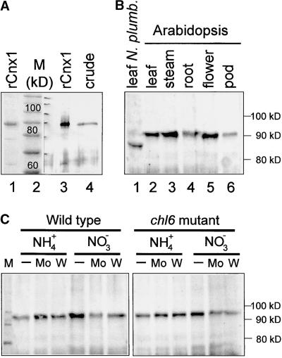
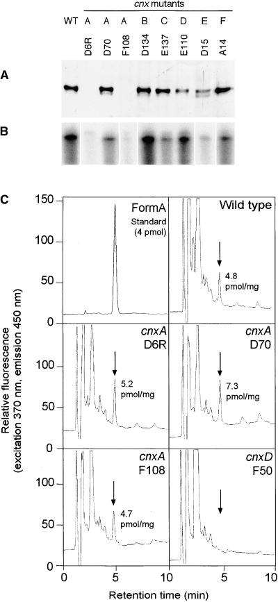
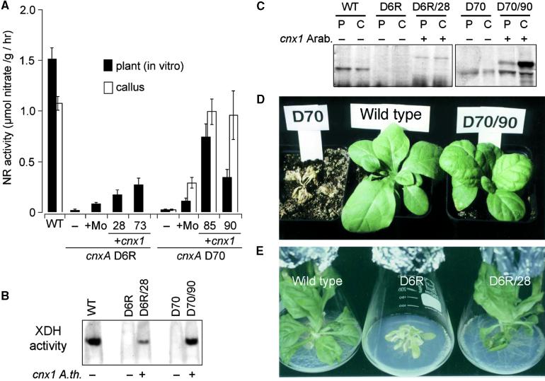
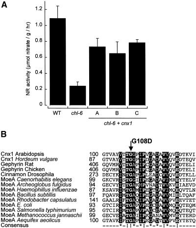
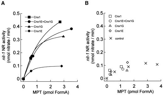
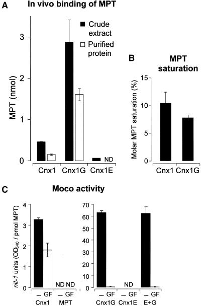
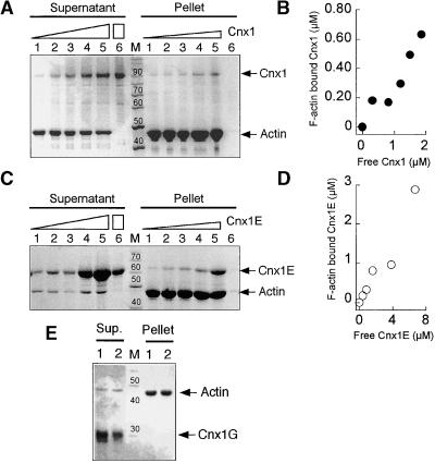
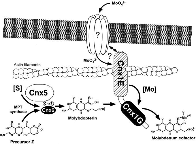
Molybdenum (Mo) plays an essential role in the active site of all eukaryotic Mo-containing enzymes. In plants, Mo enzymes are important for nitrate assimilation, phytohormone synthesis, and purine catabolism. Mo is bound to a unique metal binding pterin (molybdopterin [MPT]), thereby forming the active Mo cofactor (Moco), which is highly conserved in eukaryotes, eubacteria, and archaebacteria. Here, we describe the function of the two-domain protein Cnx1 from Arabidopsis in the final step of Moco biosynthesis. Cnx1 is constitutively expressed in all organs and in plants grown on different nitrogen sources. Mo-repairable cnxA mutants from Nicotiana plumbaginifolia accumulate MPT and show altered Cnx1 expression. Transformation of cnxA mutants and the corresponding Arabidopsis chl-6 mutant with cnx1 cDNA resulted in functional reconstitution of their Moco deficiency. We also identified a point mutation in the Cnx1 E domain of Arabidopsis chl-6 that causes the molybdate-repairable phenotype. Recombinant Cnx1 protein is capable of synthesizing Moco. The G domain binds and activates MPT, whereas the E domain is essential for activating Mo. In addition, Cnx1 binds to the cytoskeleton in the same way that its mammalian homolog gephyrin does in neuronal cells, which suggests a hypothetical model for anchoring the Moco-synthetic machinery by Cnx1 in plant cells.


Figures

Figure 1.
Figure 1.
Pathway of Moco Biosynthesis in Plants. The proposed starting compound GTP, the intermediates precursor Z, and the metal binding MPT as well as the end product Moco are shown. Cnx2 and Cnx3 (Hoff et al., 1995) are involved in opening the pyrazine ring of guanine in a cyclohydrolase-like reaction (Wuebbens and Rajagopalan, 1995), forming the sulfur-free precursor Z (Wuebbens and Rajagopalan, 1993). In the second step, sulfur has to be introduced to form the dithiolene group. This step is catalyzed by MPT synthase, a complex of Cnx6 and Cnx7 (Mendel and Schwarz, 1999; R.R. Mendel, unpublished result) that is resulfurated by the sulfurase Cnx5 (Nieder et al., 1997). Finally, Mo is activated and incorporated into MPT by the activity of Cnx1 (Stallmeyer et al., 1995).
Figure 2.
Figure 2.
Two-Domain Structure of Cnx1 and Gephyrin. (A) Schematic representation of the two highly conserved domains in Cnx1 that are fused in an inversed orientation in gephyrin. The domains are linked by an interdomain region that is enlarged in gephyrin. (B) Multiple sequence alignment of domains and proteins homologous with the G and E domains of Cnx1, performed with ClustalW (www2.ebi.ac.uk/clustalw/). The aligned protein sequences are marked with the starting and the ending amino acid in each row. Protein sequences and the organism from which they are derived are given; GenBank accession numbers are given in the order as listed for E domain homologs (Q39054, AF272663, Q03555, P39205, T20638, P45210, P12281, E70302) and G domain homologs (Q39054, AF272663, Q03555, P39205, T29649, P44645, P28694, and E703050). The consensus sequences have been calculated with a threshold of 50% (which highlights conservation within one domain). Dots were used to optimize alignment. Three motifs of homology between the E and G domains can be seen. (!), Completely conserved amino acids; (*), highly conserved residues in the consensus sequence; both are shown as white letters on a black background.
Figure 3.
Figure 3.
Genomic Structure of the cnx1 Gene from Arabidopsis. (A) The relative sizes of the exons (boxes) and introns (peaks) are shown; their lengths are indicated as numbers of base pairs. (B) The domain structure of Cnx1, encoded by 13 exons, is shown on the protein level.
Figure 4.
Figure 4.
Expression of Cnx1 in Arabidopsis and N. plumbaginifolia. (A) Expression of recombinant and endogenous Arabidopsis Cnx1. His-tagged Cnx1 (rCnx1) was separated on a 10% SDS–polyacrylamide gel and visualized by staining with Coomassie blue (lane 1, 2 μg) or by immunoblotting (lane 3, 0.1 μg). Fifty micrograms of crude protein extract from Arabidopsis plants was loaded in lane 4 (crude) and immunoblotted with polyclonal Cnx1 antibodies (Ak90cnx1; 1:2000 dilution). All other Cnx1 protein gel blots were performed under the same conditions. The discrepancy between the calculated molecular mass of Cnx1 (73 kD) and the observed migration in the gel (90 kD) reflects the greater mobility of the standard used (lane 2) and a lower SDS/protein ratio for denatured Cnx1. (B) Cnx1 protein gel blot analysis of crude protein extracts (50 μg) from different Arabidopsis organs (lanes 2 to 6) and from a N. plumbaginifolia plant (lane 1). (C) Cnx1 protein gel blot analysis of crude protein extracts from Arabidopsis wild-type and chl-6 mutant plants (B73) cultured on ammonium (NH4+)- or nitrate (NO3)-containing culture medium either otherwise unsupplemented (−) or supplemented with 0.1 mM sodium molybdate (Mo) or 0.1 mM sodium tungstate (W).
Figure 5.
Figure 5.
Biochemical Characterization of Moco-Deficient Mutants from N. plumbaginifolia. (A) Expression of Cnx1 in Moco-deficient mutants cnxA to cnxF. Shown is protein gel blot analysis of crude protein extracts from callus cultures of wild-type (WT) and mutant lines cnxA to cnxF (A to F). Equivalent amounts of total protein (50 μg) were loaded onto a 7.5% SDS–polyacrylamide gel, immunoblotted, and detected with polyclonal Cnx1 antibodies (Ak90cnx1; 1:2000 dilution). (B) RNase protection assay of total RNA preparations (50 μg) of wild-type and mutant lines cnxA to cnxF. A 0.5-kb fragment encoding for the Cnx1 G domain was used as labeled antisense RNA probe (106 cpm of 32P), hybridized with total RNA, digested, separated on a 5% acrylamide gel, and exposed to radiographic film. (C) Analysis of MPT/Moco in N. plumbaginifolia wild-type and mutant lines of cnxA and cnxD. Crude protein extracts of N. plumbaginifolia callus were oxidized with I2/KI to generate FormA-phospho from Moco and MPT. After dephosphorylation, FormA was purified further on QAE–Sephadex columns and analyzed by C18 reversed-phase HPLC with fluorescence detection. Shown is the elution profile of 4 pmol of FormA standard prepared from bovine xanthine oxidase. Arrows indicate the elution time of FormA in each chromatogram. The amount of MPT (per milligram of total protein) in the extract is given in each chromatogram.
Figure 6.
Figure 6.
Functional Complementation of Moco Biosynthesis in N. plumbaginifolia cnxA Mutants by Heterologous Expression of Arabidopsis cnx1 cDNA. (A) NR activity of the wild-type (WT) and cnxA mutant plants (WT, D6R and D70; black bars) and callus cultures (WT and D70 [line D6R was not determined]; white bars). The mutants were either untransformed (−) or grown in the presence of 1 mM sodium molybdate in the growth media (+Mo) or transformed with Arabidopsis cnx1 cDNA (+cnx1). Two representative clones of transformed D6 (D6R/28 and D6R/73) and D70 (D70/85 and D70/90) were analyzed. Before harvesting, all plants and callus cultures were grown for 6 days on nitrate-containing media. The bars represent mean values of three to eight independent measurements with the standard errors shown. (B) Xanthine dehydrogenase activity (XDH) of wild-type (WT, −) and mutant lines cnxA D6R (−) and D70 (−) as well as D6R and D70 complemented (+) with the Arabidopsis cnx1 cDNA (D6R/28 and D70/90). Crude protein extracts of callus cultures (25 μg per lane) were separated in a 5% discontinuous native gel electrophoresis system and stained for in situ enzyme activity according to Mendel and Müller (1976). (C) Protein gel blot analysis of crude protein extracts shown in (B) that were derived from plants (P) and callus cultures (C). The same amounts of total protein (50 μg) were loaded onto a 7.5% SDS–polyacrylamide gel, blotted onto a polyvinylpolypyrrolidone membrane, and detected by using polyclonal Cnx1 antibodies (Ak90cnx1; 1:2000 dilution). Samples transformed with cnx1 are indicated (+). (D) and (E) Normalization of the mutant phenotype. In (D), all plants shown were grown in sterile culture and transferred on the same day into soil fertilized with potassium nitrate as the nitrogen source. At left is the Moco-deficient cnxA mutant D70; it is unable to grow in soil and dies. At center is the wild-type N. plumbaginifolia. At right is the D70 transformant (D70/90) expressing Arabidopsis Cnx1. The wild-type–like phenotype of the D70/90 plant is representative of 21 cnx1-transformed and regenerated mutant plants. (E) In vitro culture of the wild-type N. plumbaginifolia (left), the cnxA D6R plant with small, crinkled leaves (middle), and a representative D6R transformant (D6R/28) expressing Arabidopsis Cnx1 (right).
Figure 7.
Figure 7.
Functional Complementation and Molecular Characterization of the Arabidopsis chl-6 Mutant. (A) NR activity of the wild-type (WT) and chl-6 mutant plants (B73). Shown are the untransformed mutant (chl-6) and three representative chl-6 plants (A to C) transformed with Arabidopsis cnx1 cDNA (chl-6 + cnx1) under control of the CaMV 35S core promoter (Benfey et al., 1989). Before harvest, plants were grown for 5 days on nitrate-containing media. The bars represent mean values of three to five independent measurements with the standard errors shown. (B) Alignment of the mutation surrounding the Arabidopsis chl-6 mutation in cnx1. The mutation in the Arabidopsis chl-6 mutant reflects a point mutation at the cDNA position 361 (G → A), resulting in the substitution of aspartate for the glycine 108 (G108D) located in the E domain. Various E domain–homologous sequences were aligned by using ClustalW (www2.ebi.ac.uk/clustalw/); the region surrounding G108D is shown. The numbers shown indicate the first amino acid in each sequence. Protein sequences and the organism from which they are derived are given; GenBank accession numbers are given in the order listed: Q39054, AF268595, Q03555, AF174130, P39205, T20638, A69270, P45210, E69659, AJ23848, P12281, Q56066, Q58296 and E70302. The consensus sequence has been calculated with a threshold of 70%; completely conserved amino acids (boldface letters) are marked (!), and highly conserved residues show an asterisk in the consensus sequence. Both kinds of conserved regions are given in white letters over a black background.
Figure 8.
Figure 8.
In Vitro Binding of Moco and MPT to Cnx1 and Its Domains. Shown is the nit-1 activity of MPT bound (A) to 100 nM Cnx1, G domain (Cnx1G), E domain (Cnx1E), or an equimolar mixture of both domains or of unbound MPT (B) in the presence of 5 mM sodium molybdate (50 μL reconstitution volume) (Cnx1E+Cnx1G). MPT was isolated from xanthine oxidase, and the binding mixture (500 μL) was separated by ultrafiltration (Schwarz et al., 1997). The protein-bound MPT was retained by the filter (∼250 μL), and the unbound MPT was in the flowthrough fraction (∼250 μL). Bound and unbound MPT were determined by FormA analysis, and aliquots of different dilutions were used for nit-1 reconstitution. The control consisted of free MPT not incubated with protein. NADPH-NR activities in the nit-1 assay were plotted against the amount of MPT in each sample.
Figure 9.
Figure 9.
Cnx1 Generates Active Moco from Prebound MPT in the Absence of External Molybdate. (A) Copurification of MPT with Cnx1 and its domains after recombinant expression in E. coli mogA RK5206 mutant. Cnx1 and the E and G domains were purified on small columns of nickel–trinitriloacetic acid matrix; minimal volumes of washing buffers were used to reduce dissociation of the bound MPT/Moco from the proteins. Shown are the total MPT content in crude extracts (black bars) and the total amount of MPT present in the purified protein fraction (white bars); the latter is called copurified MPT. Average values for MPT crude extracts are derived from triplicate purifications. The MPT values and standard deviations for copurified MPT were calculated from the percentage of MPT values for each purification in relation to the corresponding crude extract. (B) Molar saturation of purified Cnx1 and G domains with copurified MPT. The data shown in (A) were correlated with the amount of purified protein (data not shown) and expressed as the percentage ratio of picomoles of MPT bound per picomole of protein. (C) nit-1 reconstitutions of either the nit-1 crude extract (−) or the protein fraction of gel-filtrated nit-1 extract (GF) in the absence of external molybdate, as done with MPT bound to Cnx1, to G and E domains, or to an equimolar mixture of E and G domain (E+G), or with free MPT isolated from xanthine oxidase. The activity is given in nit-1-NR units per picomole of MPT. Standard errors were calculated from three different reconstitutions with at least three different MPT concentrations chosen from the linear range of the reconstitution assay. ND, not detectable.
Figure 10.
Figure 10.
Binding of Cnx1 to Actin Filaments. (A) Cosedimentation of actin filaments with increasing amounts of Cnx1. SDS-PAGE analysis of pellet and supernatant fractions from 4.1 μM actin and 0.5, 1.0, 1.5, 2.0, and 2.5 μM Cnx1 (lanes 1 to 5, respectively) and 2.5 μM Cnx1 without actin (lane 6). The concentration of sedimented F-actin, determined densitometrically, was 2.3 μM. Triangles denote increasing amounts of Cnx1, whereas the rectangle denotes the greatest amount of Cnx1 used in the control. (B) Cnx1 binding to F-actin. The protein concentrations of bound (pellet) and unbound (supernatant) Cnx1 were determined by densitometric scanning of the gel shown in (A). (C) Cosedimentation of actin filaments with increasing amounts of Cnx1 E domain. SDS-PAGE analysis of pellet and supernatant fractions of cosedimentation experiments with 4.2 μM actin and 0.6, 1.2, 2.4, 4.8, and 9.6 μM Cnx1 E domain (lanes 1 to 5, respectively) and 2.4 μM Cnx1 without actin (lane 6). The concentration of sedimented F-actin was determined by densitometry as 3.6 μM. Triangles denote increasing amounts of Cnx1E, whereas the rectangle denotes the greatest amount of Cnx1E used in the control. (D) Binding of Cnx1 E domain to F-actin. The protein concentrations of bound (pellet) and unbound (supernatant) E domain were determined by densitometric scanning of the gel shown in (C). (E) Cosedimentation of actin filaments with the Cnx1 G domain. SDS-PAGE analysis of pellet and supernatant fractions of cosedimentation experiments with 4 μM actin and 1 and 2 μM G domain (lanes 1 and 2, respectively). The length markers in (A), (C), and (E) = 10 kD; the numbers above the bands indicate the molecular weight of the marker bands; M, protein standard for molecular weight.
Figure 11.
Figure 11.
Proposed Model for the Function of Cnx1 in Moco Biosynthesis in Plant Cells. Cnx1 is located under the plasmalemma and is bound to an actin filament. The interaction of Cnx1 with an integral membrane protein is based on the function described for the Cnx1-homologous animal protein gephyrin in neuroreceptor anchoring and needs to be verified. We propose an unidentified molybdate transport system that interacts with Cnx1 to facilitate substrate channeling to the E domain, given that a mutation in this part of the protein results in a molybdate-repairable phenotype. The conversion of precursor Z to MPT by the MPT synthase (Cnx6 and Cnx7) and the sulfurase (Cnx5) is shown. Because MPT is highly sensitive to oxidation, we suggest that the rapid conversion of precursor Z to Moco occurs in a multienzyme complex anchored by Cnx1 on the cytoskeleton.

Similar articles

Cited by

References

    1. Bechtold, N., Ellis, J., and Pelletier, G. (1993). In planta Agrobacterium mediated gene transfer by infiltration of adult Arabidopsis thaliana plants. C.R. Acad. Sci. Paris Sci. Vie 316 1194–1199.
    1. Benfey, P., Ren, L., and Chua, N. (1989). The CaMV 35S enhancer contains at least two domains which can confer different developmental and tissue-specific expression patterns. EMBO J. 8 2195–2202. - PMC - PubMed
    1. Bevan, M. (1984). Binary Agrobacterium vectors for plant transformation. Nucleic Acids Res. 12 8711–8721. - PMC - PubMed
    1. Braaksma, F.J., and Feenstra, W.J. (1982). Isolation and characterization of nitrate reductase–deficient mutants of Arabidopsis thaliana. Theor. Appl. Genet. 64 83–90. - PubMed
    1. Calvert, C.M., Gant, S.J., and Bowles, D.J. (1996). Tomato annexins p34 and p35 bind to F-actin and display nucleotide phosphodiesterase activity inhibited by phospholipid binding. Plant Cell 8 333–342. - PMC - PubMed

Publication types

MeSH terms

LinkOut - more resources