. 2001 Jan 8;152(1):15-25.
doi: 10.1083/jcb.152.1.15.

Replication origins in Xenopus egg extract Are 5-15 kilobases apart and are activated in clusters that fire at different times

Affiliations

Replication origins in Xenopus egg extract Are 5-15 kilobases apart and are activated in clusters that fire at different times

J J Blow et al. J Cell Biol. .

Abstract

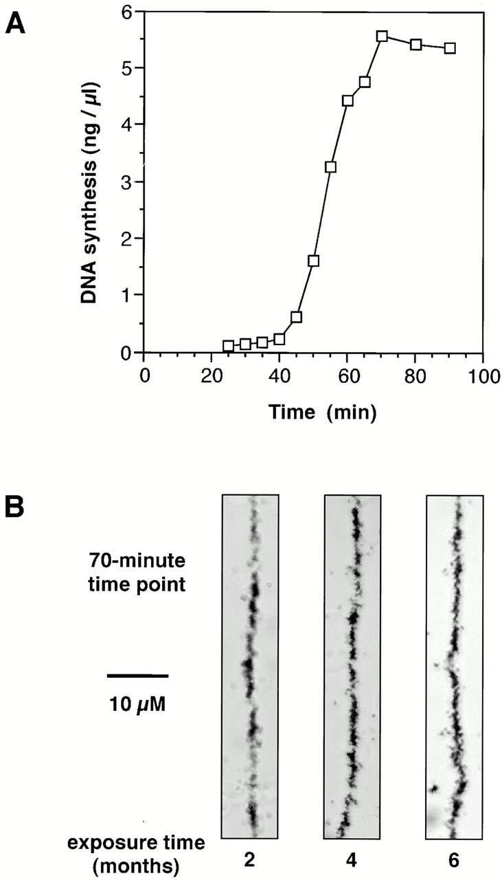
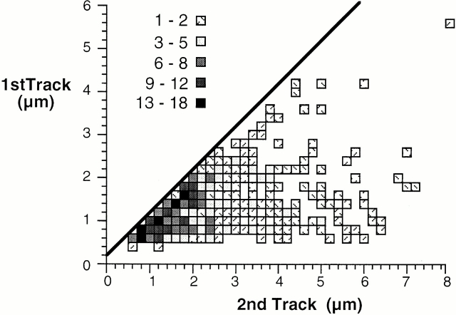
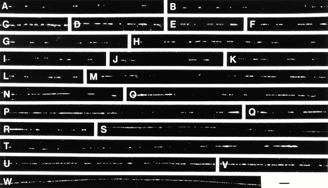
When Xenopus eggs and egg extracts replicate DNA, replication origins are positioned randomly with respect to DNA sequence. However, a completely random distribution of origins would generate some unacceptably large interorigin distances. We have investigated the distribution of replication origins in Xenopus sperm nuclei replicating in Xenopus egg extract. Replicating DNA was labeled with [(3)H]thymidine or bromodeoxyuridine and the geometry of labeled sites on spread DNA was examined. Most origins were spaced 5-15 kb apart. This regular distribution provides an explanation for how complete chromosome replication can be ensured although origins are positioned randomly with respect to DNA sequence. Origins were grouped into small clusters (typically containing 5-10 replicons) that fired at approximately the same time, with different clusters being activated at different times in S phase. This suggests that a temporal program of origin firing similar to that seen in somatic cells also exists in the Xenopus embryo. When the quantity of origin recognition complexes (ORCs) on the chromatin was restricted, the average interorigin distance increased, and the number of origins in each cluster decreased. This suggests that the binding of ORCs to chromatin determines the regular spacing of origins in this system.


Figures

Figure 1
Figure 1
Establishing conditions for DNA fiber autoradiography using Xenopus egg extracts. (A) Xenopus sperm nuclei were incubated in Xenopus egg extract supplemented with [α-32P]dATP. At the indicated times, aliquots were taken and DNA synthesis was assessed by TCA precipitation. (B) Xenopus sperm nuclei were incubated for 70 min in Xenopus egg extract supplemented with [3H]dTTP. DNA was isolated, spread on glass slides, and coated with photographic emulsion. After 2, 4, or 6 mo, slides were developed and examined by light microscopy for the continuity of fiber labeling. Bar, 10 μm.
Figure 3
Figure 3
Center-to-center distances and gap length of fiber autoradiography samples from different stages of S phase. Xenopus sperm nuclei were incubated in Xenopus egg extract supplemented with [3H]dTTP for either (A and B) 45 min, (C and D) 60 min, or (E and F) 70 min. DNA was isolated, processed for fiber autoradiography, and exposed for 6 mo. After development, fibers were examined by light microscopy to measure the center-to-center distance between adjacent tracks (A, C, and E), and to measure the length of the gaps between adjacent tracks (B, D, and F).
Figure 2
Figure 2
Representative examples of fiber autoradiography samples from early and late S phase. Xenopus sperm nuclei were incubated in Xenopus egg extract supplemented with [3H]dTTP for either (A) 45 min or (B) 60 min. DNA was isolated, spread on glass slides, and coated with photographic emulsion. After 6 mo, slides were developed and examined by light microscopy. Bar, 10 μm.
Figure 7
Figure 7
Center-to-center distances of DIRVISH samples from early and late S phase. Xenopus sperm nuclei were incubated in Xenopus egg extract supplemented with BrdUTP for either (A) 45 min or (B) 60 min. Nuclei were isolated, supplemented with SDS, spread over a glass surface, and processed for anti-BrdU immunofluorescence. Fibers were examined by immunofluorescence microscopy to measure the center-to-center distance between adjacent tracks.
Figure 4
Figure 4
Correlation between the length of adjacent tracks observed by fiber autoradiography. From the 45-min sample set described in the legend to Fig. 3, the lengths of adjacent tracks were correlated. For each adjacent pair of tracks, the larger track length is given on the horizontal axis, the shorter track length is shown on the vertical axis. Track pairs where one or more of the tracks were either >8 kb or represented by only a single silver grain have been omitted.
Figure 5
Figure 5
Visualization of replicated DNA in individual nuclei by DIRVISH. Xenopus sperm nuclei were incubated in Xenopus egg extract supplemented with BrdUTP, and after various times nuclei were isolated. Aliquots were then supplemented with SDS, spread over a glass surface and processed for anti-BrdU immunofluorescence. (A) Low power image of a single nucleus incubated for 60 min in egg extract that has almost completed S phase. (B) Higher magnification of part of the nucleus shown in A, showing DNA bundles and individual fibers. (C) Part of a nucleus incubated for 45 min in egg extract, showing many short tracks of replicated DNA. (D) Distribution of the track lengths of the sample shown in C. Bars: (A) 30 μm; (B and C) 3 μm.
Figure 5
Figure 5
Visualization of replicated DNA in individual nuclei by DIRVISH. Xenopus sperm nuclei were incubated in Xenopus egg extract supplemented with BrdUTP, and after various times nuclei were isolated. Aliquots were then supplemented with SDS, spread over a glass surface and processed for anti-BrdU immunofluorescence. (A) Low power image of a single nucleus incubated for 60 min in egg extract that has almost completed S phase. (B) Higher magnification of part of the nucleus shown in A, showing DNA bundles and individual fibers. (C) Part of a nucleus incubated for 45 min in egg extract, showing many short tracks of replicated DNA. (D) Distribution of the track lengths of the sample shown in C. Bars: (A) 30 μm; (B and C) 3 μm.
Figure 6
Figure 6
Representative examples of DIRVISH samples from different stages of S phase. Xenopus sperm nuclei were incubated in Xenopus egg extract supplemented with BrdUTP for either (A and B) 40 min, (C–L) 45 min, (M–T) 50 min, or (U–W) 60 min. Nuclei were isolated, supplemented with SDS, spread over a glass surface, and processed for anti-BrdU immunofluorescence. Bar, 3 μm.
Figure 8
Figure 8
Long fibers observed by DIRVISH. Xenopus sperm nuclei were incubated in Xenopus egg extract supplemented with BrdUTP for 50 min. Nuclei were isolated, supplemented with SDS, spread over a glass surface, and processed for anti-BrdU immunofluorescence. A, B, and C show three contiguous fibers, with arrowheads marking sites of overlap. Bar, 3 μm.
Figure 9
Figure 9
Effect of limiting XORC on origin distribution. Xenopus egg extracts were immunodepleted with anti-XOrc1 antibodies or with nonimmune antibodies. Aliquots of Xenopus sperm nuclei (400 ng DNA) were incubated in either 40 μl untreated extract, 40 μl nonimmune-depleted extract, 40 μl of a 5:1 ratio of XOrc1-depleted extract plus nonimmune-depleted extract (nonimmune-depleted extract ÷ 6), 40 μl of a 17:1 ratio of XOrc1-depleted extract plus nonimmune-depleted extract (nonimmune-depleted extract ÷ 18), 20 μl untreated extract (untreated extract ÷ 2), or 120 μl untreated extract (untreated extract × 3). (A) After 25 min, chromatin was isolated and immunoblotted for XOrc1. (B and C) After 25 min, a further 3 vol of 6-DMAP–treated extract were added to drive synchronous nuclear assembly without allowing further origin assembly. (B) [α-32P]dATP was added to the extract and total DNA synthesis at the indicated times was determined by TCA precipitation. (C) BrdUTP was added to the extract, and at 55 min (untreated extract) or 65 min (depleted extracts) nuclei were isolated and DNA was analyzed by DIRVISH. The mean center-to-center distance and mean number of origins in each cluster were determined. “Origin density” (origins per cluster ÷ mean origin spacing) is also shown.
Figure 10
Figure 10
Spacing of replication origins in the early Xenopus embryo. (A and B) Results generated by a computer program written to simulate the S phase kinetics of sperm nuclei replicating in Xenopus egg extracts. Replication origins were either positioned at random, with a mean replicon size of 9 kb (A), or were set up with a parabolic distribution over 4–14 kb and a mean replicon size of 9 kb (B). The insets show the replicon sizes obtained. Both sets of origins were then activated in asynchronous clusters (parameters based on the kinetic data presented in this paper, see Materials and Methods). The center-to-center distances of nascent DNA tracks after 15 min of S phase are shown in the main graphs. (C) Model of replication origin use in the Xenopus system. A small segment of chromosomal DNA (∼100 kb) is shown looped for convenience. (i) In late mitosis licensed origins are assembled at ∼9-kb intervals. (ii) In early S phase, two clusters initiate synchronously. As the origins are replicated, they convert to the unlicensed state. (iii) During later parts of S phase, other clusters of origins initiate replication synchronously. (iv) By G2, all DNA has replicated and all origins have converted to the unlicensed state.

References

    1. Blow J.J. Preventing re-replication of DNA in a single cell cycleevidence for a replication licensing factor. J. Cell Biol. 1993;122:993–1002. - PMC - PubMed
    1. Blow J.J., Laskey R.A. Initiation of DNA replication in nuclei and purified DNA by a cell-free extract of Xenopus eggs. Cell. 1986;47:577–587. - PubMed
    1. Blow J.J., Laskey R.A. A role for the nuclear envelope in controlling DNA replication within the cell cycle. Nature. 1988;332:546–548. - PubMed
    1. Blow J.J., Sleeman A.M. Replication of purified DNA in Xenopus egg extract is dependent on nuclear assembly. J. Cell Sci. 1990;95:383–391. - PubMed
    1. Blow J.J., Watson J.V. Nuclei act as independent and integrated units of replication in a Xenopus cell-free system. EMBO (Eur. Mol. Biol. Organ.) J. 1987;6:1997–2002. - PMC - PubMed

Publication types