. 2001 Jan 8;152(1):41-50.
doi: 10.1083/jcb.152.1.41.

Vacuolar storage proteins are sorted in the cis-cisternae of the pea cotyledon Golgi apparatus

Affiliations

Vacuolar storage proteins are sorted in the cis-cisternae of the pea cotyledon Golgi apparatus

S Hillmer et al. J Cell Biol. .

Abstract

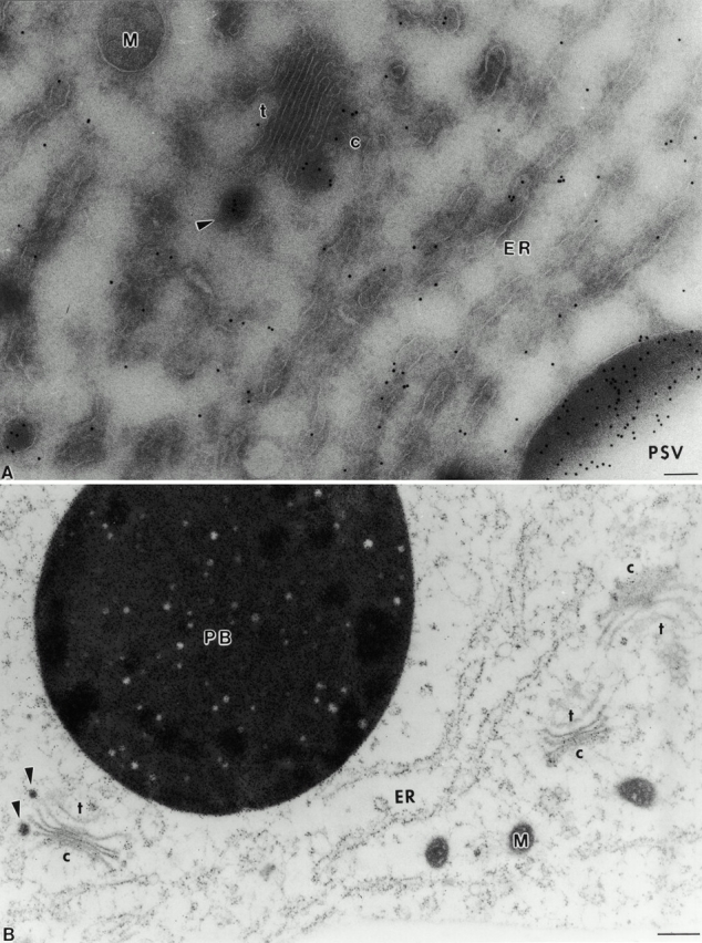
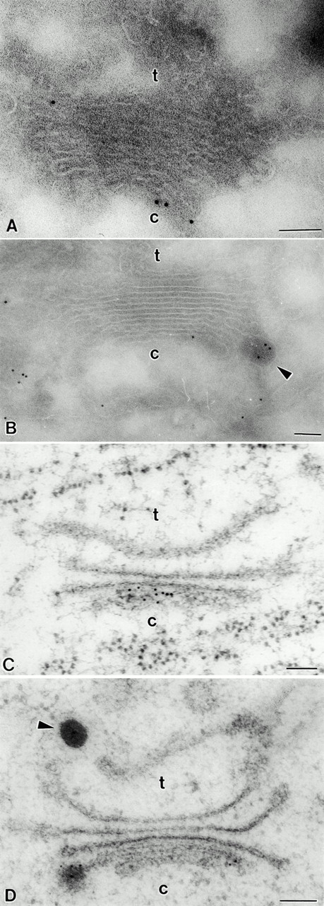
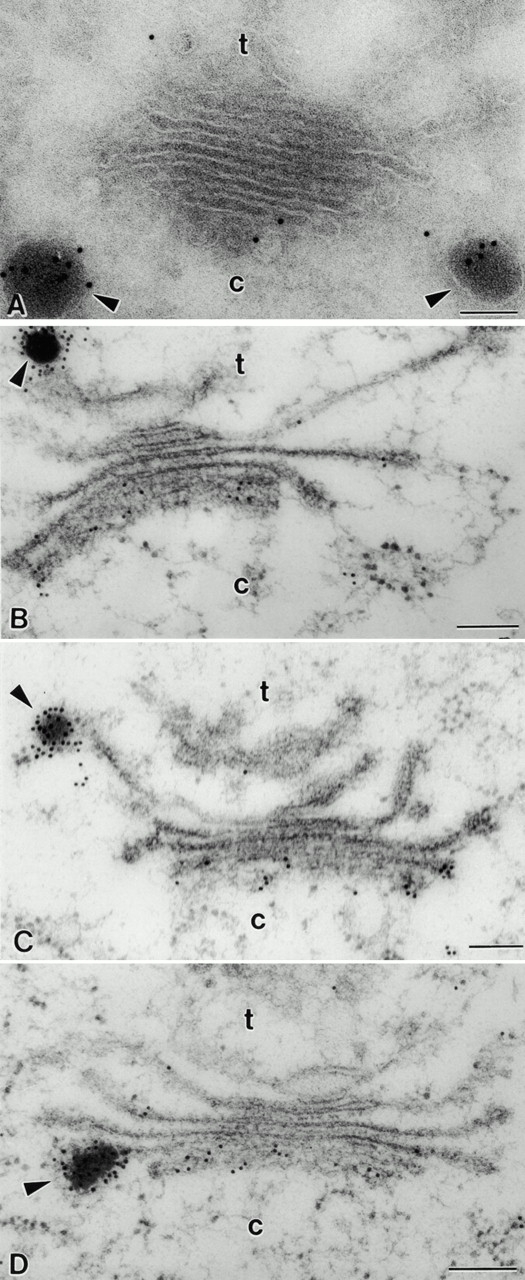
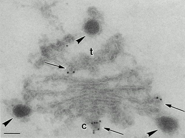
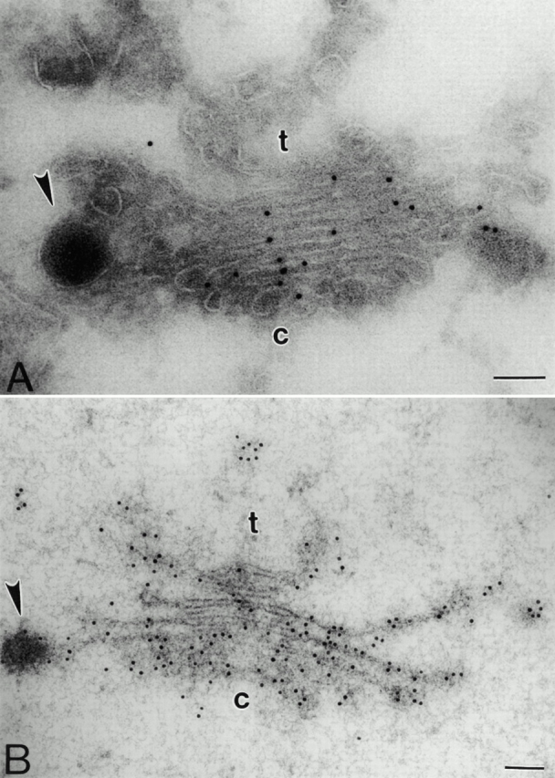
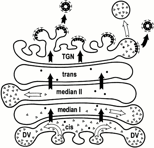
Developing pea cotyledons contain functionally different vacuoles, a protein storage vacuole and a lytic vacuole. Lumenal as well as membrane proteins of the protein storage vacuole exit the Golgi apparatus in dense vesicles rather than in clathrin-coated vesicles (CCVs). Although the sorting receptor for vacuolar hydrolases BP-80 is present in CCVs, it is not detectable in dense vesicles. To localize these different vacuolar sorting events in the Golgi, we have compared the distribution of vacuolar storage proteins and of alpha-TIP, a membrane protein of the protein storage vacuole, with the distribution of the vacuolar sorting receptor BP-80 across the Golgi stack. Analysis of immunogold labeling from cryosections and from high pressure frozen samples has revealed a steep gradient in the distribution of the storage proteins within the Golgi stack. Intense labeling for storage proteins was registered for the cis-cisternae, contrasting with very low labeling for these antigens in the trans-cisternae. The distribution of BP-80 was the reverse, showing a peak in the trans-Golgi network with very low labeling of the cis-cisternae. These results indicate a spatial separation of different vacuolar sorting events in the Golgi apparatus of developing pea cotyledons.


Figures

Figure 1
Figure 1
Distribution of the storage protein vicilin in the endomembrane system of pea cotyledons. (A) Cryosectioned sample. (B) HPF sample. M, Mitochondria; PB, protein body; ER, endoplasmic reticulum. DVs are marked by arrowheads. The cis and trans side of the Golgi are marked by c and t, respectively. c, cis; t, trans. Bars: (A) 100 nm; (B) 250 nm.
Figure 3
Figure 3
Immunogold staining of pea cotyledon Golgi stacks with antibodies against legumin. (A and B) Cryosection samples. Background labeling is very low. DVs are significantly labeled; the label is restricted to the cis-cisternae of the Golgi stack. (C and D) HPF sample. The distribution of gold particles is similar to that in A and B. c, cis; t, trans. Bars, 100 nm.
Figure 2
Figure 2
Immunogold staining of pea cotyledon Golgi stacks with antibodies against vicilin. (A) Cryosectioned sample. Background labeling is very low. DVs are heavily labeled, and the label in the Golgi stack is restricted to the cis-cisternae. (B, C, and D) HPF sample. Labeling of the cisternae reveals a steep gradient from the cis to the trans side of the Golgi. DVs are seen attached to the TGN (B), the trans-cisternae (C), and median cisternae (D). c, cis; t, trans. Bars, 100 nm.
Figure 5
Figure 5
Immunogold staining of cryosectioned pea cotyledon Golgi stacks with antibodies against Atγ-COP. Small (∼60 nm in diameter) budding COPI vesicles (arrows) are significantly labeled with the antibodies. The lumen of the cisternae is not labeled. DVs attached to the cis- and the trans-Golgi cisternae are also not labeled. c, cis; t, trans. Bar, 100 nm.
Figure 4
Figure 4
Immunogold staining of pea cotyledon Golgi stacks with antibodies directed against xylose residues (βF1). (A) Labeling of the Golgi cisternae and DVs on cryosections. Background labeling is very low. Although the Golgi cisternae are more or less uniformly labeled at a high density, labeling of the DVs is rather low. (B) Labeling of the Golgi cisternae and DVs in HPF samples. The distribution of gold particles is the same as for cryosections, but labeling density is considerably higher. c, cis; t, trans. Bars, 100 nm.
Figure 6
Figure 6
Scheme for the localization of the sorting events in the pea cotyledon Golgi stack. Vacuolar hydrolases (filled circles) are first transported through the Golgi stack (black arrows) into the TGN where they are sorted by the VSR BP-80 into CCV (closed bars). Proteins of the PSV (open circles), by contrast, are sorted mainly at the cis-half of the Golgi stack into developing DVs (white arrows). Finally, after maturation, DVs are released from the TGN.

References

    1. Ahmed S.U., Bar-Peled M., Raikhel N.V. Cloning and subcellular location of an Arabidopsis receptor-like protein that shares common features with protein-sorting receptors of eukaryotic cells. Plant Physiol. 1997;114:326–336. - PMC - PubMed
    1. Ahmed S.U., Rojo E., Kovaleva V., Venkataram S., Dombrowski J.E., Matsuoka K., Raikhel N.V. The plant vacuolar sorting receptor AtELP is involved in the transport of NH2-terminal propeptide–containing vacuolar proteins in Arabidopsis thaliana . J. Cell Biol. 2000;149:1335–1344. - PMC - PubMed
    1. Baumgarten B., Tokuyasu K.T., Chrispeels M.J. Immunocytochemical localization of reserve protein in the endoplasmic reticulum of developing bean (Phaseolus vulgaris) cotyledons. Planta. 1980;150:419–425. - PubMed
    1. Bonfanti L., Mironov A.A., Jr., Martinez-Menarguez J.A., Martella O., Fusella A., Baldassarre M., Buccione R., Geuze H.J., Mironov A.A., Luini A. Procollagen traverses the Golgi stack without leaving the lumen of the cisternaeevidence for cisternal maturation. Cell. 1998;95:993–1003. - PubMed
    1. Brigance W.T., Barlowe C., Graham T.R. Organization of the yeast Golgi complex into at least four functionally distinct compartments. Mol. Biol. Cell. 2000;11:171–182. - PMC - PubMed

Publication types