. 2001 Jan 8;152(1):51-64.
doi: 10.1083/jcb.152.1.51.

Membrane recruitment of Aut7p in the autophagy and cytoplasm to vacuole targeting pathways requires Aut1p, Aut2p, and the autophagy conjugation complex

Affiliations

Membrane recruitment of Aut7p in the autophagy and cytoplasm to vacuole targeting pathways requires Aut1p, Aut2p, and the autophagy conjugation complex

J Kim et al. J Cell Biol. .

Abstract

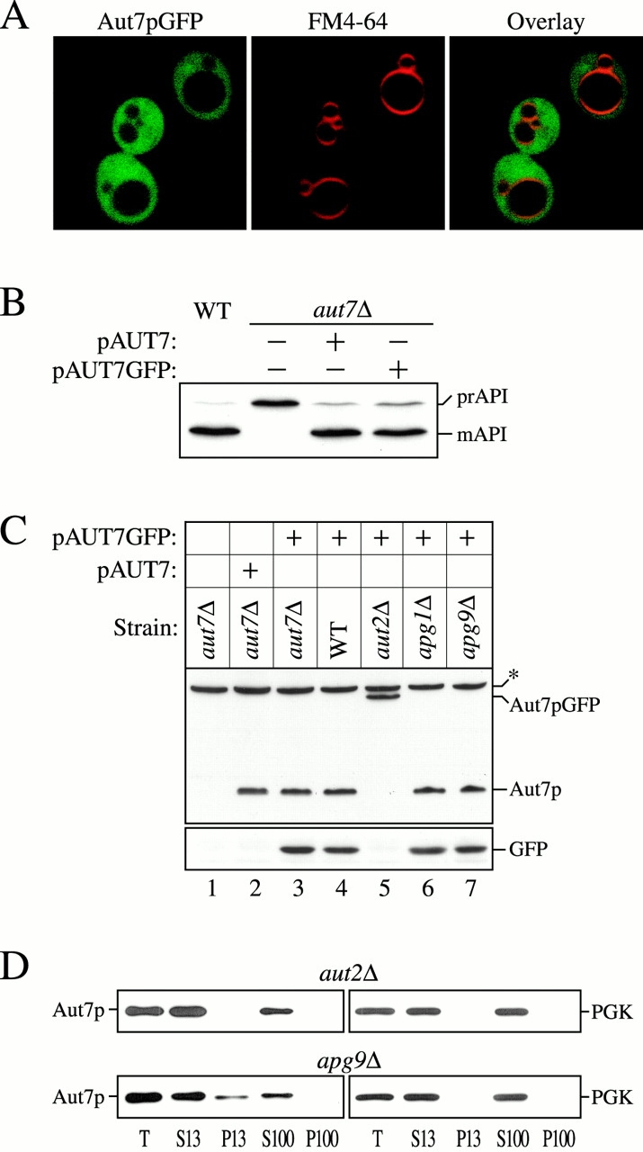
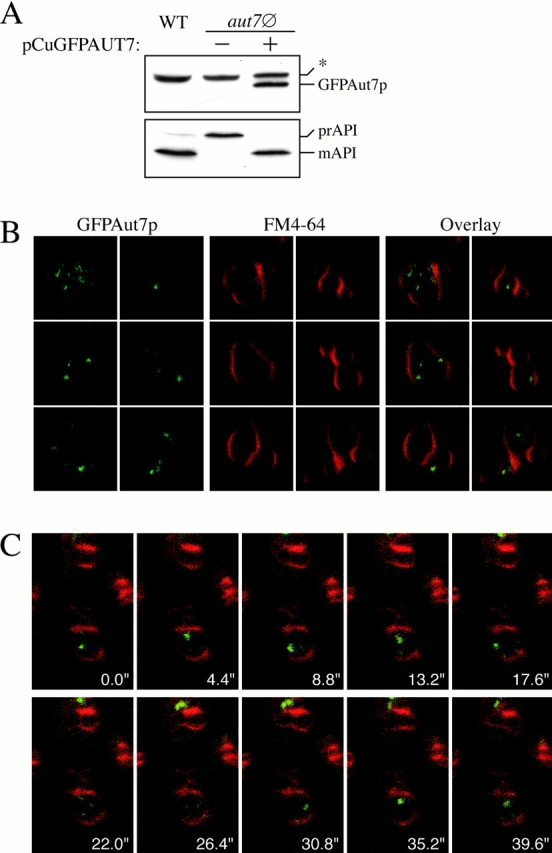
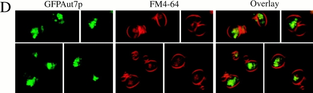
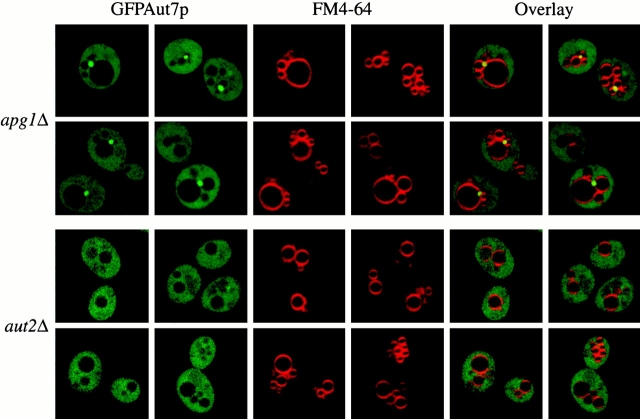
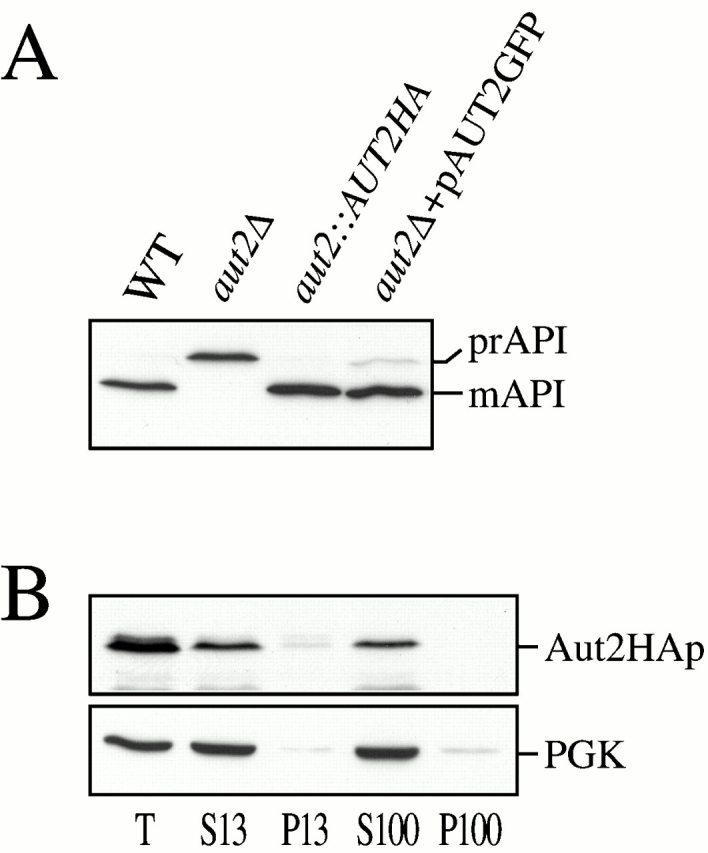
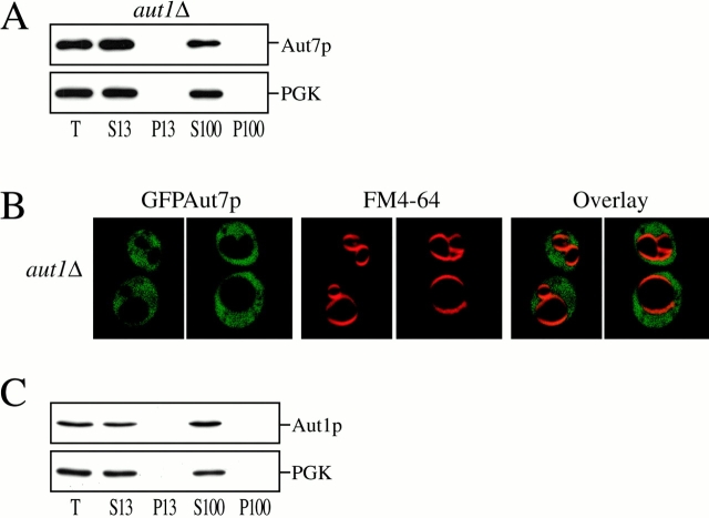
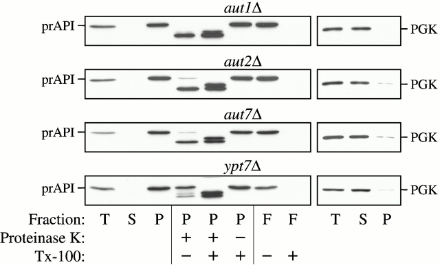
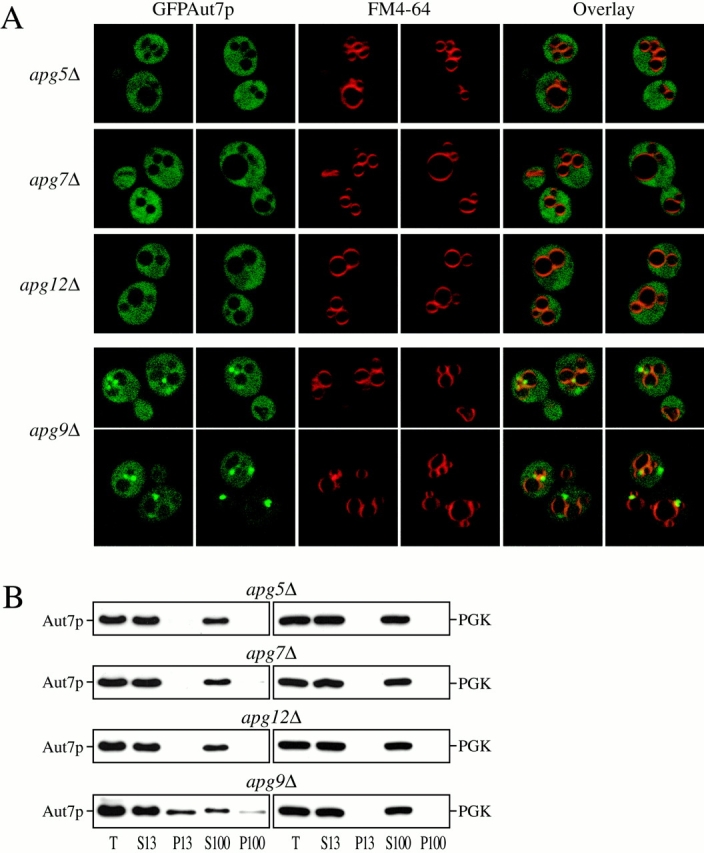
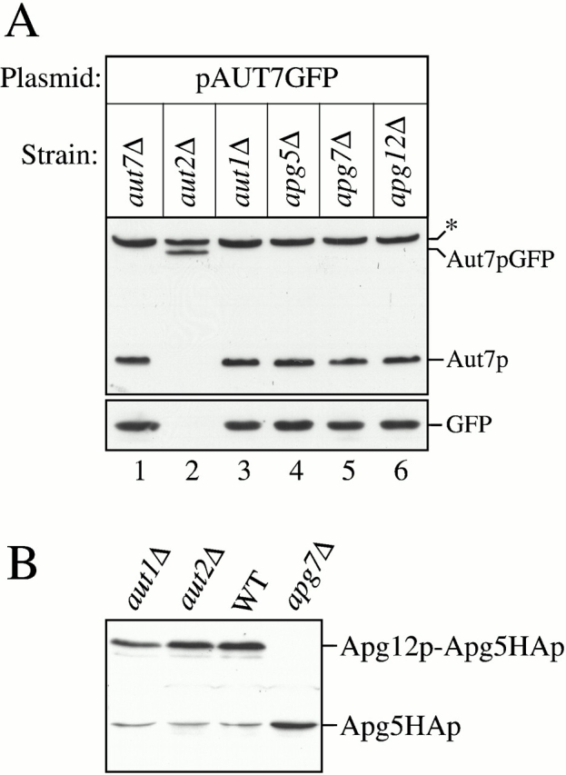
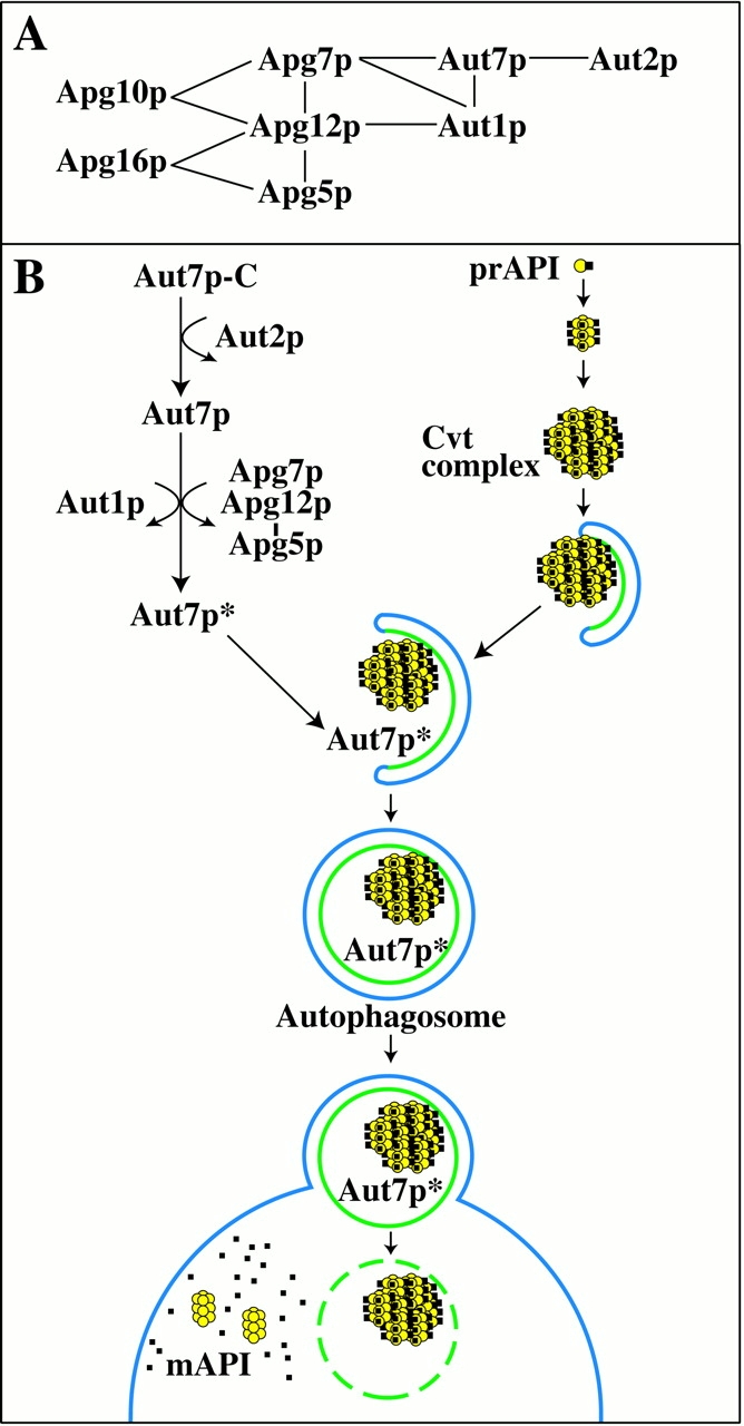
Autophagy is a degradative pathway by which cells sequester nonessential, bulk cytosol into double-membrane vesicles (autophagosomes) and deliver them to the vacuole for recycling. Using this strategy, eukaryotic cells survive periods of nutritional starvation. Under nutrient-rich conditions, autophagy machinery is required for the delivery of a resident vacuolar hydrolase, aminopeptidase I, by the cytoplasm to vacuole targeting (Cvt) pathway. In both pathways, the vesicle formation process requires the function of the starvation-induced Aut7 protein, which is recruited from the cytosol to the forming Cvt vesicles and autophagosomes. The membrane binding of Aut7p represents an early step in vesicle formation. In this study, we identify several requirements for Aut7p membrane association. After synthesis in the cytosol, Aut7p is proteolytically cleaved in an Aut2p-dependent manner. While this novel processing event is essential for Aut7p membrane binding, Aut7p must undergo additional physical interactions with Aut1p and the autophagy (Apg) conjugation complex before recruitment to the membrane. Lack of these interactions results in a cytosolic distribution of Aut7p rather than localization to forming Cvt vesicles and autophagosomes. This study assigns a functional role for the Apg conjugation system as a mediator of Aut7p membrane recruitment. Further, we demonstrate that Aut1p, which physically interacts with components of the Apg conjugation complex and Aut7p, constitutes an additional factor required for Aut7p membrane recruitment. These findings define a series of steps that results in the modification of Aut7p and its subsequent binding to the sequestering transport vesicles of the autophagy and cytoplasm to vacuole targeting pathways.


Figures

Figure 1
Figure 1
Aut2p-dependent proteolytic processing of Aut7p is required for its membrane binding. (A) Localization of Aut7pGFP. Wild-type (WT; SEY6210) cells transformed with a multicopy plasmid encoding GFP fused to the COOH terminus of Aut7p (Aut7pGFP) were grown in SMD to midlog phase and labeled with FM 4-64 to identify vacuoles. The labeled cells were viewed directly with a Leica DM IRB confocal microscope (see Materials and Methods). The cells show a diffuse cytosolic GFP pattern due to the Aut7p COOH-terminal cleavage event, which liberates GFP into the cytosol (see below). (B) WT (SEY6210) and aut7Δ (WPHYD7) strains transformed with either the centromeric AUT7 plasmid (pAUT7) or AUT7GFP plasmid were grown to midlog phase in SMD. Protein extracts were prepared and analyzed by immunoblots using antiserum to API. The positions of the precursor and mature forms of API are indicated. Both the pAUT7 and pAUT7GFP plasmids rescue the prAPI transport defect in aut7Δ. (C) WT (SEY6210), aut7Δ (WPHYD7), aut2Δ (WPHYD2), apg1Δ (NNY20), and apg9Δ (JKY007) strains transformed with either the centromeric pAUT7 or pAUT7GFP plasmids were grown to midlog phase in SMD. Protein extracts were prepared and analyzed by immunoblots using antiserum to Aut7p (top) or GFP (bottom). *Background band. The COOH-terminal processing of Aut7pGFP is dependent on Aut2p. (D) Aut7p subcellular fractionation pattern. Cells from aut2Δ and the apg9Δ control strain were grown to midlog phase in SMD, and then shifted to SD-N autophagy induction conditions for 2 h before converting them to spheroplasts. The spheroplasts were lysed in TEA osmotic lysis buffer (see Materials and Methods). After a preclearing centrifugation step at 100 g for 5 min to remove unlysed spheroplasts, the total lysate (T) was separated into low-speed supernatant (S13) and pellet (P13) fractions. The S13 fraction was further separated into 100,000 g supernatant (S100) and pellet (P100) fractions. The fractionated samples were subjected to immunoblot analysis using antiserum to Aut7p and the cytosolic marker protein PGK. The proteolytic cleavage of Aut7p is required for its subsequent association with a P13 membrane fraction.
Figure 2
Figure 2
A GFP fusion to the NH2 terminus of Aut7p (GFPAut7p) serves as a functional marker for the Cvt and autophagy pathways. (A) The aut7Δ (WPHYD7) strain was transformed with a plasmid encoding GFP fused to the NH2 terminus of Aut7p and under the control of the CUP1 copper-inducible promoter (pCuGFPAUT7). Cells were grown in SMD medium lacking copper until midlog phase, and then induced with 50 μM CuSO4 for 3 h. Protein extracts were prepared as described in Materials and Methods and examined by immunoblots with anti–Aut7p (top) and anti–API (bottom) antiserum. *Background band. GFPAut7p is expressed as a full-length fusion protein and complements the prAPI defect in aut7Δ cells. (B) GFPAut7p labels Cvt bodies inside the vacuole. The pep4Δ (TVY1) strain was transformed with pCuGFPAUT7 and grown to midlog phase in SMD medium lacking copper. GFPAut7p expression was induced with 10 μM CuSO4 for 3 h. After labeling the vacuoles with FM 4-64, the cells were examined on a Leica DM IRB confocal microscope. In nutrient-rich conditions, GFPAut7p accumulates inside Cvt bodies in pep4Δ cells. (C) Time-series examination of Cvt bodies. GFPAut7p-expressing pep4Δ (TVY1) cells were treated as in B. A single optical plane of cells was followed over the time course indicated. (D) GFPAut7p labels autophagic bodies inside the vacuole. GFPAut7p expression was induced with 50 μM CuSO4 for 3 h in pep4Δ (TVY1) cells. After labeling the vacuoles with FM 4-64, the cells were shifted to SD-N for 3 h. In starvation conditions, GFPAut7p accumulates inside autophagic bodies in pep4Δ cells.
Figure 2
Figure 2
A GFP fusion to the NH2 terminus of Aut7p (GFPAut7p) serves as a functional marker for the Cvt and autophagy pathways. (A) The aut7Δ (WPHYD7) strain was transformed with a plasmid encoding GFP fused to the NH2 terminus of Aut7p and under the control of the CUP1 copper-inducible promoter (pCuGFPAUT7). Cells were grown in SMD medium lacking copper until midlog phase, and then induced with 50 μM CuSO4 for 3 h. Protein extracts were prepared as described in Materials and Methods and examined by immunoblots with anti–Aut7p (top) and anti–API (bottom) antiserum. *Background band. GFPAut7p is expressed as a full-length fusion protein and complements the prAPI defect in aut7Δ cells. (B) GFPAut7p labels Cvt bodies inside the vacuole. The pep4Δ (TVY1) strain was transformed with pCuGFPAUT7 and grown to midlog phase in SMD medium lacking copper. GFPAut7p expression was induced with 10 μM CuSO4 for 3 h. After labeling the vacuoles with FM 4-64, the cells were examined on a Leica DM IRB confocal microscope. In nutrient-rich conditions, GFPAut7p accumulates inside Cvt bodies in pep4Δ cells. (C) Time-series examination of Cvt bodies. GFPAut7p-expressing pep4Δ (TVY1) cells were treated as in B. A single optical plane of cells was followed over the time course indicated. (D) GFPAut7p labels autophagic bodies inside the vacuole. GFPAut7p expression was induced with 50 μM CuSO4 for 3 h in pep4Δ (TVY1) cells. After labeling the vacuoles with FM 4-64, the cells were shifted to SD-N for 3 h. In starvation conditions, GFPAut7p accumulates inside autophagic bodies in pep4Δ cells.
Figure 3
Figure 3
Aut2p is required for the GFPAut7p association to punctate, perivacuolar structures. The apg1Δ (NNY20) and aut2Δ (WPHYD2) strains were transformed with the pCuGFPAUT7 plasmid. The cells were grown to midlog phase in SMD medium. GFPAut7p expression was then induced with 50 μM CuSO4 for 3 h. Cells were then labeled with FM 4-64, shifted to SD-N medium for 3 h, and examined as described in Fig. 2. In apg1Δ cells, GFPAut7p is recruited to punctate structures, whereas in the Aut7p processing–defective aut2Δ cells, GFPAut7p appears uniformly distributed in the cytoplasm.
Figure 4
Figure 4
Characterization of Aut2p. (A) API immunoblots of WT (SEY6210), aut2Δ (WPHYD2), WT with a chromosomal integration of an AUT2HA fusion at the AUT2 locus (WPHY2HA, aut2::AUT2HA), and aut2Δ transformed with a centromeric plasmid encoding GFP fused to the COOH terminus of Aut2p (pAUT2GFP). Protein extracts were prepared as described in Materials and Methods. Both Aut2HAp and Aut2pGFP are functional fusion proteins as prAPI maturation appears normal. (B) Aut2HAp subcellular fractionation pattern. Cells expressing the Aut2HAp from the AUT2 chromosomal locus were grown in SMD to midlog phase, and then shifted to SD-N for 2 h before spheroplasting. The spheroplasts were lysed in TEA osmotic lysis buffer as described in Materials and Methods. The precleared total lysate (T) was separated into low-speed supernatant and pellet fractions (S13 and P13). The S13 fraction was further resolved into high-speed supernatant and pellet fractions (S100 and P100) as described in Fig. 1 D and Materials and Methods. Immunoblot analysis was performed with antisera to HA and the cytosolic marker protein, PGK. Aut2HAp is a soluble protein and localizes to the supernatant fractions.
Figure 5
Figure 5
Characterization of Aut1p and its role in Aut7p membrane binding. (A) Aut7p subcellular fractionation pattern in aut1Δ. Cells from the aut1Δ (WPHYD1) strain were grown to midlog phase in SMD and shifted to SD-N medium for 2 h before spheroplasting. The spheroplasts were then lysed osmotically as described in Materials and Methods. The precleared total lysate (T) was separated into low-speed supernatant and pellet fractions (S13 and P13); the S13 fraction was further separated into high-speed supernatant and pellet fractions (S100 and P100). Immunoblot analysis was performed using antiserum to Aut7p and the cytosolic marker PGK. Deletion of AUT1 prevents Aut7p binding to the P13 membrane fraction. (B) Localization of GFPAut7p in aut1Δ. The aut1Δ (WPHYD1) strain was transformed with the pCuGFPAUT7 plasmid. Cells were grown to midlog phase in SMD medium, induced with 50 μM CuSO4 for 3 h, labeled with FM 4-64, shifted to SD-N medium for 3 h, and examined as described in Materials and Methods. GFPAut7p appears uniformly distributed in the cytoplasm of the aut1Δ cells. For comparison, see GFPAut7p localization in apg1Δ (Fig. 3). (C) Aut1p subcellular fractionation pattern. WT (SEY6210) cells were grown to midlog phase in SMD and shifted to SD-N medium for 2 h before spheroplasting. The subcellular fractionation procedure used to examine Aut1p was identical to the method described in A. Immunoblot analysis was performed using antisera to Aut1p and the cytosolic marker PGK. Aut1p is a soluble protein and appears in the supernatant fractions.
Figure 6
Figure 6
Protease-sensitivity and membrane-flotation analyses. The aut1Δ, aut2Δ, aut7Δ, and ypt7Δ strains were grown to midlog phase and converted into spheroplasts. The spheroplasts were lysed in PS200 osmotic lysis buffer. The total lysate (T) was resolved into supernatant (S) and pellet (P) fractions by centrifugation at 5,000 rpm. For the protease sensitivity assay, the pellet fractions were subjected to protease treatment in the absence or presence of 0.2% Triton X-100, as described in Materials and Methods. For the membrane flotation assay, the pellet fraction was subjected to centrifugation in a Ficoll step gradient with or without detergent, as described in Materials and Methods. Membrane-associated proteins were collected in the float (F) fraction. All samples were subjected to immunoblot analysis with antisera to API and PGK. The aut1Δ, aut2Δ, and aut7Δ mutants all accumulate precursor API at a protease-sensitive, membrane-associated stage.
Figure 7
Figure 7
The Apg conjugation system is required for Aut7p membrane binding. (A) The Apg conjugation mutants apg5Δ (MGY101), apg7Δ (VDY101), apg9Δ (JKY007), and apg12Δ (YNM101) strains were transformed with the pCuGFPAUT7 plasmid. GFPAut7p expression was induced at midlog stage with 50 μM CuSO4 for 3 h. Cells were then labeled with FM 4-64, shifted to SD-N medium for 3 h, and examined by confocal microscopy as described in Materials and Methods. In apg9Δ cells, GFPAut7p is recruited to punctate, perivacuolar structures, whereas GFPAut7p appears uniformly distributed in the apg5Δ, apg7Δ, and apg12Δ conjugation mutants. (B) Aut7p subcellular fractionation pattern in the Apg conjugation mutants. Cells from apg5Δ, apg7Δ, apg9Δ, and apg12Δ strains were grown to midlog phase in SMD, and then shifted to SD-N for 2 h to induce autophagy. After spheroplasting and osmotic lysis in TEA osmotic lysis buffer, the precleared total lysate (T) was separated into low-speed supernatant (S13) and pellet (P13) fractions. The S13 fraction was further separated into 100,000 g supernatant (S100) and pellet (P100) fractions. The fractionated samples were subjected to immunoblot analysis using antisera to Aut7p and PGK. The apg5Δ, apg7Δ, and apg12Δ conjugation mutants are defective in Aut7p binding to the P13 pellet fraction. Essentially identical results were seen with the apg10 and apg16Δ mutant strains (data not shown).
Figure 8
Figure 8
Aut7p COOH-terminal processing and Apg12p–Apg5p conjugate formation analyses. (A) The aut7Δ (WPHYD7), aut2Δ (WPHYD2), aut1Δ (WPHYD1), apg5Δ (MGY101), apg7Δ (VDY101), and apg12Δ (YNM101) strains were transformed with the centromeric pAUT7GFP plasmid. Cells were grown to midlog phase in SMD. Protein extracts were prepared and analyzed by immunoblots using antisera to Aut7p (top) and GFP (bottom). *Background band. Aut1p and the Apg conjugation components (Apg5p, Apg7p, and Apg12p) are not required for Aut7p COOH-terminal processing. (B) The aut1Δ, aut2Δ, wild-type (SEY6210), and apg7Δ strains were transformed with a plasmid encoding an Apg5HAp fusion protein. Protein extracts from cells grown to midlog phase were prepared and analyzed by immunoblots with antiserum to the HA epitope. The Apg12p–Apg5p conjugate can be detected in the aut1Δ, aut2Δ, and wild-type strains, but not in the apg7Δ strain.
Figure 9
Figure 9
(A) Molecular interactions between autophagy components. Apg5p, 7p, 10p, 12p, and 16p constitute the Apg conjugation system. This covalent protein-modification system is essential for the Cvt and autophagy pathways. Interactions between Apg conjugation components and Aut7p, Aut1p, and Aut2p have also been recently demonstrated. Details are discussed in the text. (B) A model of Aut7p membrane binding in the context of prAPI transport. In summary, we have defined three discrete events that lead to the membrane binding of Aut7p. First, Aut7p is synthesized in the cytosol and subsequently cleaved in an Aut2p-dependent manner. Once cleaved, Aut1p and the Apg conjugation system further interact with Aut7p to facilitate its binding to the membrane. These steps required for Aut7p membrane binding are presented in the context of prAPI import by the autophagy pathway. Details are discussed in the text.

Similar articles

Cited by

References

    1. Abeliovich H., Dunn W.A., Jr., Kim J., Klionsky D.J. Dissection of autophagosome biogenesis into distinct nucleation and expansion steps. J. Cell Biol. 2000;151:1025–1033. - PMC - PubMed
    1. Baba M., Osumi M., Scott S.V., Klionsky D.J., Ohsumi Y. Two distinct pathways for targeting proteins from the cytoplasm to the vacuole/lysosome. J. Cell Biol. 1997;139:1687–1695. - PMC - PubMed
    1. Baum P., Thorner J., Honig L. Identification of tubulin from the yeast Saccharomyces cerevisiae . Proc. Natl. Acad. Sci. USA. 1978;75:4962–4966. - PMC - PubMed
    1. George M.D., Baba M., Scott S.V., Mizushima N., Garrison B.S., Ohsumi Y., Klionsky D.J. Apg5p functions in the sequestration step in the cytoplasm-to-vacuole targeting and macroautophagy pathways. Mol. Biol. Cell. 2000;11:969–982. - PMC - PubMed
    1. Gerhardt B., Kordas T.J., Thompson C.M., Patel P., Vida T. The vesicle transport protein Vps33p is an ATP-binding protein that localizes to the cytosol in an energy-dependent manner. J. Biol. Chem. 1998;273:15818–15829. - PubMed

Publication types

MeSH terms

Substances