. 2001 Jan 8;152(1):97-110.
doi: 10.1083/jcb.152.1.97.

Targeting Pyk2 to beta 1-integrin-containing focal contacts rescues fibronectin-stimulated signaling and haptotactic motility defects of focal adhesion kinase-null cells

Affiliations

Targeting Pyk2 to beta 1-integrin-containing focal contacts rescues fibronectin-stimulated signaling and haptotactic motility defects of focal adhesion kinase-null cells

C K Klingbeil et al. J Cell Biol. .

Abstract

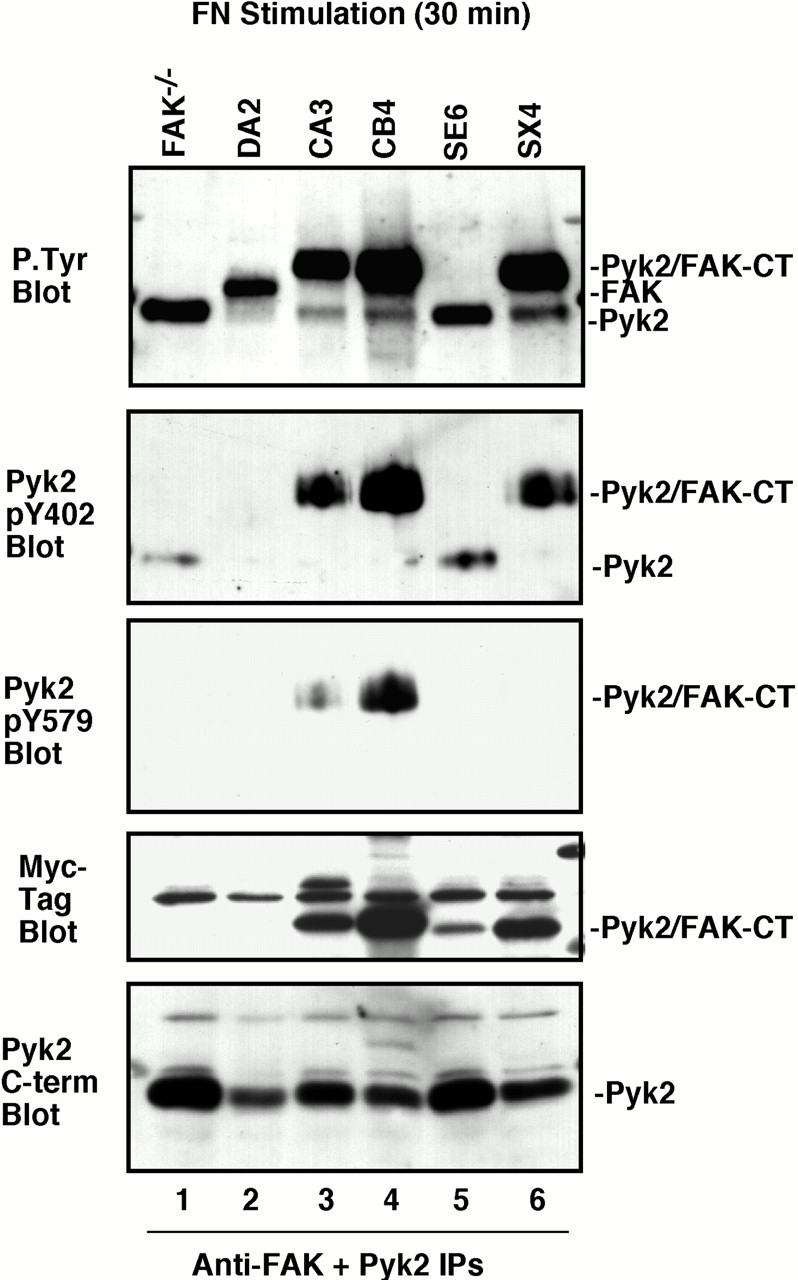
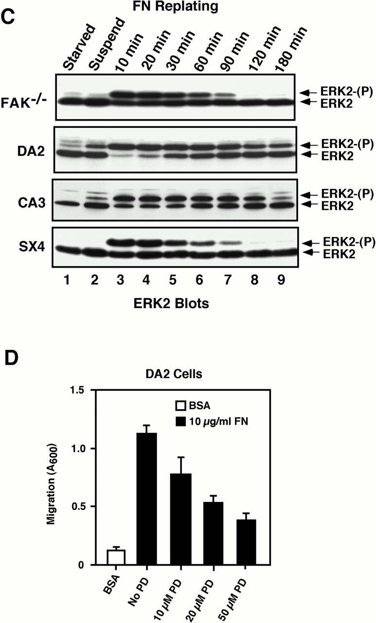
Focal adhesion kinase-null (FAK(-/-) fibroblasts exhibit morphological and motility defects that are reversed by focal adhesion kinase (FAK) reexpression. The FAK-related kinase, proline-rich tyrosine kinase 2 (Pyk2), is expressed in FAK(-/-) cells, yet it exhibits a perinuclear distribution and does not functionally substitute for FAK. Chimeric Pyk2/FAK proteins were created and expressed in FAK(-/-) cells to determine the impact of Pyk2 localization to focal contacts. Whereas an FAK/Pyk2 COOH-terminal (CT) domain chimera was perinuclear distributed, stable expression of a Pyk2 chimera with the FAK-CT domain (Pyk2/FAK-CT) localized to focal contact sites and enhanced fibronectin (FN)-stimulated haptotactic cell migration equal to FAK-reconstituted cells. Disruption of paxillin binding to the FAK-CT domain (S-1034) inhibited Pyk2/FAK-CT localization to focal contacts and its capacity to promote cell motility. Paxillin binding to the FAK-CT was necessary but not sufficient to mediate the indirect association of FAK or Pyk2/FAK-CT with a beta 1-integrin-containing complex. Both FAK and Pyk2/FAK-CT but not Pyk2/FAK-CT S-1034 reconstituted FAK(-/-) cells, exhibit elevated FN-stimulated extracellular signal-regulated kinase 2 (ERK2) and c-Jun NH(2)-terminal kinase (JNK) kinase activation. FN-stimulated FAK or Pyk2/FAK-CT activation enhanced both the extent and duration of FN-stimulated ERK2 activity which was necessary for cell motility. Transient overexpression of the FAK-CT but not FAK-CT S-1034 domain inhibited both FN-stimulated ERK2 and JNK activation as well as FN-stimulated motility of Pyk2/FAK-CT reconstituted cells. These gain-of-function studies show that the NH(2)-terminal and kinase domains of Pyk2 can functionally substitute for FAK in promoting FN-stimulated signaling and motility events when localized to beta-integrin-containing focal contact sites via interactions mediated by the FAK-CT domain.


Figures

Figure 1
Figure 1
Creation of Pyk2 and FAK chimeric proteins. (A) Schematic representation of HA-FAK, Myc-Pyk2, Pyk2/FAK-CT, and FAK/Pyk2-CT. Pyk2/FAK-CT consists of Pyk2 residues 1–682 including the Pyk2 kinase domain and FAK residues 680–1052. FAK/Pyk2-CT consists of FAK residues 1–400 including the intact Src SH2 binding site surrounding FAK Tyr-397 and Pyk2 residues 405–1009 encompassing the Pyk2 kinase and CT domains. The inclusion of either six repeated Myc tags at the NT domain or three repeated HA tags at the CT domain is indicated for each construct. (B) Analysis of FN-stimulated 32P in vitro kinase activity as measured in IPs to the transiently expressed epitope-tagged proteins. Immunoprecipitated proteins were transferred to PVDF membranes and visualized by autoradiography, followed by HA and Myc tag blotting. PBS, paxillin binding sequence.
Figure 2
Figure 2
Transient expression of Pyk2/FAK-CT localizes to focal contacts and promotes FN-stimulated FAK−/− cell motility. (A) FAK−/− cells transiently transfected with either pCDNA3 control vector, Pyk2, Pyk2/FAK-CT, or FAK/Pyk2-CT were grown on FN-coated glass slides overnight and stained with an antibody to vinculin or to the Myc epitope tag. Arrows indicate transfected cells. (B) FAK−/− cells were transiently cotransfected with 2.5 μg pCDNA3.1-lacZ and either control vector, Pyk2, FAK/Pyk2-CT, or Pyk2/FAK-CT (2.5 μg each in pCDNA3.1) and were analyzed for FN-stimulated (10 μg/ml) haptotaxis cell migration (3 h). Transfected migratory cells were identified by β-gal staining and counted. Exogenous protein expression was verified in the excess cells not used in the migration assay by Myc tag blotting. Relative fold induction of migration was calculated by normalizing transfection-induced migration to the pCDNA3.1 control transfected FAK−/− cells. Data represent the mean ± SD of four independent experiments. Bar, ∼20 μM.
Figure 3
Figure 3
Stable Pyk2/FAK-CT expression promotes fibrillar morphology of FAK−/− cells. (A) Equal total protein from WCLs of FAK−/−, FAK+/+, HA-FAK reconstituted (DA2), Pyk2/FAK-CT reconstituted (clones CA3 and CB4), or Pyk2/FAK-CT S-1034 reconstituted (clones SE6 and SX4) were resolved by SDS-PAGE and sequentially blotted with antibodies to either the FAK-CT domain, Myc tag, HA tag, p130Cas, or to the cytoplasmic domain of the β1-integrin. Panels are a compilation from two gels. The positions of FAK, HA-FAK, Pyk2/FAK-CT, and a nonspecific band are indicated in the FAK-CT blot. (B) FAK−/−, DA2, CA3, CB4, and SX4 cells were plated onto FN-coated (10 μg/ml) glass slides for 2 h in the absence of serum and stained with either affinity-purified antibodies to Pyk2, an mAb to the HA tag, or an mAb to vinculin as indicated (inset). Arrows indicate focal contact staining. Bar, ∼20 μM.
Figure 3
Figure 3
Stable Pyk2/FAK-CT expression promotes fibrillar morphology of FAK−/− cells. (A) Equal total protein from WCLs of FAK−/−, FAK+/+, HA-FAK reconstituted (DA2), Pyk2/FAK-CT reconstituted (clones CA3 and CB4), or Pyk2/FAK-CT S-1034 reconstituted (clones SE6 and SX4) were resolved by SDS-PAGE and sequentially blotted with antibodies to either the FAK-CT domain, Myc tag, HA tag, p130Cas, or to the cytoplasmic domain of the β1-integrin. Panels are a compilation from two gels. The positions of FAK, HA-FAK, Pyk2/FAK-CT, and a nonspecific band are indicated in the FAK-CT blot. (B) FAK−/−, DA2, CA3, CB4, and SX4 cells were plated onto FN-coated (10 μg/ml) glass slides for 2 h in the absence of serum and stained with either affinity-purified antibodies to Pyk2, an mAb to the HA tag, or an mAb to vinculin as indicated (inset). Arrows indicate focal contact staining. Bar, ∼20 μM.
Figure 4
Figure 4
FAK-CT but not FAK-CT S-1034 mediated targeting of the Pyk2-NT and kinase domains to focal contacts rescues FAK−/− FN-stimulated motility defects. Boyden chamber FN-stimulated haptotaxis migration assays (3 h) were performed with equal numbers of FAK−/−, FAK+/+, DA2, CA3, CB4, SE6, and SX4 fibroblasts. Migratory cells on the membrane underside were stained with Crystal Violet and the dye absorbence was quantified at 600 nm. Data represent the mean ± SD of seven independent experiments.
Figure 5
Figure 5
FAK-CT domain–mediated connections to a β1-integrin–containing complex. FAK−/− cells were transfected with expression vectors for either Myc-Pyk2, Myc–Pyk2/FAK-CT (with HA tag on CT domain), HA-FAK, HA-FAK S-1034, and the exogenous proteins were isolated with antibodies to either the Myc (lanes 1–3) or HA epitope tags (lanes 4–6). Two sets of IPs were sequentially analyzed for associated proteins by anti–β1-integrin, antitalin, anti-p130Cas, and antipaxillin blotting. Control IPs showing nonspecific protein association were performed with control vector (pCDNA) transfected cells using either Myc tag (lane 1) or HA tag (lane 4) antibodies. Combined blotting of WCLs on the adjacent gel lane was used to verify specific protein immunoreactivity (1/20 of total cell lysate) and represents an equal chemiluminescent exposure to the IP lanes. The transfected proteins were detected by either Myc or HA tag blotting of the IPs.
Figure 6
Figure 6
Differential FN-stimulated Pyk2/FAK-CT and Pyk2/FAK-CT S-1034 tyrosine phosphorylation. 2 × 10⁶ FAK−/−, DA2, CA3, CB4, SE6, or SX4 cells were plated on FN-coated dishes (2 μg/ml) for 30 min and a combination of affinity-purified antibodies to the FAK-CT or Pyk2–NT domain (2.5 μg each) were used to isolate both endogenous Pyk2 and the stably expressed HA-FAK or Pyk2/FAK-CT proteins. IPs were resolved by SDS-PAGE and sequentially blotted with antibodies to P.Tyr, phospho-specific antibodies to either the Pyk2 pY402 or pY579 phosphorylation sites, to the Myc tag, or with an mAb to Pyk2 as indicated. Panels are a compilation from two gels and the migration of Pyk2/FAK-CT, HA–FAK, or Pyk2 is indicated.
Figure 7
Figure 7
Stable FAK and Pyk2/FAK-CT expression enhance the extent and duration of FN-stimulated ERK2 activation required for cell motility. (A) Lysates from FN-stimulated cells were prepared as described in the legend to Fig. 6, and 50 μg of total cell protein/lane was resolved by SDS-PAGE and analyzed by anti-P.Tyr blotting. (B) Lysates (50 μg) from the indicated FN-stimulated cells were blotted with an mAb to activated and phosphorylated ERK1/ERK2 and then reprobed with an mAb to ERK2. (C) Lysates from either FAK−/−, DA2, CA3, or SX4 cells were prepared from either serum-starved, suspended, or cells replated onto FN-coated (10 μg/ml) dishes for the times indicated. Proteins were resolved on a 15% SDS-PAGE gel and analyzed by mAb ERK2 blotting. The slower migrating form of ERK2 represents phosphorylated and active ERK2 (ERK2-(P)). (D) Serum-starved DA2 cells, pretreated in suspension with the indicated concentrations of PD98059 for 30 min, were employed in Boyden chamber haptotactic migration assays using immobilized FN or BSA as stimulus. PD98059 was added to the lower chambers and after 3 h, cells on the lower side of the membrane were fixed and stained, and the eluted dye was quantified by absorbence. Bars represent the means from two separate experiments.
Figure 7
Figure 7
Stable FAK and Pyk2/FAK-CT expression enhance the extent and duration of FN-stimulated ERK2 activation required for cell motility. (A) Lysates from FN-stimulated cells were prepared as described in the legend to Fig. 6, and 50 μg of total cell protein/lane was resolved by SDS-PAGE and analyzed by anti-P.Tyr blotting. (B) Lysates (50 μg) from the indicated FN-stimulated cells were blotted with an mAb to activated and phosphorylated ERK1/ERK2 and then reprobed with an mAb to ERK2. (C) Lysates from either FAK−/−, DA2, CA3, or SX4 cells were prepared from either serum-starved, suspended, or cells replated onto FN-coated (10 μg/ml) dishes for the times indicated. Proteins were resolved on a 15% SDS-PAGE gel and analyzed by mAb ERK2 blotting. The slower migrating form of ERK2 represents phosphorylated and active ERK2 (ERK2-(P)). (D) Serum-starved DA2 cells, pretreated in suspension with the indicated concentrations of PD98059 for 30 min, were employed in Boyden chamber haptotactic migration assays using immobilized FN or BSA as stimulus. PD98059 was added to the lower chambers and after 3 h, cells on the lower side of the membrane were fixed and stained, and the eluted dye was quantified by absorbence. Bars represent the means from two separate experiments.
Figure 8
Figure 8
Focal contact localization of FAK or Pyk2/FAK-CT specifically promotes FN-stimulated JNK activation. (A) Lysates (50 μg) from the indicated FN-stimulated cells were blotted with affinity-purified polyclonal antibodies to activated and phosphorylated JNK and then reprobed with a polyclonal antibody to JNK-1. (B) Human 293T cells were transiently cotransfected with a flag-tagged JNK-1 reporter along with either control vector, HA-FRNK, or HA-FRNK S-1034 and lysates were prepared from either suspended (S) or FN-stimulated cells. Flag-tagged JNK-1 in vitro kinase activity was measured in flag tag IPs by the phosphorylation of GST-c-Jun and visualized by autoradiography. Expression of flag-JNK-1 was visualized by flag blotting and FRNK expression was visualized by HA-tag blotting.
Figure 9
Figure 9
FRNK expression inhibits FN-stimulated ERK2 activation and Pyk2/FAK-CT haptotactic motility. (A) Human 293T cells were transiently cotransfected with a flag-tagged ERK2 reporter along with either control vector, FAK, FAK plus FRNK, or F-397 FAK and lysates were prepared from either suspended or FN-stimulated cells. FAK and FRNK expression was verified by HA tag blotting and FAK tyrosine phosphorylation was analyzed by FAK–NT domain–directed antibody IPs followed by P.Tyr blotting. ERK2 in vitro kinase activity was measured in flag tag IPs by the phosphorylation of MBP and visualized by autoradiography. The amount of 32P incorporated into MBP was determined by Cerenkov counting, and the values represent the mean from two independent experiments. (B) CA3 cells were transiently cotransfected with 2.5 μg pCDNA3.1 Lac-Z and either control vector, FRNK, or FRNK S-1034 (5 μg in pCDNA3.1) and analyzed in FN haptotaxis assays (3 h). Transfected and migratory cells were identified by β-gal staining and counted. Data represent the mean ± SD of three independent experiments. FRNK and Pyk2/FAK-CT expression was verified by HA tag blotting of WCLs.

References

    1. Almeida E.A.C., Ilic D., Han Q., Hauck C.R., Jin F., Kawakatsu H., Schlaepfer D.D., Damsky C.H. Matrix survival signaling from fibronectin via FAK to JNK. J. Cell Biol. 2000;149:741–754. - PMC - PubMed
    1. Arthur W.T., Petch L.A., Burridge K. Integrin engagement suppresses RhoA activity via a c-Src-dependent mechanism. Curr. Biol. 2000;10:719–722. - PubMed
    1. Astier A., Avraham H., Manie S.N., Groopman J., Canty T., Avraham S., Freedman A.S. The related adhesion focal tyrosine kinase is tyrosine phosphorylated after β1-integrin stimulation in B cells and binds to p130Cas . J. Biol. Chem. 1997;272:228–232. - PubMed
    1. Avraham H., Park S.Y., Schinkmann K., Avraham S. RAFTK/Pyk2-mediated cellular signalling. Cell. Signal. 2000;12:123–133. - PubMed
    1. Calderwood D.A., Zent R., Grant R., Rees D.J., Hynes R.O., Ginsberg M.H. The talin head domain binds to integrin beta subunit cytoplasmic tails and regulates integrin activation. J. Biol. Chem. 1999;274:28071–28074. - PubMed

Publication types

MeSH terms