. 2001 Jan 8;152(1):127-40.
doi: 10.1083/jcb.152.1.127.

Calreticulin Is a receptor for nuclear export

Affiliations

Calreticulin Is a receptor for nuclear export

J M Holaska et al. J Cell Biol. .

Abstract

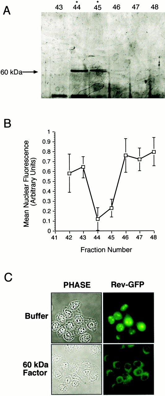
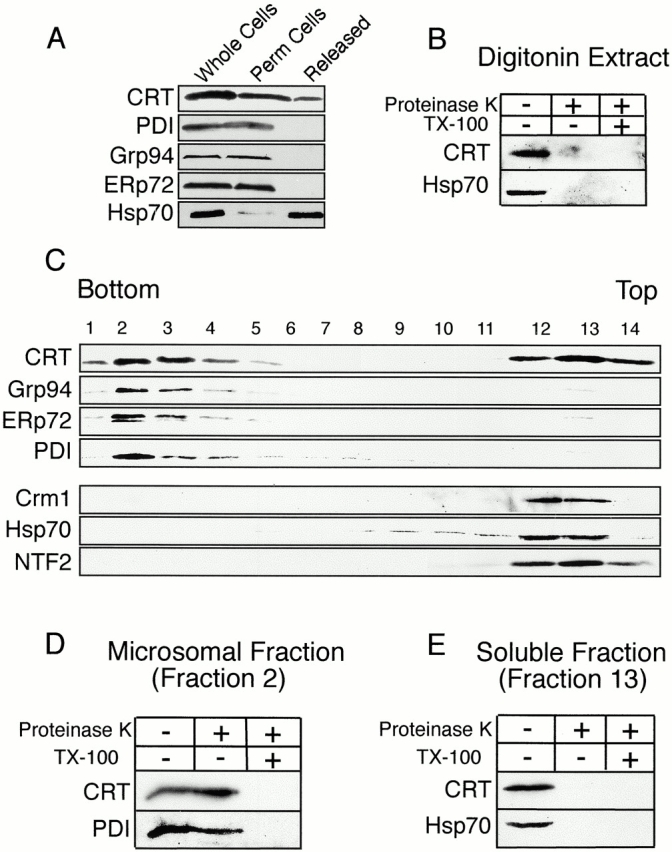
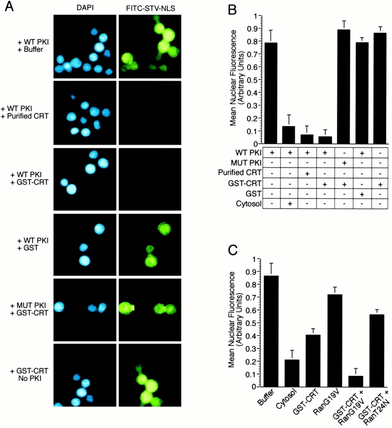
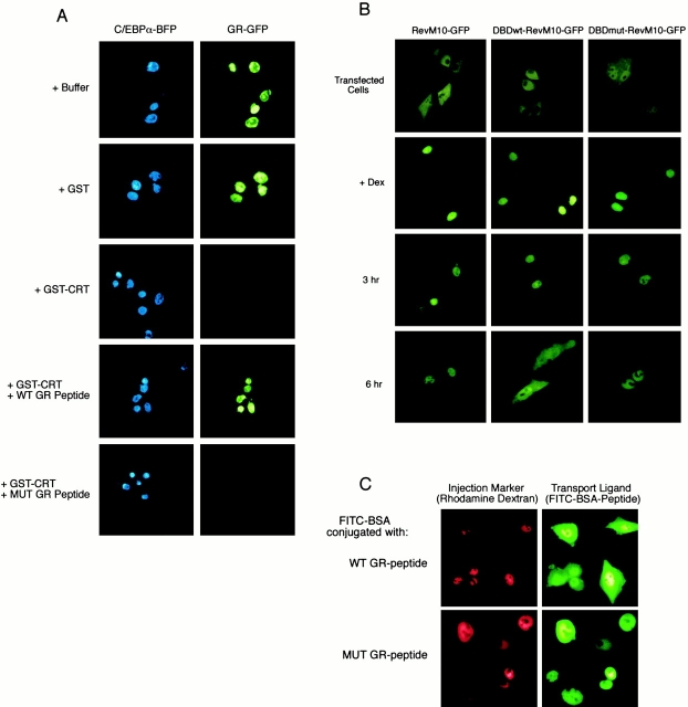
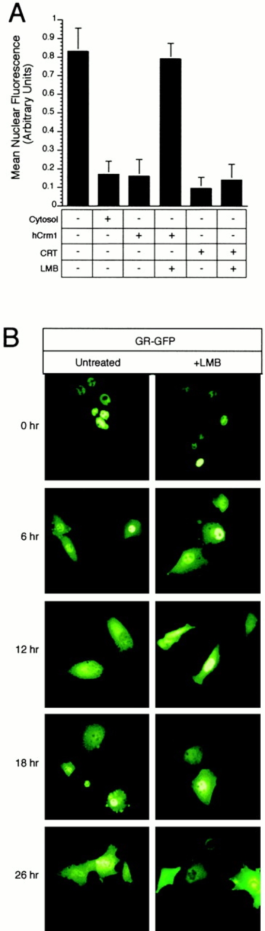
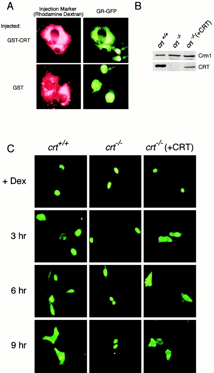
In previous work, we used a permeabilized cell assay that reconstitutes nuclear export of protein kinase inhibitor (PKI) to show that cytosol contains an export activity that is distinct from Crm1 (Holaska, J.M., and B.M. Paschal. 1995. Proc. Natl. Acad. Sci. USA. 95: 14739-14744). Here, we describe the purification and characterization of the activity as calreticulin (CRT), a protein previously ascribed to functions in the lumen of the ER. We show that cells contain both ER and cytosolic pools of CRT. The mechanism of CRT-dependent export of PKI requires a functional nuclear export signal (NES) in PKI and involves formation of an export complex that contains RanGTP. Previous studies linking CRT to downregulation of steroid hormone receptor function led us to examine its potential role in nuclear export of the glucocorticoid receptor (GR). We found that CRT mediates nuclear export of GR in permeabilized cell, microinjection, and transfection assays. GR export is insensitive to the Crm1 inhibitor leptomycin B in vivo, and it does not rely on a leucine-rich NES. Rather, GR export is facilitated by its DNA-binding domain, which is shown to function as an NES when transplanted to a green fluorescent protein reporter. CRT defines a new export pathway that may regulate the transcriptional activity of steroid hormone receptors.


Figures

Figure 1
Figure 1
Identification of a 60-kD export factor as CRT. (A and B) Purification of the export activity. Crm1-depleted cytosol was fractionated by ammonium sulfate precipitation, hydrophobic interaction, gel filtration, and MonoQ chromatography. Each fraction was tested for activity in the PKI export assay (Holaska and Paschal 1998). The final step of the purification was MonoQ chromatography. The export activity profile is plotted as the decrease in mean nuclear fluorescence, and the protein profile is shown by SDS-PAGE (7% gel, silver-stained). The maximum export activity in fractions 44 and 45 corresponds to the abundance of a 60-kD polypeptide. Mass spectrometry of tryptic fragments from the 60-kD factor revealed its identity as CRT. (C) The 60-kD factor (CRT) mediates nuclear export of Rev. A cell line expressing Rev–GFP (Love et al. 1998) was grown on coverslips, permeabilized with digitonin, and incubated with buffer or purified CRT. Nuclear export, as visualized in the fluorescence microscope, was observed in samples incubated with CRT, but not with buffer alone.
Figure 2
Figure 2
HeLa cells contain both microsomal and cytosolic pools of CRT. (A and B) CRT is released from HeLa cells by digitonin permeabilization. Suspension HeLa cells (whole cells) were permeabilized with digitonin, collected by centrifugation, and then the fractions were analyzed by immunoblotting. A pool of CRT, and most of the Hsp70, are released from cells (Released), whereas the ER lumenal proteins Grp94, ERp72, and PDI are retained (Perm cells). The CRT released by digitonin permeabilization is susceptible to proteinase K digestion, indicating it is not enclosed within membrane-bound vesicles. (C) CRT fractionates as both a microsomal and a cytosolic protein. A postnuclear supernatant from HeLa cells was fractionated over a sucrose gradient and analyzed by SDS PAGE and immunoblotting. CRT and the ER markers Grp94, Erp72, and PDI partition with microsomes (fractions 2–4). CRT also partitions with the soluble proteins Crm1, Hsp70, and NTF2 (fractions 12–14). (D) The CRT in the microsomal fraction is protected from protease digestion. Fraction 2 from the sucrose gradient was treated with proteinase K (80 μg/ml, 15 min at room temperature) in the absence and presence of detergent (1% Triton X-100); only the latter condition resulted in digestion of CRT and the ER marker protein PDI. (E) The CRT in the soluble fraction is susceptible to protease digestion. Fraction 13 from the sucrose gradient was treated with proteinase K as above. CRT and the soluble marker Hsp70 were degraded in the absence of detergent.
Figure 3
Figure 3
CRT-dependent export of PKI in digitonin permeabilized cells. (A) Nuclear export of PKI can be reconstituted by recombinant CRT, and requires a functional NES within PKI. HeLa cell nuclei were loaded with fluorescently labeled STV–NLS (FITC–STV–NLS), washed with buffer, and incubated with bPKI and soluble export factors. After the export reaction, cells were washed, pipetted onto glass slides, and viewed by fluorescence microscopy. Nuclear export of PKI/FITC–STV–NLS, visualized as the loss of nuclear fluorescence, was observed if the reaction was supplemented with HeLa cell–derived CRT (50 μg/ml) or recombinant GST–CRT (50 μg/ml). Nuclear export was not observed when the reaction was supplemented with GST (50 μg/ml), or if the NES mutant of PKI (L41,44A; MUT PKI) was used instead of WT PKI, or if PKI was omitted from the assay. Cells were stained with DAPI to show the location of the nuclei in each field. (B) Quantitation of the relative levels of nuclear export from the experiments shown in A. The level of nuclear export promoted by the addition of HeLa cell cytosol is shown for comparison. (C) CRT and Ran-dependent export of PKI. Export reactions were performed at a subsaturating concentration of CRT (5 μg/ml) and the Ran mutants G19V and T24N (each at 10 μg/ml). Export was promoted by the Ran G19V mutant and inhibited by the Ran T24N mutant. This indicates that CRT-dependent export requires the GTP form of Ran.
Figure 4
Figure 4
CRT forms a trimeric complex with the leucine-rich NES and RanGTP. A synthetic peptide containing the NES of PKI (WT or NES mutant) was immobilized on the surface of the biosensor cuvette through a biotin–STV linkage. The binding response of recombinant proteins to immobilized peptide was measured as the increase in refractive index as a function of time. (A) CRT binding to the WT NES requires the presence of RanGTP. GST–CRT (50 μg/ml) and Ran preloaded with GMP-PNP (50 μg/ml) were added alone, or in combination, to cuvettes containing immobilized WT NES or mutant NES (L41,44A). The arrow indicates the time at which buffer (PBS) was infused. Complex formation is observed in the presence of a WT NES, CRT, and RanGTP (red tracing). CRT and Ran do not form a complex on a mutant NES (light blue tracing), and only background levels of binding are observed if CRT or Ran is omitted from the assay (dark blue and green tracings, respectively). Note that the light blue and green tracings overlap at ∼40 arc s, which represents background binding. (B) Measurement of the affinity of CRT for NES in the presence of RanGTP. The association rate of CRT for NES in the presence of Ran was measured over a concentration range of CRT (1.12–22.4 nM). The dissociation rate was measured in a similar manner after infusion of buffer (beginning at 400 s), and the FASTfit program was used to calculate the affinity of CRT for NES in the presence of Ran (K D = 8.5 nM). (C) Measurement of the affinity of Crm1 for NES in the presence of RanGTP. The methods described above were used to measure the association and dissociation rates of the trimeric complex, and the FASTfit program was used to calculate the affinity (K D = 11 nM). (D) RanGTP binding to CRT and Crm1 is stimulated by the NES. His-tagged CRT and Crm1 were adsorbed to microtiter wells, and binding of Ran was measured in the absence and presence of the WT and mutant NES peptides. Since the recombinant Ran was preloaded with GTP radiolabeled on the terminal phosphate, the counts measured in the bound fraction specifically reflect the triphosphate form of Ran. Including WT NES in the binding reaction stabilizes the interaction of RanGTP with both CRT and Crm1.
Figure 5
Figure 5
CRT-dependent nuclear export of the GR is mediated by its DBD. (A) BHK cells were cotransfected with plasmids encoding C/EBPα–BFP and GR–GFP and grown for 48 h. The transfected cells were permeabilized with digitonin and export assays were performed in the presence of buffer alone, GST (50 μg/ml), or GST–CRT (50 μg/ml). Export assays were also performed with GST–CRT that was preincubated with a peptide (1 mg/ml) from within the DBD of the GR (WT GR peptide; residues 460–474: CGGGKVFFKRAVEGQHNLY). The control was a mutant peptide with two amino acid changes (MUT GR peptide; residues 460–474: CGGGKVAAKRAVEGQHNLY). CRT-dependent export of GR–GFP was blocked by preincubation with WT, but not mutant, peptide. This suggests that CRT recognition of the GR involves the DNA recognition helix within the DBD. (B) The DBD of GR is a functional NES. A plasmid encoding the NES mutant of Rev (RevM10) fused to the hormone-binding domain of GR and GFP was used to assay nuclear export. The GFP fusion undergoes agonist-dependent nuclear import (+Dex), but fails to undergo nuclear export because of the mutations (L78D,E79L) in the NES of Rev. Nuclear export is restored in a GFP fusion containing a WT DBD (DBDwt–RevM10–GFP), but not in a GFP fusion containing a mutant DBD (DBDmut–RevM10–GFP). The DBD in these constructs corresponds to residues 432–528 in human GR, and the mutations are F463,464A. (C) The DNA recognition helix in GR functions as an NES. The peptides used in the competition experiment were coupled to fluorescently labeled BSA, and analyzed by nuclear microinjection and microscopy. The WT GR-peptide, but not the MUT GR-peptide, promotes nuclear export of the fluorescent conjugate to the cytoplasm of BHK cells.
Figure 6
Figure 6
LMB-insensitive nuclear export analyzed in vitro and in vivo. (A) Nuclear export mediated by CRT is not inhibited by LMB under conditions where Crm1-dependent export is inhibited. PKI export was measured in digitonin-permeabilized cells using recombinant CRT (25 μg/ml) and purified Crm1 (50 μg/ml), pretreated with 500 nM LMB for 15 min at room temperature. The level of export promoted by HeLa cytosol is shown for comparison. (B) Nuclear export of the GR is not inhibited by LMB in living cells. BHK cells were transfected with full-length GR fused to GFP (GR–GFP). After nuclear import of GR–GFP was induced with 1 μM corticosteroid for 1 h, the cells were washed extensively with medium containing charcoal-stripped serum, and incubated at 37°C in the absence or presence of 200 nM LMB. At the indicated time points, coverslips were removed and the localization of GR–GFP recorded by fluorescence microscopy. Nuclear export of GR–GFP from the nucleus to the cytoplasm is observed in 6 h, and nuclear export is unaffected by the presence of LMB. Nuclear export mediated by Crm1 was blocked at the 6-h time point, as measured by nuclear microinjection of an NES-containing reporter protein (data not shown).
Figure 7
Figure 7
CRT mediates nuclear export of GR in vivo. (A) Cytoplasmic injection of recombinant CRT is sufficient to stimulate nuclear export of GR. BHK cells expressing GR–GFP were injected with GST–CRT or GST (0.5 mg/ml each), and the distribution of GR–GFP was examined by fluorescence microscopy after a 45 min incubation at 37°C. Uninjected cells are indicated with arrows. (B) Immunoblot showing CRT and Crm1 expression in immortalized mouse embryo fibroblasts derived from WT cells (crt +/+), CRT knockout cells (crt −/−) (Mesaeli et al. 1999), and CRT-knockout cells transfected with full-length CRT (crt −/− [+CRT]). (C) Nuclear export of GR is impaired in the absence of CRT, and is restored by CRT expression. A plasmid encoding GR–GFP was transfected into the indicated cell lines, and nuclear accumulation of the reporter was induced with dexamethasone (+Dex). After agonist removal, the cells were examined at 3 h intervals to monitor nuclear export. Nuclear export of the GR–GFP reporter was observed in WT (crt +/+) and CRT-transfected (crt −/− [+CRT]) cells, but not in CRT-deficient (crt −/−) cells.

References

    1. Arts G.J., Fornerod M., Mattaj I.W. Identification of a nuclear export receptor for tRNA Curr. Biol. 8 1998. 305 314a - PubMed
    1. Arts G.J., Kuersten S., Romby P., Ehresmann B., Mattaj I.W. The role of exportin-t in selective nuclear export of mature tRNAs EMBO (Eur. Mol. Biol. Organ.) J. 17 1998. 7430 7441b - PMC - PubMed
    1. Askjaer P., Jensen T.H., Nilsson J., Englmeier L., Kjems J. The specificity of the CRM1-Rev nuclear export signal interaction is mediated by RanGTP. J. Biol. Chem. 1998;273:33414–33422. - PubMed
    1. Askjaer P., Bachi A., Wilm M., Bischoff F.R., Weeks D.L., Ogniewski V., Ohno M., Niehrs C., Kjems J., Mattaj I.W., Fornerod M. RanGTP-regulated interactions of CRM1 with nucleoporins and a shuttling DEAD-box helicase. Mol. Cell. Biol. 1999;19:6276–6285. - PMC - PubMed
    1. Black B.E., Lévesque L., Holaska J.M., Wood T.C., Paschal B.M. Identification of an NTF2-related factor that binds Ran-GTP and regulates nuclear protein export. Mol. Cell. Biol. 1999;19:8616–8624. - PMC - PubMed

Publication types

MeSH terms