. 2001 Jan 8;152(1):157-64.
doi: 10.1083/jcb.152.1.157.

The Telomerase/vault-associated protein TEP1 is required for vault RNA stability and its association with the vault particle

Affiliations

The Telomerase/vault-associated protein TEP1 is required for vault RNA stability and its association with the vault particle

V A Kickhoefer et al. J Cell Biol. .

Abstract

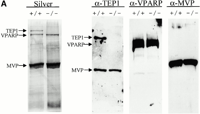
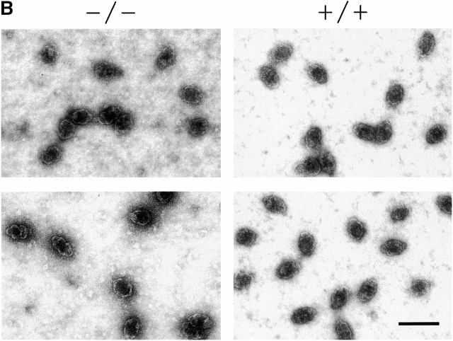
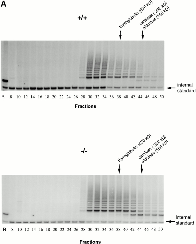
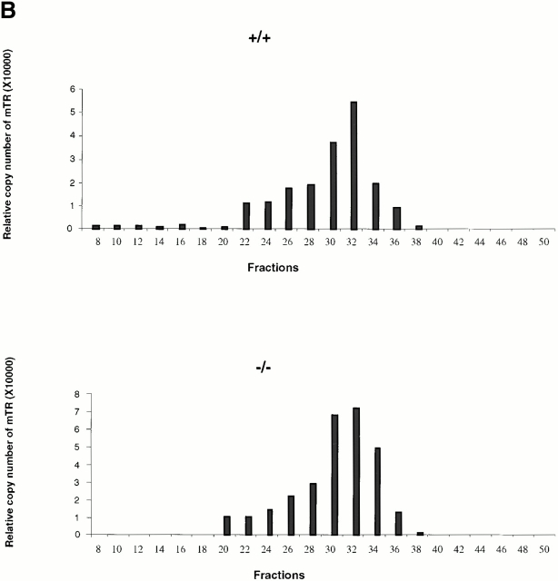
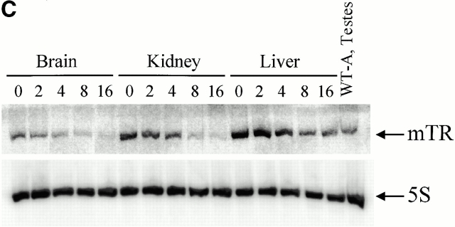
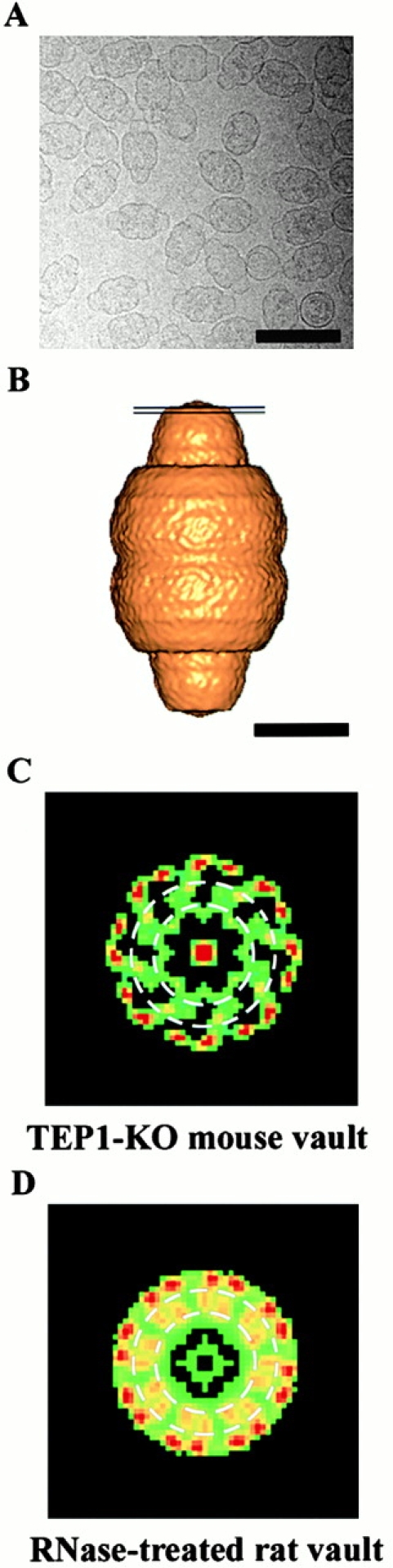
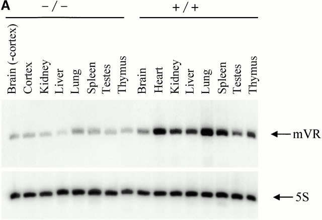
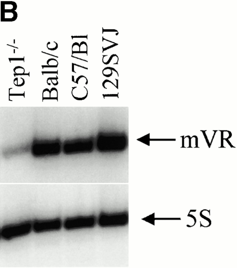
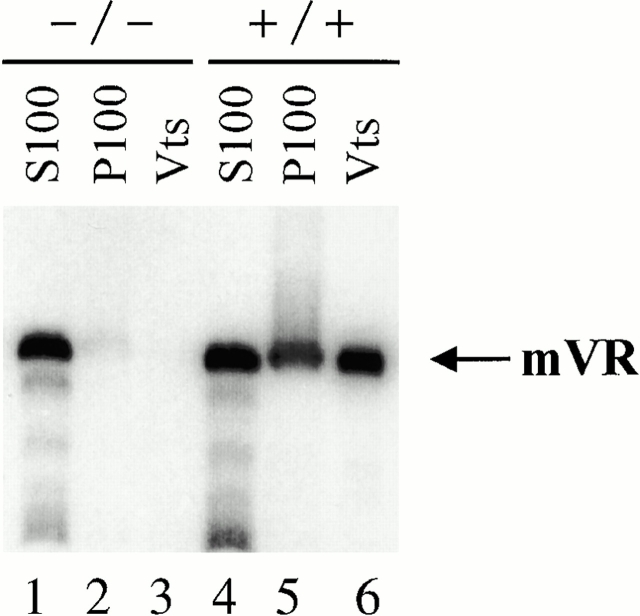
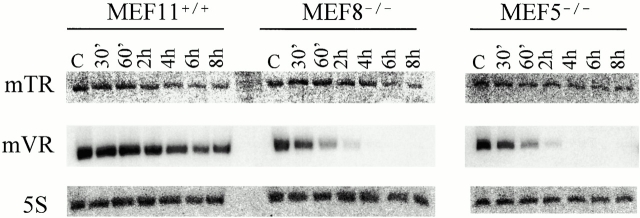
Vaults and telomerase are ribonucleoprotein (RNP) particles that share a common protein subunit, TEP1. Although its role in either complex has not yet been defined, TEP1 has been shown to interact with the mouse telomerase RNA and with several of the human vault RNAs in a yeast three-hybrid assay. An mTep1(-/-) mouse was previously generated which resulted in no apparent change in telomere length or telomerase activity in six generations of mTep1-deficient mice. Here we show that the levels of the telomerase RNA and its association with the telomerase RNP are also unaffected in mTep1(-/-) mice. Although vaults purified from the livers of mTep1(-/-) mice appear structurally intact by both negative stain and cryoelectron microscopy, three-dimensional reconstruction of the mTep1(-/-) vault revealed less density in the cap than previously observed for the intact rat vault. Furthermore, the absence of TEP1 completely disrupted the stable association of the vault RNA with the purified vault particle and also resulted in a decrease in the levels and stability of the vault RNA. Therefore, we have uncovered a novel role for TEP1 in vivo as an integral vault protein important for the stabilization and recruitment of the vault RNA to the vault particle.


Figures

Figure 2
Figure 2
Purified vaults. (A) Silver stain of vaults purified from livers of either wild-type (+/+) or mTep1-deficient (−/−) mice (arrows indicate TEP1, VPARP, and MVP). The identities of the vault proteins were confirmed by immunoblot analysis using the indicated antibodies (α-TEP1, α-VPARP, and α-MVP). Due to limited sample availability immunoblots were stripped and reprobed with the different antibodies. Consequently, the MVP antibody was not completely removed and MVP reappeared in subsequent reprobing with TEP antibodies. As expected, TEP1 is absent in the mTep1-deficient mice. The anti-VPARP antibody was made against a portion of the human VPARP protein and recognizes the mouse VPARP protein here as a smear. (B) Electron micrographs of negatively stained vaults purified from either wild-type (+/+) or mTep1-deficient (−/−) mice. Bar, 100 nm.
Figure 2
Figure 2
Purified vaults. (A) Silver stain of vaults purified from livers of either wild-type (+/+) or mTep1-deficient (−/−) mice (arrows indicate TEP1, VPARP, and MVP). The identities of the vault proteins were confirmed by immunoblot analysis using the indicated antibodies (α-TEP1, α-VPARP, and α-MVP). Due to limited sample availability immunoblots were stripped and reprobed with the different antibodies. Consequently, the MVP antibody was not completely removed and MVP reappeared in subsequent reprobing with TEP antibodies. As expected, TEP1 is absent in the mTep1-deficient mice. The anti-VPARP antibody was made against a portion of the human VPARP protein and recognizes the mouse VPARP protein here as a smear. (B) Electron micrographs of negatively stained vaults purified from either wild-type (+/+) or mTep1-deficient (−/−) mice. Bar, 100 nm.
Figure 1
Figure 1
mTR expression in mTep−/− mice. (A) Telomerase activity in the individual Sephacryl S-400 fractions of wild-type (+/+) and mTep1-deficient (−/−) ES cell lysates. TRAP was performed for 20 PCR cycles on 5 μl of each fraction of the indicated genotype. An internal PCR standard for the TRAP is shown at bottom right with an arrow. R represents the RNase A treatment of 5 μl of the fraction with the peak telomerase activity. Peak fractions of the sizing standards thyroglobulin, catalase, and aldolase are indicated above. (B) RT-PCR quantitation of mTR in the individual fractions of wild-type (+/+) and mTep1-deficient (−/−) ES cell lysates. The Taqman assay was performed on 5 μl of each fraction of the indicated genotype. The relative copies of mTR were calculated based on a standard curve using serial diluted mouse, total RNA, and/or the fraction with the peak telomerase activity. The fraction with the peak telomerase activity had no detectable mTR after the RNase A treatment (data not shown). (C) Northern blot analysis of total RNA prepared from brain, kidney, and liver tissue at the indicated postnatal developmental stages (day 0 is newborn). As a control, total RNA from adult wild-type testes was prepared. The membrane was probed with mTR (top), stripped, and reprobed with an antisense 5S oligonucleotide as a loading control (bottom).
Figure 1
Figure 1
mTR expression in mTep−/− mice. (A) Telomerase activity in the individual Sephacryl S-400 fractions of wild-type (+/+) and mTep1-deficient (−/−) ES cell lysates. TRAP was performed for 20 PCR cycles on 5 μl of each fraction of the indicated genotype. An internal PCR standard for the TRAP is shown at bottom right with an arrow. R represents the RNase A treatment of 5 μl of the fraction with the peak telomerase activity. Peak fractions of the sizing standards thyroglobulin, catalase, and aldolase are indicated above. (B) RT-PCR quantitation of mTR in the individual fractions of wild-type (+/+) and mTep1-deficient (−/−) ES cell lysates. The Taqman assay was performed on 5 μl of each fraction of the indicated genotype. The relative copies of mTR were calculated based on a standard curve using serial diluted mouse, total RNA, and/or the fraction with the peak telomerase activity. The fraction with the peak telomerase activity had no detectable mTR after the RNase A treatment (data not shown). (C) Northern blot analysis of total RNA prepared from brain, kidney, and liver tissue at the indicated postnatal developmental stages (day 0 is newborn). As a control, total RNA from adult wild-type testes was prepared. The membrane was probed with mTR (top), stripped, and reprobed with an antisense 5S oligonucleotide as a loading control (bottom).
Figure 3
Figure 3
Cryo-EM reconstruction of vaults purified from mTEP1-deficient mice. (A) A portion (640 × 640 pixels) of a digital cryoelectron micrograph of the vaults. (B) A surface representation of the final reconstruction at 27 Å resolution. The two black lines indicate the position of the density slab shown in C. (C) A two-dimensional projection of a density slab through the top of the cap of the mTep1−/− vault reconstruction. KO, knockout. (D) The corresponding density slab from the RNase-treated rat vault reconstruction (Kong et al. 2000). The dashed white circles in C and D outline the intermediate ring region in which a major difference is observed between the two reconstructions. Bars: (A) 1,000 Å; (B) 250 Å.
Figure 5
Figure 5
Comparative vault RNA expression in mTep1-deficient and wild-type mouse tissues. (A) Northern analysis of RNA prepared from the indicated tissues of wild-type (+/+) and mTep1-deficient (−/−) mice. The membrane was probed with mVR (top), stripped, and reprobed with an antisense 5S oligonucletide (bottom) to control for loading. (B) Northern analysis of RNA prepared from different mice strains as indicated, and probed as indicated above. mVR expression does not vary significantly in the different wild-type strain backgrounds.
Figure 5
Figure 5
Comparative vault RNA expression in mTep1-deficient and wild-type mouse tissues. (A) Northern analysis of RNA prepared from the indicated tissues of wild-type (+/+) and mTep1-deficient (−/−) mice. The membrane was probed with mVR (top), stripped, and reprobed with an antisense 5S oligonucletide (bottom) to control for loading. (B) Northern analysis of RNA prepared from different mice strains as indicated, and probed as indicated above. mVR expression does not vary significantly in the different wild-type strain backgrounds.
Figure 4
Figure 4
Vault RNA association with vaults. Northern analysis of RNA purified from subcellular fractions during vault purification from wild-type (+/+) and mTep1-deficient (−/−) mice. S100 (100,000 g supernatant, lanes 1 and 4), P100 (100,000 g pellet, lanes 2 and 5), and purified vaults (Vts, lanes 3 and 6).
Figure 6
Figure 6
Vault RNA expression varies in actinomycin D–treated MEFs. Northern analysis of RNA prepared from wild-type (MEF11+/+) and mTep1-deficient (MEF8−/− and MEF5−/−) cells treated with actinomycin D for the indicated times. The membrane was probed with mTR (top), stripped, and reprobed first with mVR (middle) and next with an antisense 5S oligonucleotide (bottom). Control (C) samples were not treated with drug.

References

    1. Abbondanza C., Rossi V., Roscigno A., Gallo L., Belsito A., Piluso G., Medici N., Nigro V., Molinari A.M., Moncharmont B., Puca G.A. Interaction of vault particles with estrogen receptor in the MCF-7 breast cancer cell. J. Cell Biol. 1998;141:1301–1310. - PMC - PubMed
    1. Adrian M., Dubochet J., Lepault J., McDowall A.W. Cryo-electron microscopy of viruses. Nature. 1984;308:32–36. - PubMed
    1. Beattie T.L., Zhou W., Robinson M.O., Harrington L. Reconstitution of human telomerase activity in vitro . Curr. Biol. 1998;8:177–180. - PubMed
    1. Blasco M.A., Funk W., Villeponteau B., Greider C.W. Functional characterization and developmental regulation of mouse telomerase RNA. Science. 1995;269:1267–1270. - PubMed
    1. Böttcher B., Wynne S.A., Crowther R.A. Determination of the fold of the core protein of hepatitis B virus by electron cryomicroscopy. Nature. 1997;386:88–91. - PubMed

Publication types