. 2001 Jan 8;152(1):181-95.
doi: 10.1083/jcb.152.1.181.

Cbl associates with Pyk2 and Src to regulate Src kinase activity, alpha(v)beta(3) integrin-mediated signaling, cell adhesion, and osteoclast motility

Affiliations

Cbl associates with Pyk2 and Src to regulate Src kinase activity, alpha(v)beta(3) integrin-mediated signaling, cell adhesion, and osteoclast motility

A Sanjay et al. J Cell Biol. .

Abstract

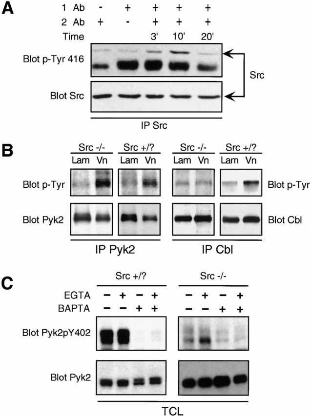
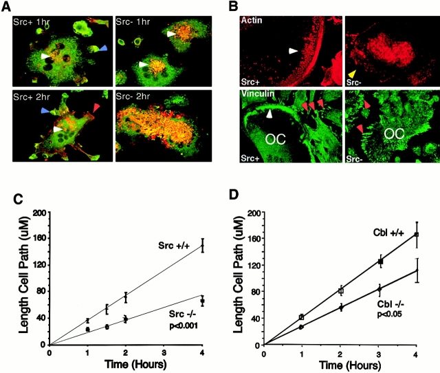
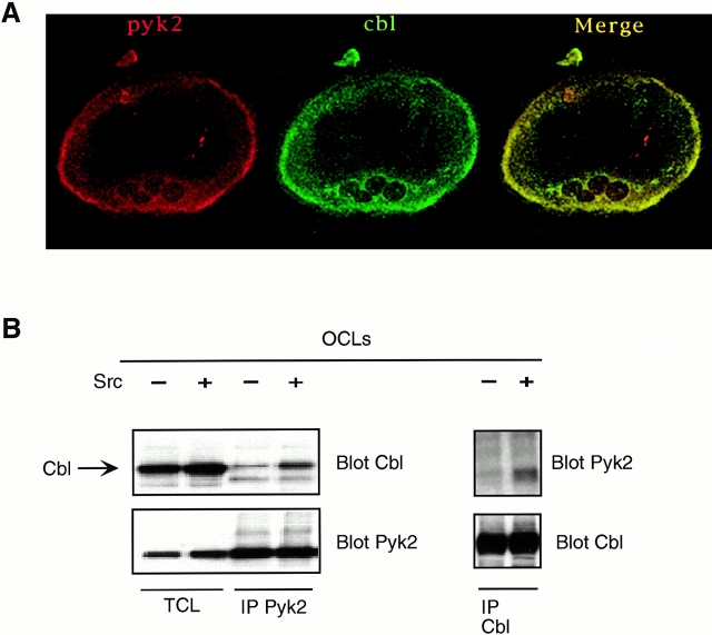
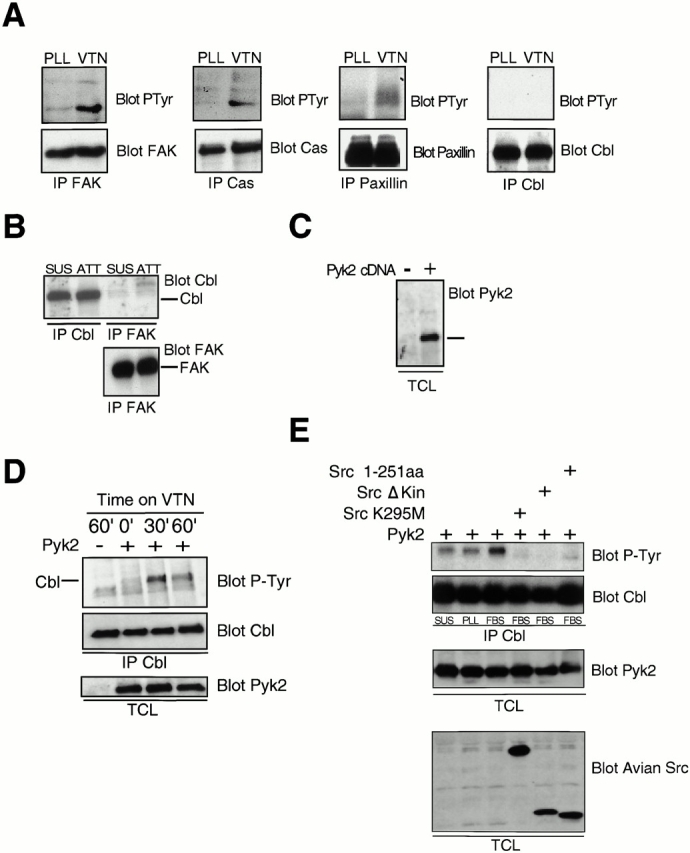
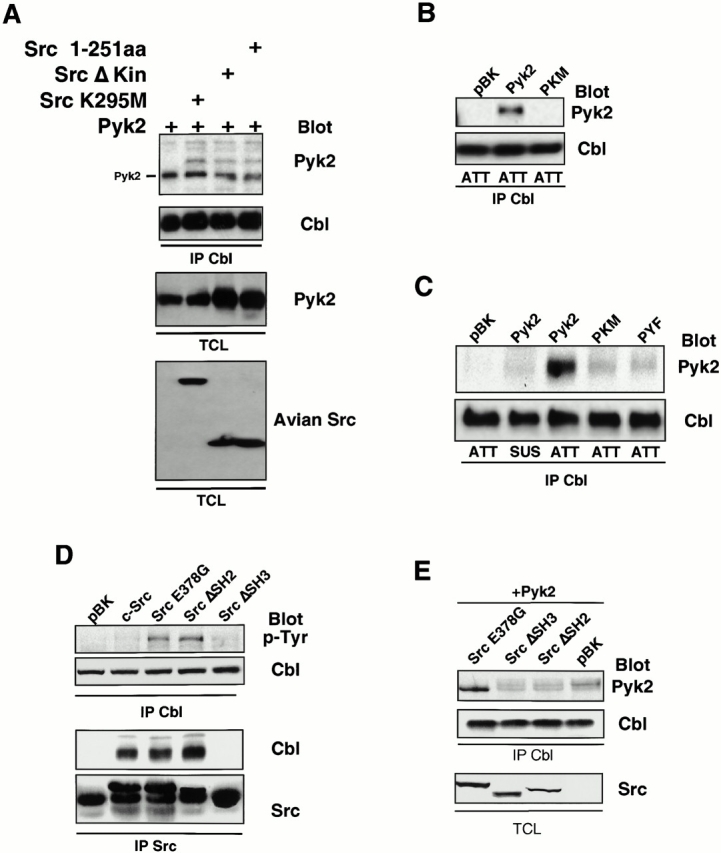
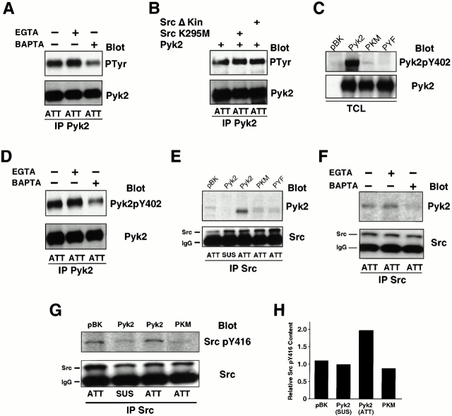
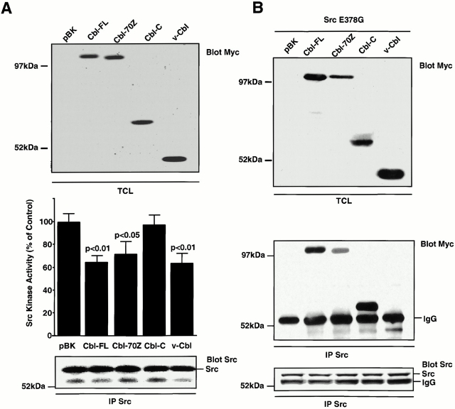
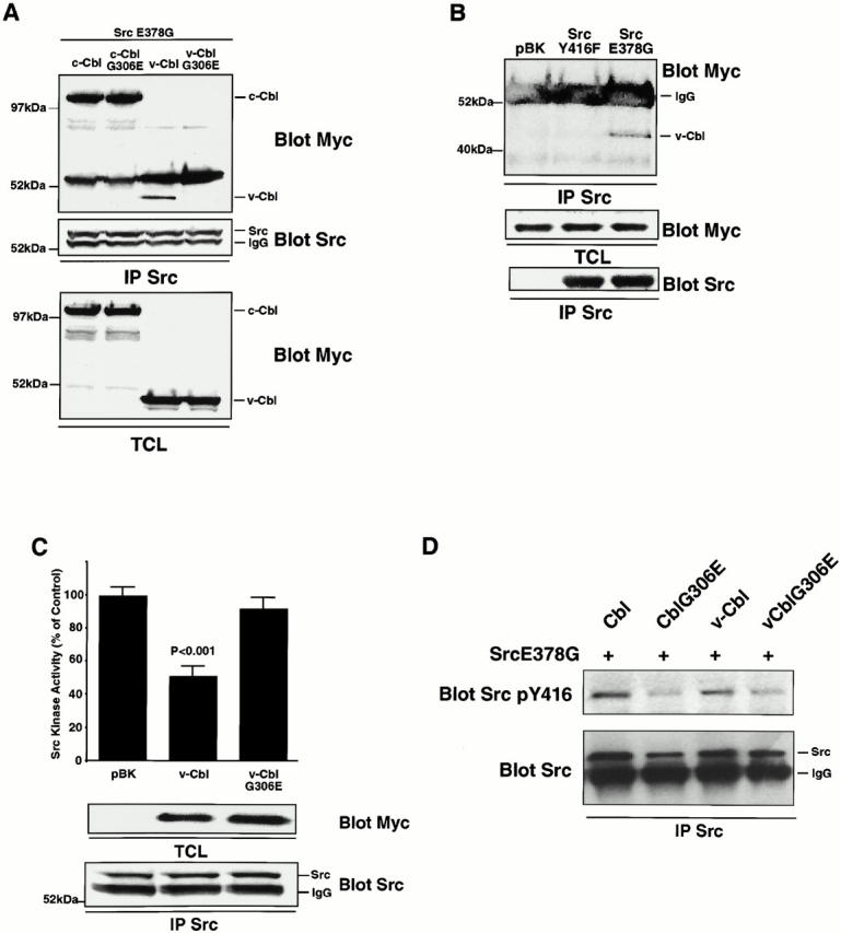
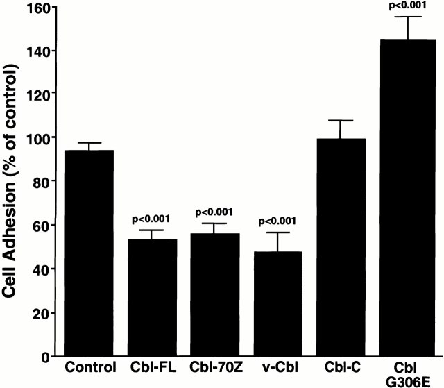
The signaling events downstream of integrins that regulate cell attachment and motility are only partially understood. Using osteoclasts and transfected 293 cells, we find that a molecular complex comprising Src, Pyk2, and Cbl functions to regulate cell adhesion and motility. The activation of integrin alpha(v)beta(3) induces the [Ca(2+)](i)-dependent phosphorylation of Pyk2 Y402, its association with Src SH2, Src activation, and the Src SH3-dependent recruitment and phosphorylation of c-Cbl. Furthermore, the PTB domain of Cbl is shown to bind to phosphorylated Tyr-416 in the activation loop of Src, the autophosphorylation site of Src, inhibiting Src kinase activity and integrin-mediated adhesion. Finally, we show that deletion of c Src or c-Cbl leads to a decrease in osteoclast migration. Thus, binding of alpha(v)beta(3) integrin induces the formation of a Pyk2/Src/Cbl complex in which Cbl is a key regulator of Src kinase activity and of cell adhesion and migration. These findings may explain the osteopetrotic phenotype in the Src(-/-) mice.


Figures

Figure 2
Figure 2
Integrin ligation activates Src and increases the tyrosine phosphorylation of a number of proteins, including c-Cbl, but not Pyk2, in a Src-dependent manner. (A) Src was immunoprecipitated from Src+ OCLs treated with VnR cross-linking antibodies and Western blotted with Src pTyr-416 antibody (top) that detects the activated form of Src. (Bottom) The same membrane stripped and reprobed with anti-Src Ab. (B) Pyk2 and Cbl immunoprecipitates from lysates of OCLs plated on laminin (Lam) or vitronectin (Vn) for 30 min were Western blotted with anti–p-Tyr antibody (top). Blots were stripped and reprobed with antibodies against Cbl or Pyk2 (bottom). (C) OCLs were pretreated with calcium chelators EGTA (100 μM) and/or BAPTA (50 μM), as described in Materials and Methods, and allowed to attach to vitronectin-coated plastic for 30 min. Total cell from lysates were Western blotted for Pyk2pY402 (top), then stripped and reprobed with anti–Pyk2 antibody (bottom).
Figure 1
Figure 1
Src deletion leads to alterations in adhesion structures and cell migration. (A) Immunofluorescence showing actin (red) and vinculin (green) colocalization (yellow) in Src+ and Src OCLs replated on vitronectin for 1 and 2 h. Micrographs are composites of both channels together. Note the formation of vinculin-rich lamellipodia (blue arrows, Src+ 1 and 2 h), which is not seen in Src OCLs, and the increased redistribution of actin from the central regions of the cell (white arrows) to the peripheral regions of the cell (red arrow, Src+ 2 h) in Src+ OCLs. These features were not observed in Src OCLs where actin and vinculin colocalization was observed to significantly increase between 1 and 2 h. (B) Immunofluorescence showing actin (red) and vinculin (green) localization in Src+ and Src authentic osteoclasts cultured on glass coverslips. Note the distinct ring-like assembly of podosomes that stain for both actin and vinculin in Src+ osteoclasts (white arrows), and the focal adhesion-like arrangement of vinculin in surrounding contaminating fibroblast-like cells (red arrows, Src+ vinculin). In Src osteoclasts, the localization of actin to the periphery of the osteoclast is not observed (yellow arrow, Src actin), and staining for vinculin demonstrates a more focal-adhesion–like arrangement (red arrow, Src vinculin) reminiscent of that seen in the previously described nonosteoclastic cells. (C and D) The lengths of cell migration paths of Src (C) and c-Cbl (D) osteoclasts and their littermate wild-type controls were measured at regular intervals as described in Materials and Methods.
Figure 3
Figure 3
Cbl associates with Pyk2 in a Src-dependent manner, and becomes tyrosine phosphorylated by Src in a reaction that also requires the presence of Pyk2. Immunocytochemistry showing Pyk2 and Cbl colocalization in Src+ authentic osteoclasts. Micrographs represent individual channels (red, Pyk2; green, Cbl) and a merging of the two channels. Pyk2 was immunoprecipitated from Src+ and Src OCLs. Immune complexes were Western blotted for Cbl (top left), and then the membranes were stripped and reprobed with Pyk2 antibodies (bottom left) Total cell lysates (TCL) were also electrophoresed along with the immunoprecipitates. Reciprocal immunoprecipitation and blotting was performed using Cbl antibodies for immunoprecipitation and Pyk2 antibodies for blotting (top right). The membrane was stripped and reprobed with Cbl antibodies (bottom right).
Figure 4
Figure 4
Cbl phosphorylation downstream of the vitronectin receptor requires Pyk2. (A) Serum-starved 293-VnR cells were replated on plastic coated with either polylysine (PLL) or vitronectin (VTN) for 30 min, and then lysed. FAK, Cas, paxillin, or Cbl were immunoprecipitated and analyzed by Western blotting for phosphotyrosine (top). The membranes were stripped and reprobed with antibodies against the immunoprecipitated antigen (bottom). (B) Cbl and FAK were immunoprecipitated from lysates of 293 VnR cells that had been replated on vitronectin-coated plastic (ATT) or kept in suspension (SUS), and the immune complexes analyzed by Western blotting for the presence of Cbl (top). (Bottom) Equal amounts of FAK in the FAK immunoprecipitates. (C) The total cell lysates (TCL) of 293 VnR cells transfected with vector control (−) or Pyk2 (+) were probed with Pyk2 antibodies to show expression of exogenous Pyk2 (+) (right). (D) 293-VnR cells transfected with 5 μg of either control vector (−) or Pyk2 (+) were harvested and either kept in suspension or plated on vitronectin-coated plastic for the indicated times. The cells were then lysed and Cbl was immunoprecipitated and Western blotted for P-Tyr (top). Membranes were then stripped and reprobed for Cbl (middle). Total cell lysates (TCL) were blotted for Pyk2 to ensure expression (bottom). (E) 293-VnR cells were transfected with a combination of 5 μg Pyk2 expression vector and 5 μg of cDNAs encoding various kinase-inactive Src mutants. The cells were harvested, and then either kept in suspension (SUS) or plated on plastic coated with polylysine (PLL) or serum (FBS) for 30 min. The cells were then lysed and Cbl was immunoprecipitated and Western blotted for p-Tyr (top). The membrane was reprobed for Cbl. Total cell lysate (TCL) blots were probed with antibodies to Pyk2 or avian Src to ensure expression.
Figure 6
Figure 6
Src acts as an adaptor molecule mediating interactions between Cbl and Pyk2. (A) Association of Cbl with Pyk2 in presence of Src kinase mutants was analyzed by probing the blot of Cbl immunoprecipitates with Pyk2 antibodies (top). The blot was stripped and reprobed with Cbl antibodies. Total cell lysates (TCL) were Western blotted with Pyk2 and avian Src antibodies to check expression of transfected proteins. (B) 293-VnR cells were transfected with Pyk2, PKM, or empty vector (pBK). Cbl was immunoprecipitated from lysates of replated cells and analyzed for the presence of phosphotyrosine (top). (C) 293-VnR cells were transfected with Pyk2 mutants. The attachment-induced association of the mutant Pyk2 proteins with Cbl (top) was analyzed by blotting Cbl IPs with anti–Pyk2 antibody. (D) Cbl IPs from lysates of 293-VnR cells transfected with 10 μg of the indicated Src expression vector were blotted with an antiphosphotyrosine antibody to demonstrate the effects of these Src mutants of Cbl phosphorylation. Src was immunoprecipitated from the same lysates and the immune complexes were blotted for the presence of Cbl. (E) 293-VnR cells were transfected with a combination of 3 μg Pyk2 and 3 μg of the indicated Src expression vector. Cbl IPs were analyzed for the presence of Pyk2 (top). (Bottom) Expression of the Src mutants in total cell lysates (TCL).
Figure 5
Figure 5
Pyk2 is autophosphorylated at Y402 after adhesion and associates with Src via the phosphorylated Y402. (A) 293-VnR cells transiently transfected with 5 μg of Pyk2 were treated with calcium chelators BAPTA and/or EGTA, and then replated on serum-coated plastic and allowed to attach to vitronectin-coated plastic for 30 min. Pyk2 IPs from lysates were Western blotted for P-Tyr (top), and then reprobed with anti–Pyk2 (bottom). (B) 293-VnR cells were transiently transfected with Pyk2 (5 μg) or cotransfected with Pyk2 (5 μg) and Src kinase-inactive mutants (10 μg). Pyk2 IPs from lysates were probed with P-Tyr antibodies (top). Membrane was stripped and reprobed with Pyk2 (bottom). (C) Pyk2 and Pyk2 mutants Y402F (PYF) and K457A (PKM) were transiently expressed in 293-VnR cells and total cell lysates (TCL) were blotted with an antibody specific for Pyk2 phosphorylated residue 402 (top). (Bottom) Transfected proteins. (D) Cells were processed as in A and Pyk2 IPs were blotted with an antibody specific for Pyk2 phosphorylated residue 402 (top). (Bottom) Pyk2 blot demonstrating equal loading. (E) 293-VnR cells were transiently transfected with wild-type Pyk2, kinase-dead Pyk2 (PKM) or Pyk2Y402F (PKF). The transfected cells were harvested, and then kept in suspension (SUS) or replated on serum-coated plastic (ATT) as in Fig. 4 E. Association of Src with mutant Pyk2 proteins was analyzed by Western blotting Src immunoprecipitates with Pyk2 antibodies (top). (Bottom) The membrane reprobed with Src antibodies. (F) Src IPs from cells treated with either EGTA or BAPTA were blotted with Pyk2 antibodies (top). (Bottom) The membrane reprobed with anti–Src. (G) 293-VnR cells transfected with 5 μg of Pyk2 or dominant-negative kinase-dead Pyk2 (PKM) were either kept in suspension or replated on vitronectin coated dishes for 30 min. Lysates were immunoprecipitated with anti–Src antibodies and probed with Src pTyr-416 antibody (top), which detects the activated form of Src. The membrane was stripped and reprobed with anti–Src (bottom). (H) Src kinase activity was quantified as an increase in pY416 Src using Scion Image1.62 C program.
Figure 7
Figure 7
Cbl inhibits Src kinase activity by binding to Src. (A) NIH3T3 cells that stably express a constitutively active Src (SrcY527F) were transfected with 10 μg of the indicated Myc-tagged Cbl cDNA and expression was demonstrated by immunoblotting using an anti–Myc antibody (top). Src was immunoprecipitated and the kinase activity was measured and expressed as a percentage of the control kinase activity levels (pBK-transfected cells). The amount of immunoprecipitated Src in the kinase assay was assessed by immunoblotting. (B) 293-VnR cells that stably express the myc-tagged Cbl constructs (top) were transfected with 10 μg of active Src (SrcE378G). Src was immunoprecipitated and the immune complex was immunoblotted to detect myc-tagged proteins (middle) and Src (bottom).
Figure 8
Figure 8
The association of the Cbl PTB domain with Src phosphotyrosine residue 416 results in an inhibition of Src kinase activity. (A) 293-VnR cells were transiently cotransfected with 5 μg Src E378G and 5 μg of normal or G306E-mutated forms of Cbl and v-Cbl. After immunoprecipitating Src, immune complexes were blotted for associated Cbl protein (top). The efficiencies of Src immunoprecipitation (middle) and Cbl expression (bottom) were determined. (B) 293-VnR v-Cbl cells were transiently transfected with active avian Src (SrcE378G) or avian Src Y416F. Avian Src was immunoprecipitated and blotted for the presence of myc-tagged v-Cbl. v-Cbl expression (middle) and efficiency of Src immunoprecipitation (bottom) were analyzed. (C) The kinase activity of immunoprecipitated Src from cells transfected with a combination of 5 μg SrcE378G and 5 μg of either v-Cbl or v-CblG306E was measured and expressed as a percentage of the control kinase activity levels (pBK-transfected cells). v-Cbl expression (middle) and efficiency of Src immunoprecipitation (bottom) were analyzed. (D) 293-Vnr cells were transiently cotransfected with 5 μg Src E378G and 5 μg normal or G306E mutated form of Cbl and v-Cbl. Src was immunoprecipitated and immune complex was immunoblotted with SrcY416 (top). The blot was reprobed with Src (bottom).
Figure 9
Figure 9
Cbl regulates vitronectin receptor–mediated 293-VnR adhesion. The adhesion of 293-VnR cells stably expressing a number of Cbl constructs was measured as described in Materials and Methods and expressed as a percentage of control (untransfected 293-VnR cells). The graph represents pooled data from two individual experiments.

References

    1. Barber D.L., Mason J.M., Fukazawa T., Reedquist K.A., Druker B.J., Band H., D'Andrea A.D. Erythropoietin and interleukin-3 activate tyrosine phosphorylation of CBL and association with CRK adaptor proteins. Blood. 1997;89:3166–3174. - PubMed
    1. Bartkiewicz M., Houghton A., Baron R. Leucine zipper-mediated homodimerization of the adaptor protein c-Cbla role in c-Cbl's tyrosine phosphorylation and its association with epidermal growth factor receptor. J. Biol. Chem. 1999;274:30887–30895. - PubMed
    1. Bonita D.P., Miyake S., Lupher M.L., Jr., Langdon W.Y., Band H. Phosphotyrosine binding domain-dependent upregulation of the platelet-derived growth factor receptor α signaling cascade by transforming mutants of Cblimplications for Cbl's function and oncogenicity. Mol. Cell. Biol. 1997;17:4597–4610. - PMC - PubMed
    1. Broome M.A., Galisteo M.L., Schlessinger J., Courtnedge S.A. The protooncogene c-Cbl is a negative regulator of DNA synthesis initiated by both receptor and cytoplasmic tyrosine kinases. Oncogene. 1999;18:2908–2912. - PubMed
    1. Chen W.T. Proteolytic activity of specialized surface protrusions formed at rosette contact sites of transformed cells. J. Exp. Zool. 1989;251:167–185. - PubMed

Publication types

MeSH terms