. 2001 Jan 8;152(1):197-212.
doi: 10.1083/jcb.152.1.197.

Mitotic spindle integrity and kinetochore function linked by the Duo1p/Dam1p complex

Affiliations

Mitotic spindle integrity and kinetochore function linked by the Duo1p/Dam1p complex

I M Cheeseman et al. J Cell Biol. .

Abstract

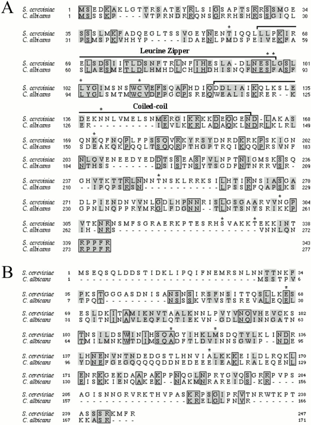
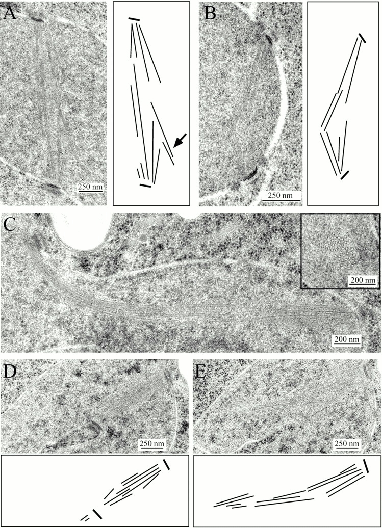
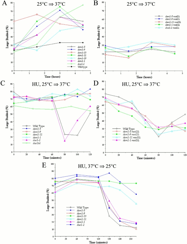
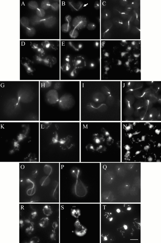
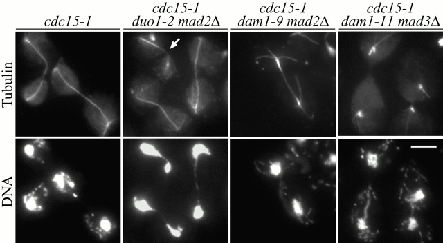
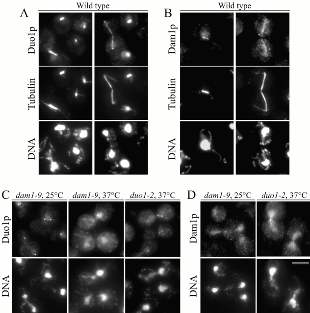
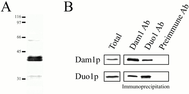
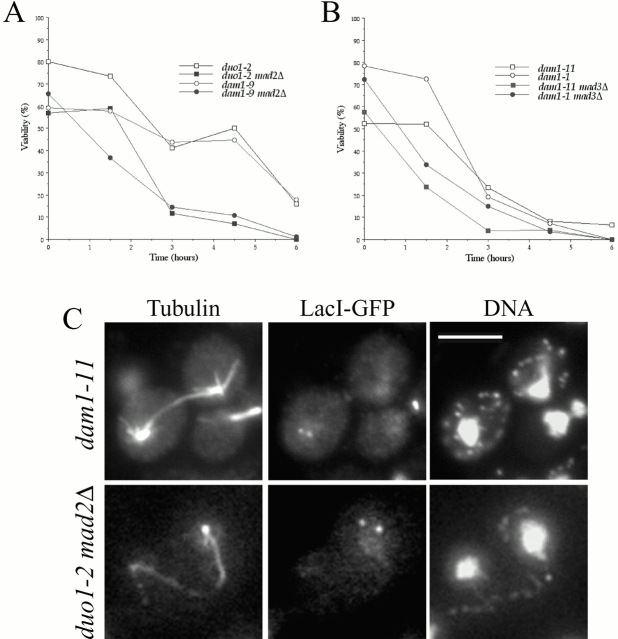
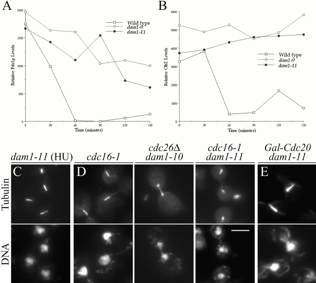
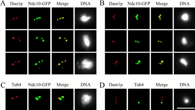
Duo1p and Dam1p were previously identified as spindle proteins in the budding yeast, Saccharomyces cerevisiae. Here, analyses of a diverse collection of duo1 and dam1 alleles were used to develop a deeper understanding of the functions and interactions of Duo1p and Dam1p. Based on the similarity of mutant phenotypes, genetic interactions between duo1 and dam1 alleles, interdependent localization to the mitotic spindle, and Duo1p/Dam1p coimmunoprecipitation from yeast protein extracts, these analyses indicated that Duo1p and Dam1p perform a shared function in vivo as components of a protein complex. Duo1p and Dam1p are not required to assemble bipolar spindles, but they are required to maintain metaphase and anaphase spindle integrity. Immunofluorescence and electron microscopy of duo1 and dam1 mutant spindles revealed a diverse variety of spindle defects. Our results also indicate a second, previously unidentified, role for the Duo1p/Dam1p complex. duo1 and dam1 mutants show high rates of chromosome missegregation, premature anaphase events while arrested in metaphase, and genetic interactions with a subset of kinetochore components consistent with a role in kinetochore function. In addition, Duo1p and Dam1p localize to kinetochores in chromosome spreads, suggesting that this complex may serve as a link between the kinetochore and the mitotic spindle.


Figures

Figure 1
Figure 1
Dam1p and Duo1p mutations and C. albicans homologues. Identical residues are boxed and conserved residues are highlighted. Residues that are mutated in duo1 or dam1 mutants are indicated with an asterisk, with the exception of mutations to a stop codon (see Table for a complete list of mutations in each allele). (A) Dam1p C. albicans homologue (orf5.6651). Both the leucine zipper region (residues 63–98) and the putative coiled coil region (residues 123–161) of Dam1p are conserved. These two proteins are 25% identical (70/277) and 53% conserved (147/277). S. cerevisiae Dam1p sequence data are available from GenBank/EMBL/DDBJ under accession no. AF280542. (B) Duo1p C. albicans homologue (orf5.4244). These two proteins are 21% identical (36/171) and 49% conserved (84/171).
Figure 5
Figure 5
Electron microscopy of duo1-2 and duo1-2 mad2Δ mutants. Thin sections (50 nm) of high pressure frozen yeast were viewed using transmission electron microscopy. (A) duo1-2 mutant after 2 h at the restrictive temperature, showing a slightly splayed spindle and corresponding diagram showing positions of the microtubules. The arrow points to microtubules that splay outwards. (B) duo1-2 mad2Δ mutant after 2 h at the restrictive temperature, showing a slightly broken spindle and corresponding diagram showing positions of the microtubules. (C) dam1-9 mad2Δ cdc15-1 mutant grown as in the legend to Fig. 4 showing a bent spindle. (C, inset) Cross section of this mutant showing a tight bundle of microtubules. (D and E) Two serial sections of dam1-10 at the permissive temperature and corresponding diagrams showing one invaginated SPB and microtubules that fail to connect with the opposite pole.
Figure 2
Figure 2
duo1 and dam1 mutants arrest as large-budded cells in a spindle assembly checkpoint–dependent manner. (A and B) Cells were grown to log phase at 25°C and shifted to 37°C at t = 0. (C and D) Cells were grown to log phase and arrested with 0.2 M HU for ∼4.5 h at 25°C. They were then released into fresh prewarmed medium lacking HU at 37°C (t = 0). (E) Cells were synchronized with alpha factor at 25°C and released into fresh prewarmed medium lacking alpha factor, but containing 0.2 M HU at 37°C. After 4 h, cells were released into fresh medium at 25°C (t = 0) with alpha factor to prevent reinitiation of the cell cycle.
Figure 3
Figure 3
duo1 and dam1 mutants display diverse spindle defects. (A–C, G–J, and O–Q) Tubulin immunofluorescence of duo1 and dam1 mutants. (D–F, K–N, and R–T) The corresponding DNA (DAPI) staining. (A and D) duo1-2; (B and E) duo1-2 mad2Δ, arrow shows broken spindle; (C and F) dam1-9; (G, K and H, L) duo1td; (I and M) dam1-11; (J and N) dam1-1; (O and R) dam1-10; (P and S) duo1-1 dam1-1; and (Q and T) dam1-19. Cells (A–N) were grown to log phase at 25°C, shifted to 37°C for 1.5 h. dam1-10 is shown at 25°C. duo1-1 dam1-1 is shown after 6 h at 37°C, though similar phenotypes are present at 25°C. dam1-19 is shown after 4.5 h at 37°C. Bar, 5 μm.
Figure 4
Figure 4
duo1 and dam1 mutants display distinct late mitotic phenotypes. Triple mutants between duo1 or dam1, a spindle assembly checkpoint mutant (mad2Δ or mad3Δ), and a temperature-sensitive allele of cdc15 were synchronized with alpha factor at 25°C and released into fresh prewarmed medium at 37°C. Samples were taken 2 and 3 h after alpha factor release and were processed for immunofluorescence and stained for tubulin (antitubulin antibody) and DNA (DAPI). Arrow shows duo1-2 mutant spindle, which is broken. Bar, 5 μm.
Figure 7
Figure 7
Duo1p and Dam1p show interdependent localization to the mitotic spindle. Wild-type, duo1, and dam1 mutant cells were grown at 25°C, shifted to 37°C for 3 h, and then processed for immunofluorescence. (A) Wild-type cells stained for tubulin (antitubulin antibody), Duo1p (anti-Duo1p antibodies), and DNA (DAPI). (B) Wild-type cells stained for tubulin (antitubulin antibody), Dam1p (anti-Dam1p antibodies), and DNA (DAPI). (C) duo1-2 and dam1-9 mutants stained for Duo1p (anti-Duo1p antibodies) and DNA (DAPI). (D) duo1-2 and dam1-9 mutants stained for Dam1p (anti-Dam1p antibodies) and DNA (DAPI). Bar, 5 μm.
Figure 6
Figure 6
Dam1p and Duo1p physically associate in vivo. (A) Immunoblot of wild-type protein extract probed with anti-Dam1p antibodies. This antibody specifically recognizes bands between 39.5 and 42 kD, near the predicted size of 38.4 kD for Dam1p. (B) Duo1p and Dam1p coimmunoprecipitate out of yeast protein extracts. Yeast protein extracts were incubated with antibodies against Duo1p, Dam1p, or preimmune serum bound to protein A beads (see Materials and Methods). Immunoprecipitated samples were run on a 12% gel and probed with antibodies against either Dam1p or Duo1p. Duo1p and Dam1p are specifically immunoprecipitated when antibodies against either Duo1p or Dam1p are used but not with preimmune serum.
Figure 8
Figure 8
duo1 and dam1 mutants show chromosome missegregation. (A and B) Viability of duo1 and dam1 mutants. Cells were grown to log phase at 25°C, then shifted to 37°C at t = 0. For each time point, cells were plated onto YPD plates at 25°C, and a sample was counted in a hemacytometer to determine the cells per milliliter of culture. (A) Viability of mutants that arrest with a short mitotic spindle and of the corresponding double mutants with mad2Δ. (B) Viability of mutants that arrest with elongated spindles and of the corresponding double mutants with mad3Δ. (C) Chromosome missegregation is observed by LacI-GFP. Anti-GFP immunofluorescence of duo1 and dam1 mutants expressing LacI-GFP in a strain with multiple repeats of the LacO sequence integrated. Cells were synchronized with alpha factor, released into fresh medium at 37°C, and fixed for immunofluorescence after 3 h. Tubulin immunofluorescence, anti-GFP immunofluorescence to show the LacI-GFP fusion protein (each dot indicates a single chromatid), and DNA (DAPI) staining are shown for dam1-11 and duo1-2 mad2Δ. The frequency of chromosome missegregation is tabulated in Table . Bar, 5 μm.
Figure 9
Figure 9
Some dam1 mutants undergo anaphase-like events while under a metaphase arrest. (A and B) Pds1p and Clb2 levels after HU release. Cells were synchronized with alpha factor, released into medium containing 0.15 M HU but lacking alpha factor, and incubated for ∼4.5 h at 25°C. The cells were then released into fresh prewarmed medium lacking HU at 37°C (t = 0). Protein samples were taken every 30 min and probed with anti-myc antibodies (9E10) against Pds1-myc18 or anti-Clb2 antibodies. Relative levels of Pds1p (A) or Clb2 (B) were quantified using a densitometer and were compared with β-tubulin levels as a loading control. (C) duo1 and dam1 mutants were synchronized with alpha factor at 25°C and released into fresh prewarmed medium at 37°C containing 0.2 M HU. They were then processed for immunofluorescence and stained with the YOL134 antibody against alpha tubulin. dam1-11 is shown, but wild-type and other duo1 and dam1 mutants look identical. (D) Double mutants between dam1 alleles and temperature-sensitive alleles of the APC (cdc16-1 or cdc26Δ) were synchronized with alpha factor at 25°C and released into fresh prewarmed medium at 37°C. They were then processed for immunofluorescence as described above. (E) Double mutants between dam1 and a galactose-inducible CDC20 were grown in galactose and shifted to glucose for 2 h at 25°C to arrest the cells in metaphase. They were then shifted to 37°C for 2.5 h and processed as above. Bar, 5 μm.
Figure 10
Figure 10
Duo1p and Dam1p localize to kinetochores. Cells expressing an Ndc10-GFP fusion protein were prepared for chromosome spreads as described (Loidl et al. 1998). They were then processed for immunofluorescence and stained with (A) anti-GFP antibodies to localize Ndc10-GFP (green) and antibodies against Duo1p (red), (B) anti-GFP (green) and anti-Dam1p (red) antibodies, (C) anti-GFP (green) and anti-Tub4p (red) antibodies, and (D) anti-Dam1p (red) and anti-Tub4p (green) antibodies. Bar, 5 μm.

References

    1. Ayscough K.R., Drubin D.G. Immunofluorescence microscopy of yeast cells. In: Celis J., editor. Cell BiologyA Laboratory Handbook. Vol. 2. Academic Press; New York: 1998. pp. 477–485.
    1. Belmont L.D., Drubin D.G. The yeast V159N actin mutant reveals roles for actin dynamics in vivo. J. Cell Biol. 1998;142:1289–1299. - PMC - PubMed
    1. Biggins S., Severin F.F., Bhalla N., Sassoon I., Hyman A.A., Murray A.W. The conserved protein kinase Ipl1 regulates microtubule binding to kinetochores in budding yeast. Genes Dev. 1999;13:532–544. - PMC - PubMed
    1. Botstein D., Amberg D., Mulholland J., Huffaker T., Adams A., Drubin D., Stearns T. The yeast cytoskeleton. In: Pringle J.R., Broach J.R., Jones E.W., editors. The Molecular and Cellular Biology of the Yeast Saccharomyces. Cold Spring Harbor Laboratory Press; Cold Spring Harbor, NY: 1997. pp. 1–90.
    1. Chen R.-H., Brady D.M., Smith D., Murray A.W., Hardwick K.G. The spindle checkpoint of budding yeast depends on a tight complex between the Mad1 and Mad2 proteins. Mol. Biol. Cell. 1999;10:2607–2618. - PMC - PubMed

Publication types

MeSH terms