. 2001 Jan 16;98(2):473-8.
doi: 10.1073/pnas.98.2.473. Epub 2001 Jan 9.

Direct evidence for the participation of gap junction-mediated intercellular communication in the transmission of damage signals from alpha -particle irradiated to nonirradiated cells

Affiliations

Direct evidence for the participation of gap junction-mediated intercellular communication in the transmission of damage signals from alpha -particle irradiated to nonirradiated cells

E I Azzam et al. Proc Natl Acad Sci U S A. .

Abstract

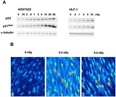
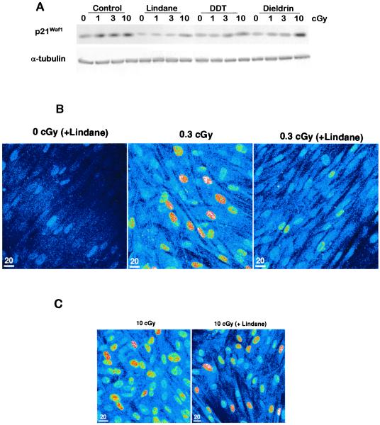
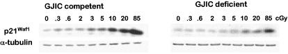
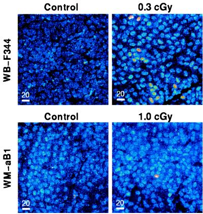
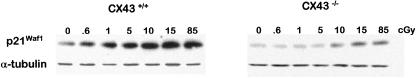
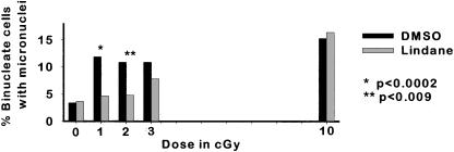
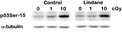
It has generally been considered that important biological effects of ionizing radiation arise as a direct consequence of DNA damage occurring in irradiated cells. We have examined this hypothesis by exposing cells to very low fluences of alpha-particles, similar to those emitted by radon gas, such that as few as 1% of the cells in a population are traversed by a particle and thus receive any radiation exposure. By using the endpoints of changes in gene expression and induction of DNA damage, we show that nonirradiated "bystander" cells participate in the overall response of confluent density-inhibited populations of cultured fibroblast and epithelial cells. By in situ immunofluorescence techniques and the use of cells genetically compromised in their ability to perform gap junction intercellular communication, we present direct evidence for the involvement of connexin43-mediated intercellular communication in the transmission of damage signals to nonirradiated cells. Induction of the stress-inducible p21(Waf1) protein in aggregates of neighboring cells far exceeding the fraction of cells whose nucleus has been traversed occurred in gap junction-competent cells only. These changes in p21(Waf1) expression correlated with both the induction of DNA damage (as measured by micronucleus formation) as well as increased Ser-15 phosphorylation of p53.


Figures

Figure 1
Figure 1
(A) Western blot analysis of p53 and p21Waf1 expression levels in low fluence α-particle exposed normal human fibroblasts. Confluent density-inhibited AG1522 skin or HLF-1 lung fibroblasts were exposed to α-particles doses ranging from 0.16 to 85 cGy and held at 37°C for 3 h. Cell lysates from irradiated and control nonexposed cultures were prepared and examined. (B) In situ immunofluorescence detection of p21Waf1 in control and α-particle exposed (0.3 cGy) AG1522 density-inhibited cultures where induction is seen to occur in aggregates of cells. About 1 cell in 50 would be traversed by an α-particle at this mean dose.
Figure 2
Figure 2
(A) Western analysis of p21Waf1 expression in α-particle-irradiated AG1522 human fibroblasts in the presence or absence of lindane (40 μM), DDT, or dieldrin. At 10 cGy, by which about 50–60% of the cells' nuclei are traversed by an α-particle track, p21Waf1 is induced in the presence or absence of the inhibitors. At 1 cGy, by which only 7% of the cells in the culture would have their nuclei traversed by an α-particle, p21Waf1 induction is not detected in the presence of any of the inhibitors. (B). In situ immunofluorescence detection of p21Waf1 expression in nonirradiated lindane-treated (40 μM), and irradiated AG1522 cultures exposed to 0.3 cGy α-particles in the presence or absence of lindane. The absence of induced aggregates of cells in the irradiated and lindane-pretreated cultures indicates that GJIC contributes to the bystander response. (C) In situ immunofluorescence detection of p21Waf1 expression in AG1522 cultures exposed to 10 cGy in the presence or absence of lindane.
Figure 3
Figure 3
(A) Transfer of the fluorescent dye Lucifer yellow through gap junction in AG1522 confluent density-inhibited cultures, and (B) inhibition of its transfer to adjacent cells by 40 μM lindane.
Figure 4
Figure 4
(A) Transfer of Lucifer yellow through gap junction in WB-F344 confluent density inhibited cultures, and (B) inhibition of its transfer by 40 μM lindane. (C) Inability to transfer Lucifer yellow to adjacent cells in gap junction-deficient WM-aB1 cells.
Figure 5
Figure 5
Expression of p21Waf1 in protein lysates from gap junction-competent WB-F344 or gap junction-deficient WM-aB1 confluent cultures after exposure to α-particles. Cells were harvested 4 h after the exposure and proteins were examined by Western blot analyses.
Figure 6
Figure 6
(Upper) In situ immunofluorescence detection of p21Waf1 expression in control nonirradiated WB-F344 cultures and cultures exposed to 0.3 cGy of α-particles. (Lower) Expression of p21Waf1 in control nonirradiated and 1 cGy exposed cultures of GJIC-deficient WM-aB1 cells.
Figure 7
Figure 7
Western analyses of p21Waf1 expression in lysates from isogenic wild-type or connexin43−/− cultures exposed to α-particles. Cells were harvested 4 h after the exposure.
Figure 8
Figure 8
Micronucleus formation in α-particle-exposed AG1522 cultures in the presence or absence of lindane. P values were determined by the χ2 test.
Figure 9
Figure 9
Detection of Ser-15 phosphorylation in p53 after exposure of confluent AG1522 cultures to α-particles in the presence or absence of the gap junction-inhibitor lindane.

References

    1. Nagasawa H, Little J B. Cancer Res. 1992;52:6394–6396. - PubMed
    1. Deshpande A, Goodwin E H, Bailey S M, Marrone B L, Lehnert B E. Radiat Res. 1996;145:260–267. - PubMed
    1. Nagasawa H, Little J B. Radiat Res. 1999;152:552–557. - PubMed
    1. Zhou H, Randers-Pehrson G, Waldren C A, Vannais D, Hall E J, Hei T K. Proc Natl Acad Sci USA. 2000;97:2099–2104. - PMC - PubMed
    1. Prise K M, Belyakov O V, Folkard M, Michael B D. Int J Radiat Biol Relat Stud Phys Chem Med. 1998;74:793–798. - PubMed

Publication types

MeSH terms