

doi: 10.1073/pnas.98.2.615.
Epub 2001 Jan 9.
Extreme hydrops fetalis and cardiovascular abnormalities in mice lacking a functional Adrenomedullin gene
Affiliations
- PMID: 11149956
- PMCID: PMC14636
- DOI: 10.1073/pnas.98.2.615
Item in Clipboard
Extreme hydrops fetalis and cardiovascular abnormalities in mice lacking a functional Adrenomedullin gene
Proc Natl Acad Sci U S A.
.
Abstract
Adrenomedullin, a recently identified potent vasodilator, is expressed widely and has been suggested to have functions ranging from reproduction to blood pressure regulation. To elucidate these functions and define more precisely sites of Adm expression, we replaced the coding region of the Adm gene in mice with a sequence encoding enhanced green fluorescent protein while leaving the Adm promoter intact. We find that Adm(-/-) embryos die at midgestation with extreme hydrops fetalis and cardiovascular abnormalities, including overdeveloped ventricular trabeculae and underdeveloped arterial walls. These data suggest that genetically determined absence of Adm may be one cause of nonimmune hydrops fetalis in humans.
Figures
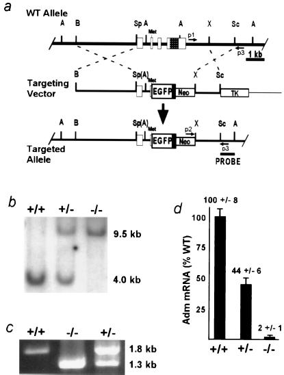
Generation of Adm−/− animals by homologous
recombination. (a) Strategy to disrupt the
Adm gene. (Top) Endogenous
wild-type (WT) allele. (Middle) Linearized targeting
vector. (Bottom) Targeted allele after homologous
recombination. Open boxes represent exons 1–4; the checkered region in
exon 4 codes for the mature Adm peptide; the initiator methionine of
the propeptide is indicated (Met). The solid black box depicts a bovine
growth hormone poly(A) addition sequence downstream of the EGFP cDNA.
The locations of primer sequences for PCR (p1, p2, and p3) are shown by
small arrows. The sequence used as a probe for the Southern-based
detection strategy is indicated by a labeled line (PROBE). Restriction
sites: A, AvrII; B, BamHI; Sc,
SacI; Sp, SpeI; X, XhoI.
Parentheses denote sites destroyed during cloning. (b)
Detection of the targeted allele by Southern blot analysis. Genomic DNA
from E9.5 embryos was digested with AvrII and probed
with the SacI/AvrII fragment (PROBE)
depicted in the bottom line of a. (c)
Detection of the targeted allele by PCR. Genomic DNA from embryos was
amplified with the primer sequences depicted in a.
(d) Measurement of Adm RNA in total RNA extracts from
E9.5 embryos by real-time quantitative reverse transcription–PCR. The
relative quantity of Adm RNA in Adm+/− and
Adm−/− embryos is expressed as a
percentage of total Adm RNA in Adm+/+
embryos. Error bars represent SEM.
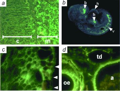
Sites of Adm expression determined by EGFP fluorescence.
(a) Frozen section of adrenal gland from an adult
Adm+/−. Note intense fluorescence in
adrenal medulla (m) and the absence of fluorescence in adrenal cortex
(c). (b) Confocal microscopy of an E9.5
Adm+/− embryo. Moderate expression is seen
in the heart (arrowhead h), and strong expression in the developing
vasculature (arrowhead v) and forebrain (arrowhead b); a speck of
nonspecific fluorescence has been covered with a black box.
(c) Frozen section of an E13.5
Adm+/− embryo, showing expression in the
developing left ventricle. Arrowheads indicate the pericardial surface
of the left ventricle. (d) Frozen section of an E16.5
Adm+/− embryo, showing expression in the
aorta (a), esophagus (oe), and thoracic duct (td).
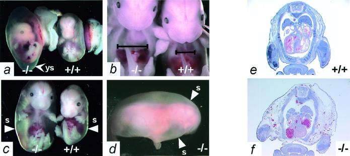
Adm−/− embryos have massive generalized edema.
(a) Appearance of E14.5
Adm−/− (Left) and
Adm+/+ (Right) embryos in
their yolk sacs shortly after dissection from the uterus. Note that the
yolk sac (arrowhead ys) of the Adm−/−
embryo is distended with fluid, and its blood vessels are thinner than
those of its wild-type littermate. (b) The thoracic
cavity of E13.5 Adm−/− embryos is
considerably enlarged (Left) compared with a wild-type
littermate (Right). The black lines indicate the width
of the thoracic cavity just above the diaphragm. (c)
Severe hydrops fetalis is apparent in the E14.5
Adm−/− embryos (Left),
readily visible after their yolk sacs are removed. The arrowheads (s)
indicate the outer skin layer of the embryos. (d) The
extreme hydrops of the Adm−/− embryos,
dissected away from their yolk sacs, is completely general, as
indicated by the uniformly swollen back and head. The arrowheads (s)
again indicate the outer skin layer. (e and
f) H&E stain of transverse sections through
E14.5 Adm+/+ (e) and
Adm−/− (f) embryos.
The Adm−/− embryo has a fluid-filled
thoracic cavity, and the tissues external to the rib cage are markedly
swollen (e and f, ×1).
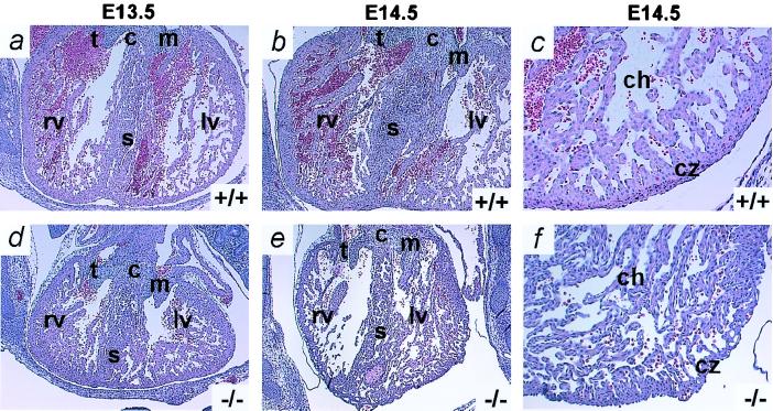
Adm−/− embryos have developmental heart defects.
Transverse sections through the hearts of
Adm+/+ (Upper) and
Adm−/− (Lower) embryos were
stained with H&E. (a) E13.5
Adm+/+ littermate. (d) E13.5
Adm−/− embryo. The heart of the
Adm−/− embryo is smaller than the heart of
its wild-type littermate. (b) E14.5
Adm+/+ littermate. (e) E14.5
Adm−/− embryo. The heart of the
Adm−/− embryo is still smaller, and the
left ventricle is significantly occluded with increased trabeculae
compared with its Adm+/+ littermate.
(c) E14.5 Adm+/+ littermate.
(f) E14.5 Adm−/−
embryo. Higher-power magnification of the left ventricle at E14.5 shows
an increase in the number of trabeculae, a thinner, convoluted compact
zone (cz), and a generally delaminated appearance of the
Adm−/− heart compared with wild type. c,
endocardial cushion; s, septum; t, tricuspid valve; m, mitral valve;
rv, right ventricle; lv, left ventricle; ch, chamber; cz, compact zone
(a, b, d, and e, ×2; c
and f, ×10).
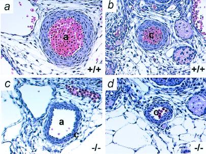
Adm−/− embryos have thin arterial walls. Transverse
sections through vessels of Adm+/+
(Upper) and Adm−/−
(Lower) embryos at E13.5 were stained with H&E.
(a and c) aorta; (b and
d) carotid artery. Note the thin vascular walls
(approximately three cells thick) of the
Adm−/− vessels compared with wild-type
vessels (approximately five cells thick). a, aorta; c, carotid artery
(×10).
References
-
- Sakata J, Shimokubo T, Kitamura K, Nakamura S, Kangawa K, Matsuo H, Eto T. Biochem Biophys Res Commun. 1993;195:921–927. - PubMed
-
- Hinson J P, Kapas S, Smith D M. Endocr Rev. 2000;21:138–167. - PubMed
-
- McLatchie L M, Fraser N J, Main M J, Wise A, Brown J, Thompson N, Solari R, Lee M G, Foord S M. Nature (London) 1998;393:333–339. - PubMed
-
- Kapas S, Martinez A, Cuttitta F, Hinson J P. J Endocrinol. 1998;156:477–484. - PubMed
Publication types
MeSH terms
Substances
Grants and funding
LinkOut - more resources
Full Text Sources
Other Literature Sources
Molecular Biology Databases
