. 2001 Jan 1;21(1):45-52.
doi: 10.1523/JNEUROSCI.21-01-00045.2001.

Blockade of NGF-induced neurite outgrowth by a dominant-negative inhibitor of the egr family of transcription regulatory factors

Affiliations

Blockade of NGF-induced neurite outgrowth by a dominant-negative inhibitor of the egr family of transcription regulatory factors

Y Levkovitz et al. J Neurosci. .

Abstract

Although it is well established that members of the Egr family of transcription regulatory factors are induced in many neuronal plasticity paradigms, it is still unclear what role, if any, they play in this process. Because NGF stimulation of pheochromocytoma 12 cells elicits a robust induction of Egr family members, we have investigated their role in mediating long-term effects elicited by NGF in these cells by using the Egr zinc finger DNA-binding domain as a selective antagonist of Egr family-mediated transcription. We report that expression of this Egr inhibitor construct suppresses neurite outgrowth elicited by NGF but not by dibutyryl cAMP. To check that this Egr inhibitor construct does not act by blocking the MEK/ERK pathway, which is known to mediate NGF-induced neurite outgrowth, we confirmed that the Egr inhibitor construct does not block NGF activation of Elk1-mediated transcription, a response that is dependent on this pathway. Conversely, inhibition of MEK does not impair Egr family-mediated transcription. Thus, we conclude (1) that induction of Egr family members and activation of the MEK/ERK pathway by NGF are mediated by separate signaling pathways and (2) that both are required to trigger neurite outgrowth induced by NGF.


Figures

Fig. 1.
Fig. 1.
Characterization of the Egr inhibitor construct Δ(1-249)Egr3. A, Top, Thebar shows a schematic diagram of Egr3 that contains three zinc finger motifs near its C terminal that mediate its interaction with the ERE, two distinct activation domains, A1 and A2, and a modulatory domain, R1, that serves as a binding site for NAB1 and NAB2. Bottom, The bar illustrates the Egr inhibitor construct Δ(1-249)Egr3 designed to retain the DNA-binding domain without the upstream activation or modulatory domains.B, The autoradiogram illustrates that the Egr inhibitor construct forms a gel-shift complex with the ERE oligonucleotide probe. Formation of this complex is blocked by addition of unlabeled ERE [wild-type ERE (wt ERE)] but not a mutant ERE (mut ERE) in which a single base pair has been changed. (Wild-type and mutant ERE oligonucleotide sequences are provided in Materials and Methods.) Unlabeled ERE oligonucleotides were added at the same concentration as the ERE probe (∼0.5 nm). In addition, neither AP-1 nor Sp1 oligonucleotides inhibit binding of the Egr inhibitor construct to the ERE, when added at 500-fold higher concentration than the ERE probe. C, HEK293 cells were transfected with a luciferase reporter plasmid containing a tandem repeat of ERE sites in its promoter as well as with the other expression plasmids listed under eachcolumn of the bar graph [Egr3 plasmid, 0.5 μg/well; Δ(1-249)Egr3, 0.5 μg/well]. The ability of Egr3 to increase luciferase activity in extracts from these cells was blocked by the Egr inhibitor construct. As expected, the Egr inhibitor construct was unable to increase luciferase activity compared with that in control cells that only received the ERE luciferase reporter plasmid.D, HEK293 cells were transfected with a luciferase reporter plasmid containing a tandem repeat of GAL4 response elements in its promoter as well as with the other expression plasmids listedunder each column. GAL4/Egr3(1-104) refers to a chimeric protein generated by fusing the GAL4 DNA-binding domain with the A1 activation domain of Egr3. In contrast to its ability to block stimulation of reporter gene expression by full-length Egr3, the Egr inhibitor construct [Δ(1-249)Egr3; 0.5 μg/well] does not block the increase in luciferase activity driven by the GAL4/Egr3(1-104) construct (0.5 μg/well) acting on a GAL4 reporter plasmid. In C and D and the figures that follow, “Δ 1-249” refers to Δ(1-249)Egr3. Error bars shown in this and subsequent figures represent the SEM. Data shown inC and D are presented in relative luciferase units (RLU).
Fig. 2.
Fig. 2.
The Egr inhibitor construct selectively blocks ERE-mediated transcription in PC12 cells. A, PC12 cells were transfected with a luciferase reporter plasmid (TG5) that is driven by a segment of the TGFβ1 promoter containing an ERE site. As found in HEK293 cells, the Egr inhibitor construct blocks the increase in luciferase activity induced by Egr3. B, The Egr inhibitor construct does not block the increase in luciferase activity displayed by cells transfected with the GAL4/Egr3(1-104) expression construct and the GAL4 reporter plasmid. C, In PC12 cells transfected with the ERE reporter, NGF induces a strong increase in luciferase activity that is suppressed by the Egr inhibitor construct. Cells were harvested 6 hr after addition of NGF.D, PC12 cells were cotransfected with a GAL4 reporter plasmid and an expression plasmid encoding a chimeric protein generated by fusing the GAL4 DNA-binding domain with the C-terminal activation domain of Elk1 (Path Detect Elk Trans-Reporting System, Stratagene). Stimulation of these cells with NGF (6 hr) triggers a robust increase in luciferase activity that is not blocked by cotransfection with a third plasmid encoding the Egr inhibitor construct.
Fig. 3.
Fig. 3.
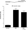
Selectivity of the Egr inhibitor construct.Top, PC12 cells were transfected with a luciferase reporter plasmid driven by a segment of the Egr1 promoter that contains four SRE sites. Bottom, NGF treatment increases luciferase activity in extracts prepared from these cells. This increase is not blocked by cotransfection with the Egr inhibitor construct.
Fig. 4.
Fig. 4.
The Egr inhibitor construct selectively blocks neurite outgrowth induced by NGF. PC12 cells were transfected with a GFP expression plasmid with or without Δ(1-249)Egr3, and cell morphology was monitored by fluorescence microscopy.Top, The typical small, round, and flat morphology found in undifferentiated PC12 cells (left; control) is shown. After treatment with NGF (right), cells elaborate one or more neurites. Middle, The Egr inhibitor construct, which does not induce neurite outgrowth (left), blocks neurite outgrowth triggered by NGF [right; NGF + Δ(1-249)Egr3]. Bottom, Neurite outgrowth can also be induced by db-cAMP (left) that is not affected by cotransfection with the Egr inhibitor construct [right; db-cAMP + Δ(1-249)Egr3]. Images shown were obtained 2 d after addition of NGF or db-cAMP.
Fig. 5.
Fig. 5.
Quantitative analysis of effects of the Egr inhibitor construct on neurite outgrowth. A, PC12 cells expressing GFP were scored as either neurite bearing or not. Exposure to NGF elicits neurite outgrowth in ∼80% of cells, indicating that expression of the GFP construct does not alter this response to NGF. Cotransfection with the Egr inhibitor construct markedly reduced the percentage of neurite-bearing cells observed after NGF treatment (NGF +Δ1-249). B, The Egr inhibitor construct does not reduce the percentage of neurite-bearing cells induced by exposure to db-cAMP (dbcAMP + Δ1-249).C, GFP-positive cells were grouped according to the number of neurites present after NGF treatment. Cotransfection with the Egr inhibitor construct produces a marked shift toward lower neurite number. D, The Egr inhibitor construct does not affect the distribution of neurite number after db-cAMP treatment.Values shown in C differ slightly from those in A because they are based on independent counts. Data presented in this figure are from cells counted 2 d after NGF or db-cAMP treatment. Over 500 cells were scored for each experimental condition.
Fig. 6.
Fig. 6.
Parallel effects of the Egr inhibitor construct in blocking neurite outgrowth and ERE-mediated transcription by NGF.A, The amount of Egr inhibitor plasmid used for transfection was varied from 0 to 1000 ng/well. To keep the total amount of transfected DNA constant, we added appropriate amounts of pCB6 plasmid. The bar graph presents the increase in luciferase activity detected in cell extracts harvested 6–8 hr after treatment with NGF. B, The effect of varying the amount of Egr inhibitor plasmid on the percentage of cells scored as neurite bearing after NGF exposure is presented in this bar graph. In the absence of NGF, 9 ± 6% (mean ± SEM) of cells were scored as bearing neurites.
Fig. 7.
Fig. 7.
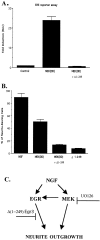
Effect of MEK inhibition on neurite outgrowth and transcriptional responses stimulated by NGF. A, This schematic diagram depicts a scenario in which Egr induction is located downstream of MEK activation by NGF. In this model, UO126, an MEK inhibitor, and the Egr inhibitor construct Δ(1-249)Egr3 block at sequential steps in the signaling pathway linking NGF receptor activation to neurite outgrowth. B, The bar graph presents the effect of pretreating cells with UO126 (15 μm) on neurite outgrowth induced by NGF.C, The bar graph shows the effect of UO126 (15 μm) on the ability of NGF to stimulate ERE-mediated and Elk1-mediated transcription.
Fig. 8.
Fig. 8.
MEK stimulation of ERE-mediated transcription: role in neurite outgrowth. A, The ERE reporter plasmid was used to monitor the effects of a constitutively active MEK1 construct [MEK(DD); 0.5 μg/well] on ERE-mediated transcription in PC12 cells. Cotransfection with the activated MEK construct increased luciferase activity in extracts from these cells. This increase is abolished when the MEK construct is cotransfected with the Egr inhibitor construct. B, Transfection with the MEK(DD) expression plasmid elicits neurite outgrowth that is blocked by cotransfection with the Egr inhibitor construct. C, The schematic diagram presents a model of signaling pathways mediating NGF-induced neurite outgrowth that incorporates the results presented in this report. A key feature of this model is that separate signaling pathways link NGF receptor stimulation to induction of Egr family members and activation of MEK. This inference is based on our finding that NGF stimulation of ERE-mediated transcription is not blocked by inhibition of MEK. These findings suggest that both of these responses to NGF are required for NGF to trigger neurite outgrowth, because blockade of either pathway suppresses the ability of NGF to elicit neurite outgrowth. However, at first glance, the assertion that both MEK activation and Egr family induction are required for neurite outgrowth appears to be at odds with the observation that constitutively activated MEK is sufficient to induce neurite outgrowth. This ostensible discrepancy is explained by our observation that the constitutively active MEK construct stimulates ERE-mediated transcription, allowing it to comply with both requirements for neurite outgrowth stipulated by this model. Further confirmation of this scenario is provided by the ability of the Egr inhibitor construct to block neurite outgrowth induced by the constitutively active MEK construct. However, it appears that induction of Egr family members is not sufficient to induce neurite outgrowth because transfection of these cells with Egr1 does not trigger neurite outgrowth. Therefore, the model shown includes a second arrow emanating from MEK leading to neurite outgrowth.

Similar articles

Cited by

References

    1. Abraham WC, Otani S. Macromolecules and the maintenance of long-term potentiation. In: Morrell F, editor. Kindling and synaptic plasticity, the legacy of Graham Goddard. Birkhauser; Boston: 1991. pp. 92–109.
    1. Ahn S, Ginty DD, Linden DJ. A late phase of cerebellar long-term depression requires activation of CaMKIV and CREB. Neuron. 1999;23:559–568. - PubMed
    1. Chapman NR, Perkins ND. Inhibition of the RelA(p65) NF-kB subunit by Egr-1. J Biol Chem. 2000;275:4719–4725. - PubMed
    1. Chen C, Okayama H. High-efficiency transformation of mammalian cells by plasmid DNA. Mol Cell Biol. 1987;7:2745–2752. - PMC - PubMed
    1. Christy B, Nathans D. DNA binding site of the growth factor-inducible protein Zif268. Proc Natl Acad Sci USA. 1989a;86:8737–8741. - PMC - PubMed

Publication types

MeSH terms

LinkOut - more resources