. 2001 Jan 1;21(1):59-66.
doi: 10.1523/JNEUROSCI.21-01-00059.2001.

Presynaptic kainate receptors regulate spinal sensory transmission

Affiliations

Presynaptic kainate receptors regulate spinal sensory transmission

G A Kerchner et al. J Neurosci. .

Abstract

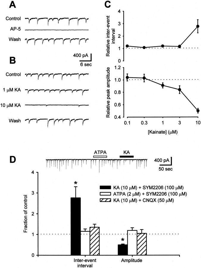
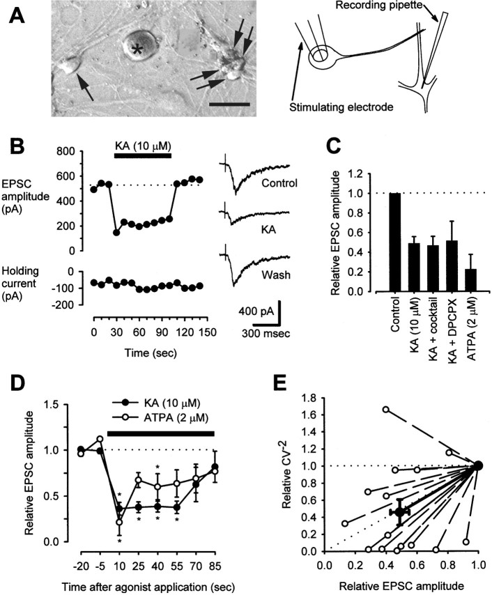
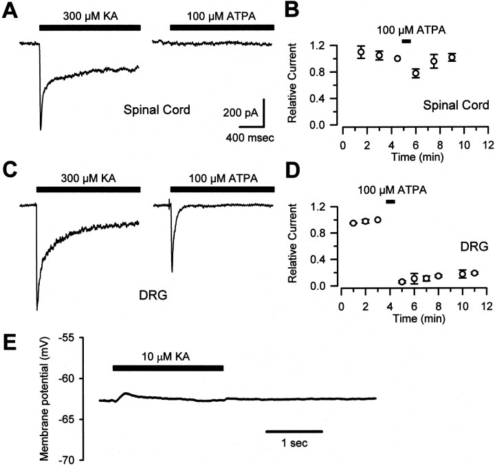
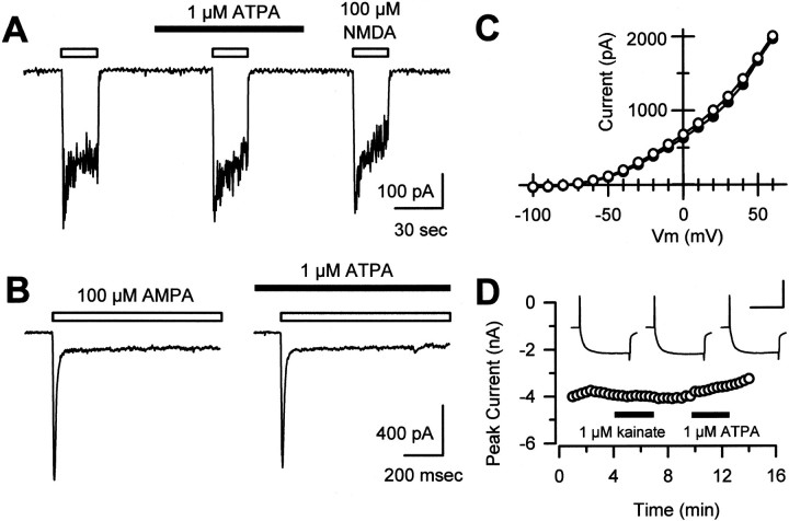
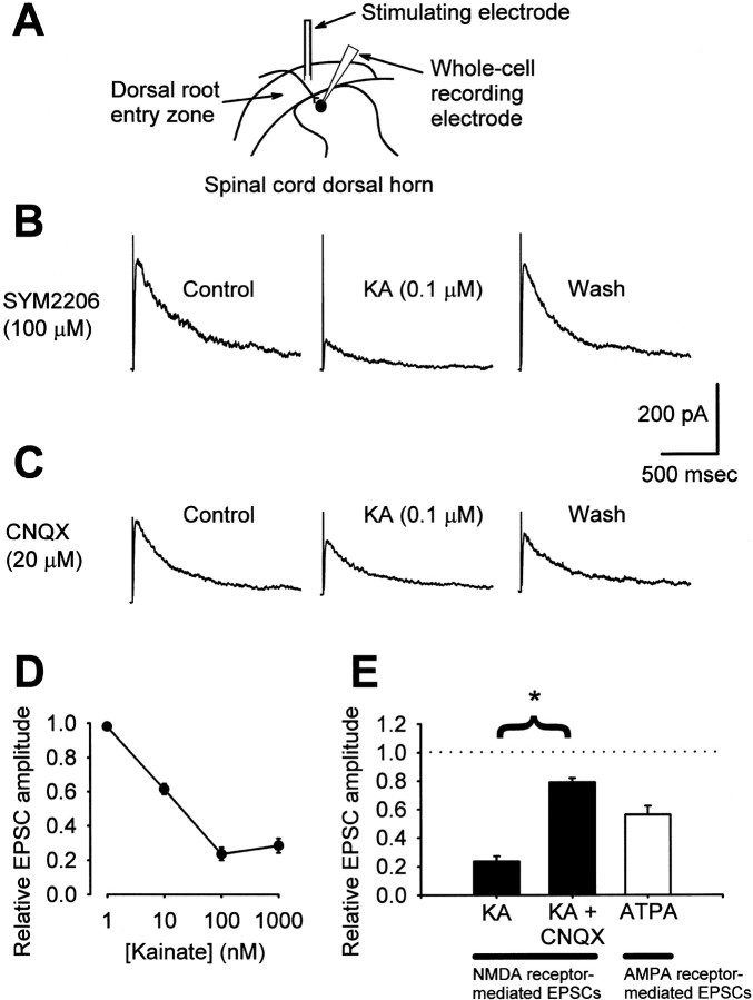
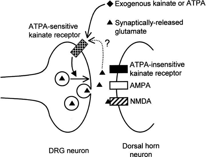
Small diameter dorsal root ganglion (DRG) neurons, which include cells that transmit nociceptive information into the spinal cord, are known to express functional kainate receptors. It is well established that exposure to kainate will depolarize C-fiber afferents arising from these cells. Although the role of kainate receptors on sensory afferents is unknown, it has been hypothesized that presynaptic kainate receptors may regulate glutamate release in the spinal cord. Here we show that kainate, applied at low micromolar concentrations in the presence of the AMPA-selective antagonist (RS)-4-(4-aminophenyl)-1, 2-dihydro-1-methyl-2-propyl-carbamoyl-6,7-methylenedioxyphthalazine++ +, suppressed spontaneous NMDA receptor-mediated EPSCs in cultures of spinal dorsal horn neurons. In addition, kainate suppressed EPSCs in dorsal horn neurons evoked by stimulation of synaptically coupled DRG cells in DRG-dorsal horn neuron cocultures. Interestingly, although the glutamate receptor subunit 5-selective kainate receptor agonist (RS)-2-alpha-amino-3-(3-hydroxy-5-tert-butylisoxazol-4-yl) propanoic acid (ATPA) (2 micrometer) was able to suppress DRG-dorsal horn synaptic transmission to a similar extent as kainate (10 micrometer), it had no effect on excitatory transmission between dorsal horn neurons. Agonist applications revealed a striking difference between kainate receptors expressed by DRG and dorsal horn neurons. Whereas DRG cell kainate receptors were sensitive to both kainate and ATPA, most dorsal horn neurons responded only to kainate. Finally, in recordings from dorsal horn neurons in spinal slices, kainate and ATPA were able to suppress NMDA and AMPA receptor-mediated EPSCs evoked by dorsal root fiber stimulation. Together, these data suggest that kainate receptor agonists, acting at a presynaptic locus, can reduce glutamate release from primary afferent sensory synapses.


Figures

Fig. 1.
Fig. 1.
Kainate suppresses spontaneous excitatory transmission between cultured dorsal horn neurons. A, In the presence of 100 μm SYM2206 and no Mg2+, sEPSCs were recorded from dorsal horn neurons in mass culture. These events were silenced in the presence of 25 μm AP-5. B, Addition of kainate altered the characteristics of sEPSCs. At 1 μm, kainate reduced sEPSC amplitude without affecting frequency. Both parameters were decreased by 10 μm kainate. C, Summary of the effects of various doses of kainate on sEPSC interevent interval and peak amplitude (0.1 μm KA,n = 2 cells; 0.3 μm ka,n = 6; 1 μm ka, n= 8; 3 μm ka, n = 6; 10 μm ka; n = 8). D, Whereas 10 μm kainate altered both the amplitude and frequency of spontaneous NMDA receptor-mediated EPSCs (n = 7 cells), neither 2 μm ATPA (n = 5) nor 10 μm kainate plus 50 μm CNQX (n = 3) had any effect. *p < 0.05 indicates significant difference from control; two-way ANOVA with Tukey's test for post hoccomparison.
Fig. 2.
Fig. 2.
Kainate suppresses evoked excitatory transmission between DRG neurons and dorsal horn neurons in coculture.A, A photograph of cocultured DRG (asterisk) and dorsal horn neurons (arrows) and a diagram showing placement of recording and stimulating electrodes illustrate the experimental system. Scale bar, 20 μm. B, In a representative neuron, 10 μm kainate reversibly reduced NMDA receptor-mediated EPSC amplitude. Traces at the right show the shapes of EPSCs before, during, and after KA treatment. This recording was from a neuron treated with the antagonist cocktail described inC. The dotted line indicates the baseline level. C, Pooled data illustrate the relative amplitudes of NMDA receptor-mediated EPSCs in control conditions and after treatment with 10 μm KA (n = 8) or 2 μm ATPA (n = 6) alone; reduction of EPSC amplitude in both cases was statistically significant (p < 0.05; paired t test comparing absolute EPSC amplitudes in control and test conditions). In addition, the effect of KA was tested in some cells that were exposed continuously to a cocktail (containing 50 μm atropine, 100 μm naloxone, 2 mm AIDA, 500 μm CPPG, and 10 μm CGP55845;n = 4) or 1 μm DPCPX (n = 3). The effect of KA in these instances was indistinguishable from and did not differ statistically from the effect of KA alone (one-way ANOVA, comparing the percentage of EPSC depression in each condition). D, The effects of a continuous administration of 10 μm kainate (n = 8) or 2 μm ATPA (n = 6) on EPSC amplitude are plotted relative to baseline values. *p < 0.05 indicates significant difference between relative EPSC amplitude in kainate- or ATPA-treated cells at the indicated time points compared with baseline; Kruskal–Wallis one-way ANOVA on ranks with Dunn's test for post hoccomparison. The dotted line indicates the baseline level. E, Relative values for the statistic CV−2 (see Materials and Methods) are plotted against relative values for mean EPSC amplitude in cells before and after treatment with 10 μm KA. Data are included from all 15 KA-treated cells described in C, including experiments using various receptor antagonists. The bold line and bold points represent the mean relationship. The diagonal dotted line indicates the predicted relationship if only presynaptic phenomena underlie a change in EPSC amplitude; the horizontal dotted line indicates the relationship predicted by purely postsynaptic effects (Bekkers and Stevens, 1990; Clements, 1990; Malinow and Tsien, 1990).
Fig. 3.
Fig. 3.
ATPA activates kainate receptors on DRG but not dorsal horn neurons. A, In cultured spinal cord dorsal horn neurons, fast application of 300 μm kainate but not 100 μm ATPA evoked a fast, incompletely desensitizing current. B, A 30 sec application of ATPA exerted little or no effect on the amplitude of peak currents evoked by 300 μm KA in spinal neurons (n = 11).C, In cultured DRG neurons, fast currents were elicited by both 300 μm KA and 100 μm ATPA.D, In experiments performed as in B, but using DRG neurons, ATPA caused a prolonged desensitization of peak kainate-evoked currents (n = 9). E, In a representative current-clamp recording from a cultured DRG neuron, 10 μm KA induced little somatic depolarization.
Fig. 4.
Fig. 4.
ATPA and kainate do not affect AMPA- or NMDA-evoked currents in postsynaptic cells or affect Ca2+ or K+ channels in presynaptic cells. A, In cultured dorsal horn neurons, application of 100 μm NMDA and 1 μm glycine evoked inward currents that were insensitive to the presence of 1 μm ATPA. B, As in A, ATPA did not affect currents evoked by 100 μm AMPA.C, In cultured DRG neurons, the I–Vrelationship of voltage-gated K+ channel currents was identical in the absence (open circles) or presence (filled circles) of 1 μm ATPA. Steady-state currents are plotted at each potential. Similar results were obtained in 13 other cells. D, Voltage-gated Ca2+ channel currents in DRG neurons were not affected by application of 1 μm kainate (98 ± 1% of control; n = 23) or 1 μm ATPA (98 ± 1% of control; n = 10). Peak current evoked by stepping from −80 to 0 mV is plotted as a function of time.Inset, Individual traces recorded in control, kainate, and ATPA. Calibration: 4 nA, 50 msec.
Fig. 5.
Fig. 5.
Kainate receptor activation suppresses primary afferent neurotransmission in spinal cord slices. A, A diagram illustrates the placement of stimulating and recording electrodes in the dorsal horn of a spinal slice. B, NMDA receptor-mediated EPSCs were isolated at a holding potential of +40 mV in the presence of 100 μm SYM2206. Traces from a representative neuron show reversible inhibition of EPSCs by 0.1 μm KA. C, When 20 μm CNQX replaced SYM2206 in experiments, as in B, 0.1 μm KA had less of an effect. D, Kainate exerted a dose-dependent inhibition of NMDA receptor-mediated EPSCs (n = 3–4 cells per concentration).E, Summarized data showing inhibition of NMDA receptor-mediated EPSCs by 0.1 μm KA in the presence of 100 μm SYM2206 (KA; n= 3) that was sensitive to 20 μm CNQX (KA+ CNQX; n = 3). Treatment with 100 μm CNQX (n = 3) afforded no additional blockade of the effect of kainate (data not shown). In addition, AMPA receptor-mediated EPSCs were suppressed by application of 2 μm ATPA (n = 5). ATPA- and KA-induced EPSC suppression were both statistically significant (p < 0.05; paired t test comparing absolute EPSC amplitudes in control and test conditions). *p < 0.05 indicates a significant difference in EPSC reduction by KA in the presence of SYM2206 versus CNQX;t test comparing the percentage of EPSC suppression in each condition.
Fig. 6.
Fig. 6.
Kainate autoreceptors at primary afferent synapses. A model illustrates the hypothesis that kainate receptors, in addition to residing in the postsynaptic membrane of dorsal horn neurons, are also present on the presynaptic terminals of DRG neurons. Presynaptic and postsynaptic kainate receptors could be distinguished in this study by their sensitivity to the GluR5-selective agonist ATPA, and activation of presynaptic kainate receptors, at least by exogenous agonists, inhibited evoked release of glutamate. Unresolved is whether the presynaptic receptors can be activated by synaptically released glutamate (dotted line), and, if so, whether subsequent glutamate release would be inhibited or enhanced.

Similar articles

Cited by

References

    1. Agrawal SG, Evans RH. The primary afferent depolarizing action of kainate in the rat. Br J Pharmacol. 1986;87:345–355. - PMC - PubMed
    1. Ault B, Hildebrand LM. Activation of nociceptive reflexes by peripheral kainate receptors. J Pharmacol Exp Ther. 1993;265:927–932. - PubMed
    1. Bekkers JM, Stevens CF. Presynaptic mechanism for long-term potentiation in the hippocampus. Nature. 1990;346:724–729. - PubMed
    1. Bortolotto ZA, Clarke VR, Delany CM, Parry MC, Smolders I, Vignes M, Ho KH, Miu P, Brinton BT, Fantaske R, Ogden A, Gates M, Ornstein PL, Lodge D, Bleakman D, Collingridge GL. Kainate receptors are involved in synaptic plasticity. Nature. 1999;402:297–301. - PubMed
    1. Chergui K, Bouron A, Normand E, Mulle C. Functional GluR6 kainate receptors in the striatum: indirect downregulation of synaptic transmission. J Neurosci. 2000;20:2175–2182. - PMC - PubMed

Publication types

MeSH terms

LinkOut - more resources