. 2001 Jan 1;21(1):75-83.
doi: 10.1523/JNEUROSCI.21-01-00075.2001.

Cholinergic modulation of excitatory synaptic transmission in the CA3 area of the hippocampus

Affiliations

Cholinergic modulation of excitatory synaptic transmission in the CA3 area of the hippocampus

K E Vogt et al. J Neurosci. .

Abstract

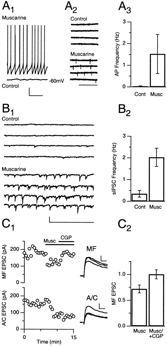
Cholinergic innervation of the hippocampus has been implicated in memory formation and retrieval. Here we study cholinergic modulation of excitatory transmission in the CA3 area of the rat hippocampus. We used a combination of optical measurements of presynaptic calcium and electrophysiological measurements of synaptic currents to study associational-commissural (A/C) and mossy fiber (MF) synapses in brain slices. Direct synaptic modulation mediated by ACh receptors is only evident at the A/C synapse, where synaptic inhibition primarily reflects presynaptic calcium channel inhibition mediated by muscarinic receptors. MF synapses can, however, be indirectly modulated by muscarinic receptor activation. Muscarine elevates the firing rate of inhibitory cells, which increases GABA release and inhibits MF synapses by activating presynaptic GABA(B) receptors. Muscarine also depolarizes dentate granule cells and elevates their rate of firing. This leads to synaptic enhancement when combined with the use-dependent facilitation of MF synapses. In addition we were unable to evoke an increase in presynaptic calcium levels in MF boutons with local application of nicotinic receptor agonists. This finding does not support a leading hypothesis for MF modulation in which activation of presynaptic nicotinic receptors enhances transmission directly by elevating presynaptic calcium levels. However, indirect synaptic modulation could arise from nicotinic excitation of inhibitory neurons. Thus, to understand cholinergic modulation within the CA3 region, it is necessary to take into account secondary actions on synapses arising from other chemical messengers released by other cell types and to consider effects on firing patterns of presynaptic cells, which in turn influence release via use-dependent synaptic plasticity.


Figures

Fig. 1.
Fig. 1.
Measurements of presynaptic calcium transients in MFs. A, Schematic showing the placement of the local superfusion system for labeling hippocampal mossy fibers. Arectangle marks the area shown in B.B, Hippocampal CA3 region of a slice in which MFs were labeled with magnesium green. A bright-field image (1), an image of magnesium green fluorescence converted to gray scale with regions of intense fluorescence appearing dark(2), and an overlay of the two (3) are shown. Scale bar, 100 μm. C, D, Fluorescence transients evoked by extracellular MF stimulation in slices in which MFs were labeled with fura-2 (C) and magnesium green (D). A single stimulus was used (1), and fluorescence transients were evoked by a stimulus pair 40 msec apart (2). The 380 nm-evoked fura-2 fluorescence has been inverted for clarity in this and the next figure. Calibration: vertical, 1% ΔF/F; horizontal, 500 msec (1), 50 msec (2).str, Stratum.
Fig. 2.
Fig. 2.
Measurements of presynaptic calcium transients in associational–commissural fibers. A, Schematic showing the labeling of A/C fibers. The area shown inB is marked with a rectangle.B, High-magnification image of the stratum radiatum in a magnesium green-labeled slice. Fluorescence signals are converted to a gray scale image with dark structures representing high-fluorescence intensity. Scale bar, 10 μm. C, D, Fluorescence transients evoked by extracellular stimulation in the stratum radiatum in slices labeled with fura-2 (C) and magnesium green (D). Fluorescence transients were evoked by a single stimulus (1) and a stimulus pair 40 msec apart (2). Calibration: vertical, 0.25% ΔF/F; horizontal, 500 msec (1), 50 msec (2).
Fig. 3.
Fig. 3.
The effect of external calcium on presynaptic calcium influx and synaptic strengths. A, Reduction in peak magnesium green ΔF/F during application of 1 mm Cae (horizontal line) for MF (left) and A/C (right) fibers. The control solution contained 3 mm Cae. Insets, Averages of 10–20 ΔF/F transients before, during (smallest trace), and after the application of 1 mmCae. Calibration: vertical, 0.2% ΔF/F; horizontal, 10 msec. B, Relationship between peak ΔF/F and Cae in MF and A/C fibers. The relative change in ΔF/F for different Caevalues was measured and normalized to that of control conditions.C, Reduction in NMDAR-mediated EPSC amplitude for MF and A/C fibers for the same reduction in Cae.Insets, Averages of 10–20 NMDAR-mediated EPSCs before, during, and after the application of 1 mmCae. Calibration: vertical, 20 pA; horizontal, 20 msec. D, E, The relationships between EPSC amplitude and peak ΔF/F normalized to control conditions and plotted on linear scales (D) and on log scales (E). Fits in D andE are to Equation 1 with EPSCmax,KD, and n being ≫50, 19.6, and 3.63, respectively, for the MF synapse and 14.2, 0.99, and 3.86, respectively, for the A/C synapse.
Fig. 4.
Fig. 4.
The effect of presynaptic modulators on presynaptic calcium influx and synaptic strengths.Traces were normalized to control. Drug applications are indicated by horizontal lines. A, B, Response of peak magnesium green ΔF/F(left) and NMDAR-mediated EPSC amplitude (right) to baclofen, l-CCG-I, and muscarine in the MF system (A) and in the A/C system (B). Insets, Averages of 10–20 ΔF/F transients (left) and NMDAR-mediated EPSCs (right) before and during (smallest trace) drug application. Calibration: vertical, 0.2% ΔF/F (A), 20 pA (B); horizontal, 10 msec (A), 20 msec (B).C, Log plots of the EPSC amplitudes versus peak ΔF/F changes for the indicated experimental conditions for MF (C1) and A/C (C2) responses. The solid lines in C are the fits described in Figure 3. Means are of 4–10 experiments for each manipulation.Baclo, Baclofen; Musc, muscarine.
Fig. 5.
Fig. 5.
Muscarine depolarizes CA3 interneurons and causes heterosynaptic depression of MF release.A1, Current-clamp whole-cell recording of a CA3 interneuron before and after the application of muscarine. Calibration: vertical, 10 mV; horizontal, 1 sec.A2, Example of a cell-attached recording of a CA3 interneuron under control conditions and during the application of muscarine. Calibration: horizontal, 1 sec.A3, Summary of the effect of muscarine on interneuronal firing in cell-attached recordings (N = 4). B1, Example of sIPSCs in a whole-cell recording from a CA3 pyramidal cell under control conditions and during the presence of muscarine. Calibration: vertical, 50 pA; horizontal, 1 sec.B2, Average sIPSC frequency under control conditions and during the presence of muscarine (N = 4). C1, Example of the MF (top) and A/C (bottom) NMDAR-mediated EPSC amplitude response to the application of muscarine and the subsequent addition of the GABAB antagonist CGP.Insets, Average traces for the respective conditions. Calibration: vertical, 20 pA; horizontal, 20 msec.C2, Summary of the relative effects of muscarine alone (N = 4) and muscarine and CGP (N = 10) on MF EPSC amplitude. AP, Action potential; Cont, Control.
Fig. 6.
Fig. 6.
Muscarine elevates firing rates of granule cells and causes mossy fiber synaptic enhancement.A1, Example of the effect of muscarine on the firing rate of a granule cell. Inset, Representative traces in control conditions and in the presence of muscarine. Calibration: horizontal, 2 sec.A2, Summary of the effect of muscarine on granule cell-firing frequency (N = 5).B1, Example of muscarine-mediated increase of NMDAR-mediated MF EPSC amplitude. Muscarine andl-CCG-I were applied as indicated (horizontal lines). Inset, Average EPSCs for the respective conditions. Calibration: vertical, 50 pA; horizontal, 20 msec.B2, Average effect of muscarine on MF EPSC amplitude in intact slices (N = 4) and when the MF tract was cut (N = 6).
Fig. 7.
Fig. 7.
Mossy fiber terminals are not responsive to the nicotinic agonists nicotine and choline. A, Fluorescence transient from an MF tract labeled with fura-2. ΔF/Ftraces at 350 and 380 nm excitation wavelengths as a response to extracellular stimulation are shown. B, MF fura-2 ΔF/F transients at 380 and 350 nm as a response to focal application of KCl. C, Focal application of nicotine (20 μm) and recording of ΔF/Ftransients at 350 and 380 nm. C1, Application at a distance similar to the one used in B. C2, Application of nicotine at close range. D, Focal application of the nicotinic acetylcholine receptor agonist choline (3 mm) to a fura-2-labeled MF tract. D1, Placement of application pipette similar to that in B. D2, Application pipette in close proximity to the labeled tract. Calibration (A–D): vertical, 1%; horizontal, 1 sec.Traces are averages of 10–20 trials. E, Whole-cell currents from an interneuron in voltage clamp as a response to focal choline application. E1, Amplitude response under control conditions and during MLA application.E2, Averages of five currenttraces under the respective conditions. Calibration: vertical, 10 pA; horizontal, 0.5 sec.

References

    1. Albuquerque EX, Pereira EF, Castro NG, Alkondon M, Reinhardt S, Schroder H, Maelicke A. Nicotinic receptor function in the mammalian central nervous system. Ann N Y Acad Sci. 1995;757:48–72. - PubMed
    1. Albuquerque EX, Pereira EF, Alkondon M, Schrattenholz A, Maelicke A. Nicotinic acetylcholine receptors on hippocampal neurons: distribution on the neuronal surface and modulation of receptor activity. J Recept Signal Transduct Res. 1997;17:243–266. - PubMed
    1. Alkondon M, Pereira EF, Cortes WS, Maelicke A, Albuquerque EX. Choline is a selective agonist of alpha7 nicotinic acetylcholine receptors in the rat brain neurons. Eur J Neurosci. 1997;9:2734–2742. - PubMed
    1. Alkondon M, Braga MF, Pereira EF, Maelicke A, Albuquerque EX. Alpha7 nicotinic acetylcholine receptors and modulation of gabaergic synaptic transmission in the hippocampus. Eur J Pharmacol. 2000;393:59–67. - PubMed
    1. Amaral DG, Dent JA. Development of the mossy fibers of the dentate gyrus. I. A light and electron microscopic study of the mossy fibers and their expansions. J Comp Neurol. 1981;195:51–86. - PubMed

Publication types

MeSH terms

LinkOut - more resources