. 2001 Jan 1;21(1):98-108.
doi: 10.1523/JNEUROSCI.21-01-00098.2001.

Vitamin D hormone confers neuroprotection in parallel with downregulation of L-type calcium channel expression in hippocampal neurons

Affiliations

Vitamin D hormone confers neuroprotection in parallel with downregulation of L-type calcium channel expression in hippocampal neurons

L D Brewer et al. J Neurosci. .

Abstract

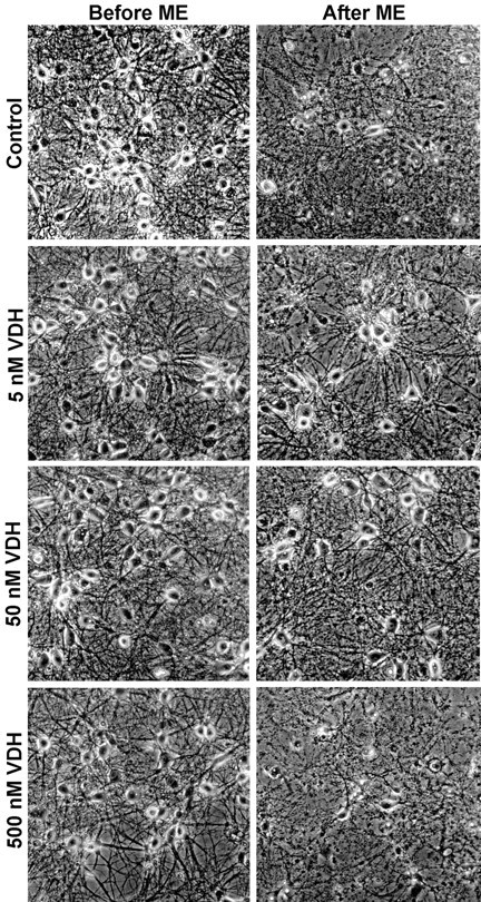
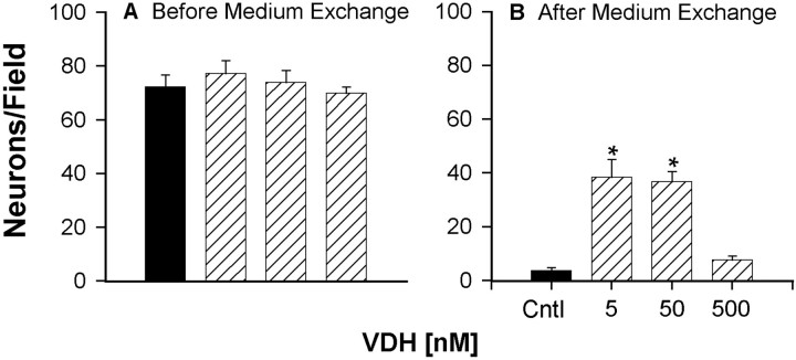
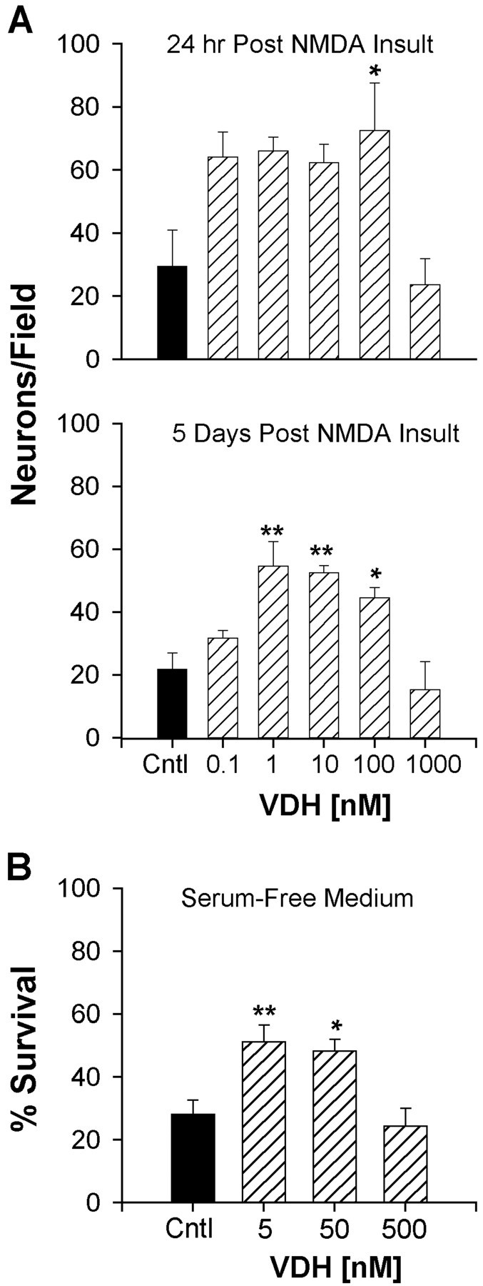
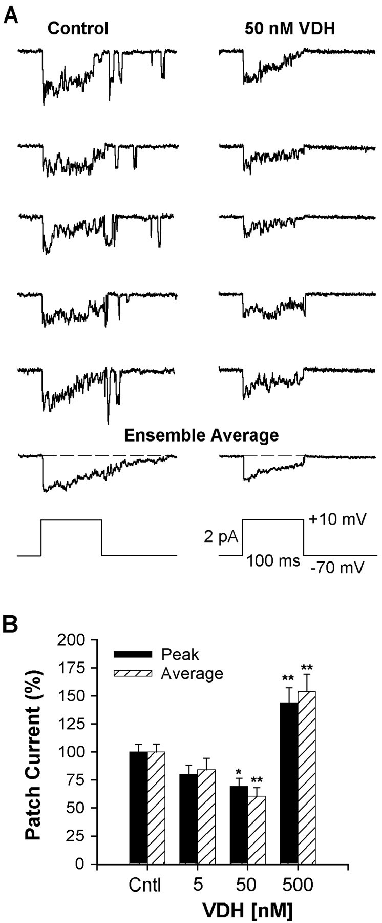
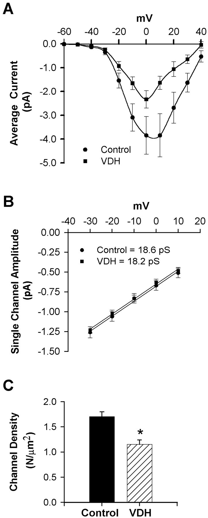
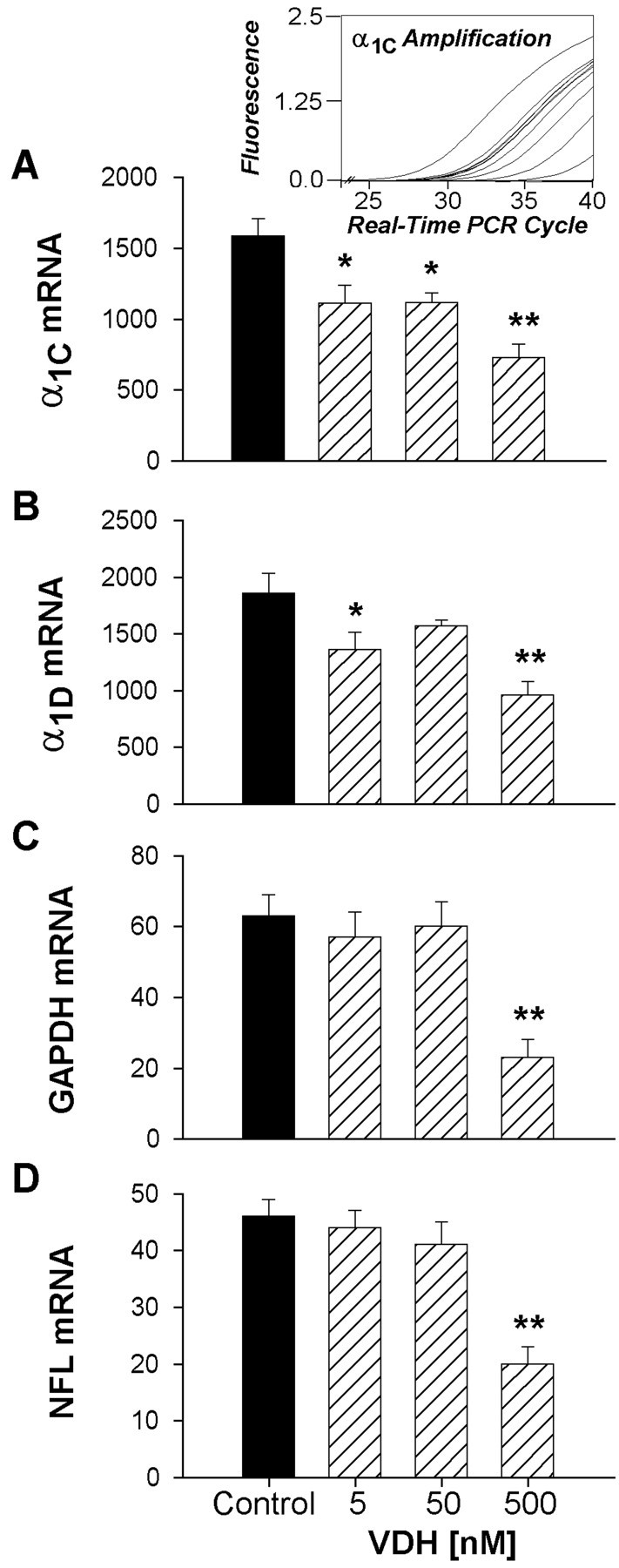
Although vitamin D hormone (VDH; 1,25-dihydroxyvitamin D(3)), the active metabolite of vitamin D, is the major Ca(2+)-regulatory steroid hormone in the periphery, it is not known whether it also modulates Ca(2+) homeostasis in brain neurons. Recently, chronic treatment with VDH was reported to protect brain neurons in both aging and animal models of stroke. However, it is unclear whether those actions were attributable to direct effects on brain cells or indirect effects mediated via peripheral pathways. VDH modulates L-type voltage-sensitive Ca(2+) channels (L-VSCCs) in peripheral tissues, and an increase in L-VSCCs appears linked to both brain aging and neuronal vulnerability. Therefore, we tested the hypothesis that VDH has direct neuroprotective actions and, in parallel, targets L-VSCCs in hippocampal neurons. Primary rat hippocampal cultures, treated for several days with VDH, exhibited a U-shaped concentration-response curve for neuroprotection against excitotoxic insults: lower concentrations of VDH (1-100 nm) were protective, but higher, nonphysiological concentrations (500-1000 nm) were not. Parallel studies using patch-clamp techniques found a similar U-shaped curve in which L-VSCC current was reduced at lower VDH concentrations and increased at higher (500 nm) concentrations. Real-time PCR studies demonstrated that VDH monotonically downregulated mRNA expression for the alpha(1C) and alpha(1D) pore-forming subunits of L-VSCCs. However, 500 nm VDH also nonspecifically reduced a range of other mRNA species. Thus, these studies provide the first evidence of (1) direct neuroprotective actions of VDH at relatively low concentrations, and (2) selective downregulation of L-VSCC expression in brain neurons at the same, lower concentrations.


Figures

Fig. 1.
Fig. 1.
Photomicrographs of VDR immunoreactivity in hippocampal cultures (7–14 DIV). A, Two representative pyramidal-shaped neurons positively stained for the VDR with Ab4707 (phase contrast optics). B, No cellular staining was observed when the VDR antibody was preincubated with its epitope.C, Cell culture double-labeled with antibodies to VDR and GFAP to determine cell-specific labeling of VDR. Most cells expressing the VDR (brown stain) were neurons, as indicated by the morphology and the lack of co-labeling with the glial marker GFAP (violet stain). D, Nuclei of astrocytes (arrow) showed either weak or no staining for the VDR. A pyramidal-shaped neuron in the field stained positively for VDR (arrowhead) is shown for comparison. Scale bars:A, B, D, 50 μm; C, 100 μm.
Fig. 2.
Fig. 2.
Representative photomicrographs of hippocampal neurons before an ME insult and again 24 hr after insult at 15 and 16 DIV, respectively. Cultures were chronically treated either with control vehicle (0.05% ethanol) or VDH (5, 50, and 500 nm) at 3, 6, 8, and 14 DIV. Few neurons survived the ME insult in control cultures; however, cultures treated with 5 and 50 nm VDH were partially protected from the insult. No protection was observed in cultures treated with the highest concentration of VDH, 500 nm.
Fig. 3.
Fig. 3.
VDH-treated hippocampal neurons were partially protected from ME insult. Quantitative results from the cultures in Figure 2 after ME insult of control (Cntl) and VDH-treated cells are shown. Survival was assessed from photographs taken at 15 DIV before ME insult and again 24 hr after insult.A, Chronic treatment with VDH did not alter the number of neurons before ME insult. B, The number of surviving neurons after ME insult was significantly greater in wells pretreated with 5 and 50 nm VDH (*p < 0.001 vs control). Results are mean ± SEM; n = 15–18 culture wells per group.
Fig. 4.
Fig. 4.
Survival of hippocampal neurons after insult with NMDA or glutamate in VDH-treated cultures. A, top panel, Cultures were treated with VDH at 3, 6, and 8 DIV and subjected to NMDA (100 μm) insult 6 hr after the last VDH treatment. Survival 24 hr after NMDA insult was significantly greater in cultures treated with 100 nm VDH (p < 0.05 vs control). A, bottom panel, Survival was reassessed in the same cultures 5 d after NMDA insult. Enhanced long-term survival of hippocampal neurons was found in cultures previously treated with 1, 10, and 100 nm VDH (*p < 0.05; **p < 0.01 vs control). Results are mean ± SEM; n = 4 culture wells/group. B, VDH added to cultures maintained in serum-free conditions (B27, lacking VDH). Hippocampal neurons in VDH-treated wells (5 and 50 nm) were partially protected from glutamate insult similar to experiments in serum-maintained cultures. Cultures were treated with VDH at 15 or 16 DIV for 24 hr and again 4–6 hr before glutamate insult (5 μm for 5 min). Survival was assessed 24 hr after insult. VDH treatment for 24–30 hr is sufficient to confer protection (*p < 0.05; **p < 0.01 vs control). Results are mean ± SEM;n = 8–12 culture wells per group.
Fig. 5.
Fig. 5.
VDH reduced L-VSCC activity. A,Five representative traces of L-VSCC activity recorded in the cell-attached patch mode from a control (0.05% ethanol) and from a VDH-treated (50 nm, 24–30 hr) hippocampal neuron. Multichannel activity was evoked by depolarizations fromVh = −70 mV toVc = +10 mV. The ensemble average of the full 15 traces from each neuron is shown below the 5 traces. The voltage protocol is shown at the bottom.B, Relationship between VDH concentration and L-VSCC activity recorded from multichannel patches. Effects of VDH on peak and average current activity were determined as percentage of control. Treatment with 50 nm VDH for 24 hr significantly reduced both peak and average current activities (*p < 0.05; **p < 0.01, respectively). Reduction in current by 5 nm VDH was not significant. The highest concentration (500 nm) exerted an opposite action and increased L-VSCC activity. Results are mean ± SEM;n = 89 control neurons and 19–44 VDH-treated neurons.
Fig. 6.
Fig. 6.
A, I–V series of average patch current from multichannel patches obtained from control or VDH-treated (50 nm, 24–30 hr) neurons. VDH reduced mean current amplitude without altering voltage dependence. Results are mean ± SEM B, Slope conductance of L-VSCCs was unaffected by VDH. Current amplitudes of clearly resolvable single-channel openings (of at least 3 msec duration) were measured from the I–V series. Mean slope conductance for each group was calculated from the average of individual slope conductances.C, Membrane density of functionally available L-VSCCs was reduced by VDH (p < 0.05). Channel density (N per square micrometer) was calculated using the method of maximal simultaneous openings. The membrane area (square micrometers) of a patch was calculated from pipette resistance (see Materials and Methods).
Fig. 7.
Fig. 7.
Low concentrations of VDH (5 and 50 nm) selectively reduced L-VSCC mRNA expression of the pore-forming α1C and α1D subunits. Quantitative real-time PCR was used to determine mRNA expression.A, inset, representative real-time PCR amplification plots for one row of α1C subunit samples over the entire kinetic curve. A–D, Quantitative results (mean ± SEM) for the four mRNA target species from control and VDH-treated cell culture samples estimated from a standard curve based on serial dilution of each mRNA species. Nonselective reduction of all messages was observed at the highest VDH concentration (500 nm). *p < 0.05; **p < 0.01 vs control; n = 10 samples per group.

Similar articles

Cited by

References

    1. Alexianu ME, Robbins E, Carswell S, Appel SH. 1α,25 Dihydroxyvitamin D3-dependent up-regulation of calcium binding proteins in motoneurons cells. J Neurosci Res. 1998;51:58–66. - PubMed
    1. Ankarcrona M, Dypbukt JM, Bonfoco E, Zhivotovsky B, Orrenius S, Lipton SA, Nicotera P. Glutamate-induced neuronal death: a succession of necrosis or apoptosis depending on mitochondrial function. Neuron. 1995;15:961–973. - PubMed
    1. Banker GA, Cowan WM. Rat hippocampal neurons in dispersed cell culture. Brain Res. 1977;126:397–425. - PubMed
    1. Bean BP. Classes of calcium channels in vertebrate cells. Annu Rev Physiol. 1989;51:367–384. - PubMed
    1. Bito H, Deisseroth K, Tsien RW. Ca2+-dependent regulation in neuronal gene expression. Curr Opin Neurobiol. 1997;7:419–429. - PubMed

Publication types

MeSH terms

LinkOut - more resources