. 2001 Jan 1;21(1):125-35.
doi: 10.1523/JNEUROSCI.21-01-00125.2001.

The contribution of dendritic Kv3 K+ channels to burst threshold in a sensory neuron

Affiliations

The contribution of dendritic Kv3 K+ channels to burst threshold in a sensory neuron

A J Rashid et al. J Neurosci. .

Abstract

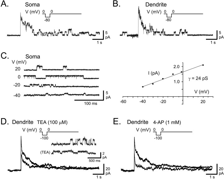
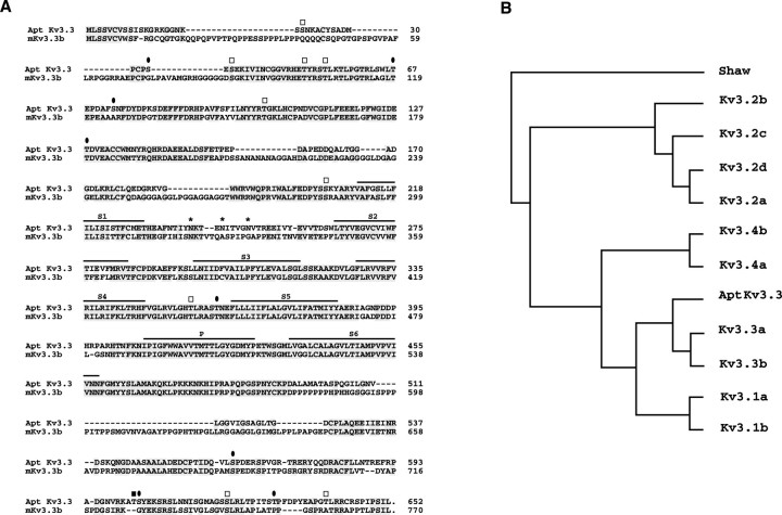
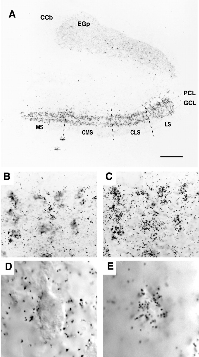
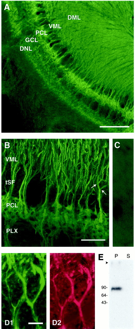
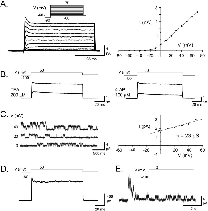
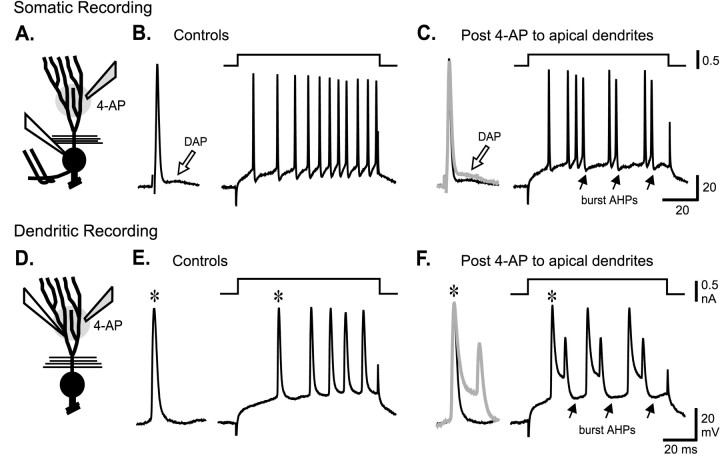
Voltage-gated ion channels localized to dendritic membranes can shape signal processing in central neurons. This study describes the distribution and functional role of a high voltage-activating K(+) channel in the electrosensory lobe (ELL) of an apteronotid weakly electric fish. We identify a homolog of the Kv3.3 K(+) channel, AptKv3.3, that exhibits a high density of mRNA expression and immunolabel that is distributed over the entire soma-dendritic axis of ELL pyramidal cells. The kinetics and pharmacology of native K(+) channels recorded in pyramidal cell somata and apical dendrites match those of AptKv3.3 channels expressed in a heterologous expression system. The functional role of AptKv3.3 channels was assessed using focal drug ejections in somatic and dendritic regions of an in vitro slice preparation. Local blockade of AptKv3.3 channels slows the repolarization of spikes in pyramidal cell somata as well as spikes backpropagating into apical dendrites. The resulting increase in dendritic spike duration lowers the threshold for a gamma-frequency burst discharge that is driven by inward current associated with backpropagating dendritic spikes. Thus, dendritic AptKv3.3 K(+) channels influence the threshold for a form of burst discharge that has an established role in feature extraction of sensory input.


Figures

Fig. 1.
Fig. 1.
AptKv3.3 channels in ELL pyramidal cells.A, B, Outside-out patch recordings of K+ channels isolated from a pyramidal cell soma (A) and an apical dendrite 150 μm from the soma (B) in the presence of normal extracellular aCSF. A 7 sec depolarizing step from −80 to 0 mV produces a fast activation of K+ channels that subsequently inactivate over 1–2 sec to reveal a unitary conductance level for channels in the patch. C, An outside-out patch recording obtained from somatic membrane and stepped from a holding potential of −90 mV to the indicated potentials reveals K+ channels with a conductance of 24 pS. D, E, A macropatch K+ current in an outside-out recording isolated from a pyramidal cell apical dendrite (100 μm from soma) evoked by a depolarizing step from −100 to 0 mV for 7 sec. Outward current was blocked by 100 μm TEA (D).Inset shows single-channel recordings from another dendritic outside-out patch recording at 0 mV (100 μm from soma) with a substantial reduction of single-channel conductance by 100 μm TEA. E, After washout of TEA, outward current from the same dendritic patch in D is blocked by perfusion of 1 mm 4-AP. Currents in A,B, D, and E were leak-subtracted, and capacitance artifacts were removed by digital subtraction.
Fig. 2.
Fig. 2.
Molecular characterization of AptKv3.3.A, Alignment of predicted amino acid sequence of AptKv3.3 with that of the murine splice isoform mKv3.3b. The six transmembrane domains (S1–S6) and the pore domain (P) are indicated above the sequence. Also indicated are consensus sites for N-linked glycosylation (asterisks) and phosphorylation by protein kinase C (open squares), protein kinase A (filled square), and calcium/calmodulin-dependent protein kinase (filled ovals). Amino acid identities areshaded. B, Phylogenetic comparison of AptKv3.3 to members of the mammalian Kv3 family. The sequences used for comparison included splice isoforms of each Kv3 subtype and, with the exception of murine Kv3.3a and Kv3.3b, were from rat (see Materials and Methods). The phylogenetic tree demonstrates that AptKv3.3 is most related to mammalian Kv3.3. Analysis by the parsimony method was performed using the PROTPARS program in the Phylogeny Inference Package (PHYLIP) (Felsenstein, 1989). In this algorithm, the DrosophilaShaw K+ channel was used as the outgroup.
Fig. 3.
Fig. 3.
The expression of AptKv3.3 mRNA in the hindbrain. Tissue sections from hindbrain were hybridized with AptKv3.3 RNA probe. The distribution of silver grains over pyramidal and granule cell layers is indicated in A. B andC show that AptKv3.3 is expressed in all of the pyramidal cells. D and E illustrate localization of silver grains over the pyramidal cell somata.A, In situ hybridization of AptKv3.3 mRNA in the hindbrain at low power demonstrates prominent expression in the ELL and lighter expression in the adjacent caudal lobe of the cerebellum (eminentia granularis posterior; EGp) and the corpus cerebelli (CCb). The four topographic maps of the ELL are indicated: MS, medial segment;CMS, centromedial segment; CLS, centrolateral segment; LS, lateral segment. Label is dense over the entire extent of the ELL pyramidal cell (PCL) and granule cell (GCL) layers. Scale bar, 200 μm. B, A section of the pyramidal cell layer showing a cluster of pyramidal cell somata viewed under DIC optics. Scale bar, 25 μm. C, The position of silver grains in the micrograph shown in B when viewed at the plane of emulsion illustrates dense labeling positioned over individual pyramidal cell somata. D, A pyramidal cell viewed under DIC optics at higher magnification. Scale bar, 10 μm.E, Corresponding image as that shown in Dviewed at the plane of emulsion illustrates the restriction of grains to the somatic region of a pyramidal cell.
Fig. 4.
Fig. 4.
AptKv3.3 protein is localized to somata and dendrites of ELL pyramidal cells. Tissue sections of hindbrain were stained with α-AptKv3.3 antibody (A–D) or double-labeled with MAP-2 monoclonal antibody (D). The specificity of the α-AptKv3.3 antibody is indicated in a Western blot (E) and tissue section (C). A andB show that AptKv3.3 protein is distributed throughout the pyramidal cell somatic and dendritic domains. Colocalization of AptKv3.3 with MAP2 in apical dendrites is shown for pyramidal cells from the medial segment (D). A, Low-power micrograph of α-AptKv3.3-immunolabeled hindbrain. Intense label is seen throughout the ELL, particularly the pyramidal cell layer (PCL), granule cell layer (GCL), and deep neuropil layer (DNL). Note also the dense label in the ventral (VML) and dorsal (DML) molecular layers that overlie the PCL and contain pyramidal cell apical dendritic projections. Scale bar, 400 μm. B, Higher-magnification confocal image of the ELL pyramidal cell layer in the centrolateral segment illustrating immunolabeling of pyramidal cell somata and apical dendrites. Labeling of apical dendrites remains constant in intensity past primary and secondary branchpoints (indicated with arrows). Note the lack of immunolabel in the tractus stratus fibrosum (tSF) and plexiform (PLX) layers, which both contain dense axonal fascicles. Scale bar, 100 μm. C, A control section from centrolateral segment viewed at the magnification used forB. This section was treated with α-AptKv3.3 that had been preadsorbed with the AptKv3.3 fusion protein. Only a diffuse background signal is detected. D, High-magnification image of two medial segment apical dendrites colabeled with α-AptKv3.3 (D1) and MAP-2a,b (D2) antibodies. AptKv3.3 protein appears to be localized exclusively to MAP-2-containing dendritic structures. Scale bar, 10 μm.E, Western blot analysis demonstrates that α-AptKv3.3 recognizes a single protein of ∼87 kDa in the brain membrane fraction (P) but not the corresponding soluble protein fraction (S).
Fig. 5.
Fig. 5.
Expression of AptKv3.3 currents in HEK tsA201 cells. A, Whole-cell recording of currents expressed 1 d after AptKv3.3 cDNA transfection reveals an outward rectifying K+ current for command steps from −90 to 70 mV (10 mV steps after a 10 msec prepulse to −90 mV). AptKv3.3 K+ current exhibits a fast activation that reaches a characteristic early peak of ∼5 msec duration that subsequently relaxes to a steady-state current and fast deactivation on stepping back to −60 mV. Little steady-state inactivation is apparent after 60 msec. The current–voltage relationship indicates outward rectification, a high threshold for initial activation of −10 mV, and little saturation for steps up to 70 mV. B, Whole-cell AptKv3.3 currents are highly sensitive to micromole concentrations of externally applied TEA and 4-AP. C, An outside-out patch recording of AptKv3.3 single channels stepped to different steady-state potentials reveals a single channel slope conductance of 23 pS. D, An outside-out macropatch recording of AptKv3.3 current reveals a very similar response as found for whole-cell currents >100 msec (compare with A).E, An outside-out patch recording of AptKv3.3 single channels indicates a fast and maximal activation of channels over the initial 200 msec followed by inactivation during a 7 sec depolarizing command step from −100 to 0 mV. Capacitance artifacts inD and E were digitally subtracted.
Fig. 6.
Fig. 6.
Somatic and dendritic spike repolarization is highly sensitive to TEA and 4-AP. A, B, Schematic diagrams of pyramidal cells are shown to indicate the placement of a stimulating electrode in the plexiform layer for antidromic activation (double wires), intracellular recording electrodes (white fill), and pressure electrodes for focal drug ejections (shaded fill) in an in vitro ELL slice preparation. Focal pressure ejection of either TEA or 4-AP (1 mm) in the immediate region of separate (A) somatic or (B) dendritic recordings slows the rate of repolarization and increases the duration of antidromic spikes. Control and test responses are shown superimposed, with test responses identified as thick gray traces.
Fig. 7.
Fig. 7.
Dendritic spike repolarization controls burst threshold. AC, The effects of dendritic 4-AP ejection on somatic spike discharge. A, Schematic diagram of a pyramidal cell to indicate the placement of a stimulating electrode for antidromic activation (double wires), an intrasomatic recording electrode (white fill), and a pressure electrode for focal drug ejection of 2 mm 4-AP (shaded fill) in anin vitro ELL slice preparation. B,C, The effects of dendritic 4-AP ejection on somatic spike discharge. B, Control intrasomatic recordings of antidromic spike discharge and associated DAP (open arrow) and current-evoked spike discharge when set below threshold for generating oscillatory spike bursts. C, Focal ejection of 4-AP to apical dendrites selectively enhances the somatic DAP (open arrow), as shown by superimposition of the control and test antidromic response (test response shown bygray trace). The lack of any change in somatic spike repolarization confirms that the drug was restricted to the dendritic region. This is sufficient to convert cell output from a tonic to bursting pattern, as indicated by a repeating series of spike bursts. Burst period is indicated by solid arrows designating the occurrence of burst afterhyperpolarizations (burst AHPs). D, Schematic diagram of a pyramidal cell indicates an intradendritic recording in another pyramidal cell (white filled electrode) and pressure electrode for focal ejection of 2 mm 4-AP (shaded fill). E, F, The effects of dendritic 4-AP ejection on dendritic spike discharge.E, Control intradendritic recordings showing current-evoked spike discharge set below threshold for generating spike bursts. Insets to the left inE and F show expanded views of the first current-evoked spikes in control and test recordings (asterisks). F, Focal ejection of 4-AP in the dendritic region broadens the dendritic spike by slowing spike repolarization and shifts cell output from a tonic to bursting pattern.Solid arrows indicate the occurrence of burst AHPs that terminate each spike burst. Inset shows the control and test responses superimposed (test response shown by gray trace). Excitation after dendritic 4-AP results in burst discharge composed of a repeating series of spike doublets.

Similar articles

Cited by

References

    1. Berman NJ, Maler L. Neural architecture of the electrosensory lateral line lobe: adaptations for coincidence detection, a sensory searchlight and frequency-dependent adaptive filtering. J Exp Biol. 1999;202:1243–1253. - PubMed
    1. Bottai D, Dunn RJ, Ellis W, Maler L. N-methyl-d-aspartate receptor 1 mRNA distribution in the central nervous system of the weakly electric fish Apteronotus leptorhychus. J Comp Neurol. 1997;389:65–80. - PubMed
    1. Bottai D, Maler L, Dunn RJ. Alternative RNA splicing of the NMDA receptor NR1 mRNA in the neurons of the teleost electrosensory system. J Neurosci. 1998;18:5191–5202. - PMC - PubMed
    1. Chandy KG, Gutman GA. Voltage gated channels. In: North RA, editor. Handbook of receptors and channels: ligand-gated and voltage-gated ion channels. CRC; Boca Raton, FL: 1995. pp. 1–71.
    1. Chen WR, Midtgaard J, Shepherd GM. Forward and backward propagation of dendritic impulses and their synaptic control in mitral cells. Science. 1997;278:463–467. - PubMed

Publication types

MeSH terms

Associated data

LinkOut - more resources