. 2001 Jan 1;21(1):159-68.
doi: 10.1523/JNEUROSCI.21-01-00159.2001.

The external granule layer of the developing chick cerebellum generates granule cells and cells of the isthmus and rostral hindbrain

Affiliations

The external granule layer of the developing chick cerebellum generates granule cells and cells of the isthmus and rostral hindbrain

J C Lin et al. J Neurosci. .

Abstract

The external granule layer (EGL) on the dorsal surface of the developing cerebellum consists of neural progenitors originating from the rostral rhombic lip (RRL). The RRL and the EGL were thought to give rise exclusively to the granule neurons of the cerebellum (Alder et al., 1996). To study the fate of individual RRL cells, we used a retroviral library to mark clones in the chick embryo at Hamberger-Hamilton stages 10-12. RRL clones comprised the EGL and cerebellar granule cells, as expected. Surprisingly, however, as many as 50% of the RRL clones also contained cells ventral to the cerebellum proper. Ventral derivatives were found in clones with a medial origin, as well as in those with a lateral origin along the RRL. Some of the ventral progeny appeared to be in the process of migration, whereas others appeared to be differentiating neurons in the isthmus and the rostral hindbrain region, including the locus coeruleus (LC) and pontine reticular formation. Furthermore, the Phox2a marker of LC precursors was detected in the EGL within the anterior aspect of the cerebellum. A stream of cells originating in the EGL and expressing Phox2a was observed to terminate ventrally in the LC. These data demonstrate that single RRL progenitor cells are not restricted to producing only cerebellar granule cells; they produce both cerebellar granule cells and ventral derivatives, some of which become hindbrain neurons. They also suggest that some progeny of the EGL escape the cerebellum via the anterior aspect of the cerebellar peduncles, to contribute to the generation of ventral structures such as the LC.


Figures

Fig. 1.
Fig. 1.
An RRL/EGL clone (JZ2-10-3, harvested at E8; see Table 1) with several sibling cells in the rostral hindbrain region ventral to the cerebellum. A, Dorsal view of the whole mount of the cerebellum of embryo JZ2, with two sets of black lines bracketing the levels of sections shown inB–F and an arrow showing the clonal origin in the medial rhombic lip. B–F, Six micrometer coronal sections of embryo JZ2 at the rostral portion of the cerebellum in rostral-to-caudal order. Red arrows indicate the clonally related cells defined by PCR sequencing analysis. Dorsal is at the top, and the midline is to the leftin B–F. This clone contained a few cells in the hindbrain close to the presumptive locus coeruleus (B, D, F, bottom red arrows), as well as numerous migrating EGL cells in the cerebellum (C, D, F, top red arrows). No AP+ hindbrain siblings were found in the more caudal levels (data not shown). Cb, Cerebellum; Cp, cerebellar peduncle; Hb, hindbrain.
Fig. 2.
Fig. 2.
Two RRL/EGL clones (JZ10-5-4 and JZ10-5-19; see Table 1) contained several cells in the region ventral to the cerebellum at E8. A, Dorsal view of the cerebellar area of embryo JZ10 shown as a whole mount, with a series of black lines indicating the levels of sections shown inB–G. The rostral aspect of the cerebellum is at thetop, and the midline is indicated by a black arrow in A. B–G, Serial 60 μm coronal sections of embryo JZ10 at the levels indicated inA. Dorsal is at the top, and the midline is to the left. The PCR-defined sibling cells in clone JZ10-5-4 are indicated by red arrows, and those in clone JZ10-5-19 are indicated by blue arrows. Black arrows in C–F indicate a distinct, unrelated hindbrain clone with radial glia in the VZ of the fourth ventricle (IV). Both clones JZ10-5-4 and JZ10-5-19 contained sibling cells in the hindbrain area (probably the prospective pontine reticular formation and the locus coeruleus; B–E, red, blue arrows; F, bottom-most blue arrow) and many cells in the cerebellar anlage (F, G, arrows). Some siblings in the rostral hindbrain region appeared to be migrating cells (C, arrows), and others appeared to be differentiating neurons (B, D–F, arrows) in the hindbrain. In addition, many cellular processes, probably those of the migrating cells, were observed in the cerebellar peduncle (B–G). Considerably more AP+ hindbrain siblings of clones JZ10-5-4 and JZ10-5-19 were observed in the rostral than in the caudal sections (compare B–E with F–G; some data not shown). Two separate clusters of AP+ cells, most likely the clonal origins, were found in the medial portion of the posterior margin of the RRL (the midline indicated in A, black arrow). Thered arrow in A indicates the origin of JZ10-5-4 as confirmed by PCR sequencing, whereas the origin of JZ10-5-19 cannot be labeled because of the failure of PCR amplification of the presumptive origin.
Fig. 3.
Fig. 3.
An RRL/EGL clone (JL36-13-6-3; see Table 1) contained several sibling cells in the region ventral to the cerebellum at E9. A, B, Dorsal oblique view of the whole-mount cerebellar area of embryo JL36-13 in two distinct focal planes, with a series of white lines indicating the levels of sections shown in C–K. The anterior aspect of the cerebellum is at the top. The midline is not in view but is to the right in A and B. C–K, Sixty micrometer coronal sections of the same embryo at each level indicated in panel A. F–H,L–N, High-power views of the framed areas in C–E and I–K, respectively. Dorsal is at the top inC–N. Red arrows andarrowheads indicate the PCR-defined sibling cells in this clone (JL36-13-6-3). This clone contained cells in the rostral hindbrain area (corresponding approximately to the prospective vestibular nuclei; F–H, red arrows), cells in the cerebellar peduncle (L, M, red arrows), and cells in the EGL (E, M, red arrowheads; also see A, B, black arrows, for whole-mount views). Some of the siblings ventral to the cerebellar anlage appeared to be in the process of migration with a typical bipolar morphology (G, H, L, M, red arrows), whereas others appeared to be differentiating neurons (F, M, red arrows). No AP+ hindbrain siblings were observed in the more caudal sections (K; some data not shown). This clone originated from the lateral edge of the RRL (B, black arrowhead; also see K, N for sections).
Fig. 4.
Fig. 4.
An RRL/EGL clone (JL33-30-3-8; see Table 1) contained several sibling cells in the region ventral to the cerebellum at E11–E12. A–D, Sixty micrometer coronal sections of embryo JL33-30 in a rostral-to-caudal order. The blue arrows indicate the clonally related cells defined by PCR sequencing. Dorsal is at the top, and midline is to theright. E, Dorsal oblique view of the cerebellar region of this embryo stained by whole-mount AP histochemistry, with a series of black lines indicating the levels of sections shown in A–D. The orientation inE is indicated by a frame of reference (a, anterior; p, posterior;m, medial; l, lateral; d, dorsal). This clone contained cells in the hindbrain area (possibly the prospective vestibular nuclei and locus coeruleus; A, blue arrow,B, two bottom blue arrows) and many cells in the EGL and the IGL (B–D, top blue arrows). No AP+ hindbrain siblings were observed in the more caudal sections (D; some data not shown). This clone originated from the lateral edge of the posterior margin of the RRL (E, black arrow).
Fig. 5.
Fig. 5.
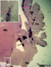
An RRL/EGL clone (JL35-17-9-12; see Table 1) at E18 contained a sibling cell in the isthmus region. This is a 60 μm parasagittal section of the midbrain and cerebellar region of embryo JL35-17. Rostral is to the left, and dorsal is at the top. Clone JL35-17-9-12, indicated by the red arrows, contained migrating EGL cells in the cerebellum and a neuron in the isthmus region, specifically in the parvocellular isthmic nucleus (bottom, red arrow within theframed area). The tentative neuronal morphology of this isthmic sibling cell is better appreciated at the high-power view in the top left inset. The blue arrowsindicate a distinct, unrelated clone containing many glial cells in the cerebellar cortex. Isth, Isthmus; Mb, midbrain.
Fig. 6.
Fig. 6.
Several models are proposed for the development of RRL/EGL clones with ventral progeny in the isthmus and the rostral hindbrain. Each panel is a three-dimensional schematic diagram of the midbrain–hindbrain area. The orientation in A–C is indicated by a frame of reference in C(A, anterior; D, dorsal;L, lateral). A, It has been well established that the cells from the RRL migrate rostrally to give rise to EGL cells in the cerebellum (solid red arrows), whereas the cells of the caudal rhombic lip migrate ventrally to give rise to the pontine and inferior olivary nuclei (green arrows). Model 1 (A) suggests that a progenitor splits early in development into two separate clusters of cells, with one giving rise to EGL cells of the cerebellum (solid red arrows) and the other producing hindbrain cells (dotted red arrows). This model does not invoke any ventral migration of the cells from the RRL, and it predicts the existence of a separate clonal origin in the VZ of the fourth ventricle. Model 2 (B) suggests that cells originating from the transitional zone between the rostral and the caudal rhombic lip migrate rostrally as well as ventrally (blue arrows). This model predicts that the origins of these clones cluster around the lateral aspect of the RRL. Model 3 (C) suggests that EGL progenitor cells produce some progeny that migrate ventrally into the isthmus and the rostral hindbrain via the anterior aspect of the cerebellar peduncle. These clones can originate from either the medial (red arrows) or the lateral (blue arrows) portion of the RRL.D, Frontal perspective of model 3, with its orientation indicated by the frame of reference at the bottom right corner. EGL cells migrate rostrally away from the rhombic lip and transversely across the midline (solid red arrows). A subset of the progeny of an EGL clone migrates ventrally to the hindbrain and the isthmus only via the anterior aspect of the cerebellar peduncle (dotted red arrows).CRL, Caudal rhombic lip.
Fig. 7.
Fig. 7.
Some monoaminergic neurons in the ventral region share a similar axonal morphology with cerebellar granule cells, and they may originate from the EGL. A, Schematic diagram depicting the axonal morphologies of the cerebellar granule cells (black), the mossy fibers (light blue), the climbing fibers (dark blue), and the monoaminergic neurons in the ventral regions (red). A subset of the serotonergic neurons in the pontine reticular formation and the noradrenergic neurons in the locus coeruleus project axons to the cerebellar cortex. The monoaminergic cerebellopetal axons generally exhibit morphology distinct from that of the mossy fibers or of the climbing fibers. Some of them terminate diffusely in the cerebellar cortex (red dotted lines), whereas others assume a T-shaped bifurcation in the molecular layer (red solid line), very similar to the granule cell axons. Adopted and modified from Chan-Palay (1975). B, In situ hybridization of the Zic-1 probe on a 10 μm coronal section of the E8 chick brain at the level of the LC. Zic-1 is a cerebellar granule cell marker, and a high level of Zic-1 mRNA expression was observed in the EGL but not the cerebellar peduncle.C, D, Anti-TH antibody staining on 25 μm coronal sections of E7–E8 chick cerebellar and hindbrain regions. AlthoughB–D were derived from different embryos of the same age, coronal sections at a similar level of the rostral hindbrain were used for comparison. TH is a marker for catecholamine-producing cells, including the noradrenergic neurons in the locus coeruleus. In addition to the cluster of TH-positive cells in the presumptive LC, some scattered TH-positive cells were observed in the cerebellar anlage and the cerebellar peduncles (C, D, arrows). A few TH-positive cells in the cerebellar peduncle exhibited a morphology consistent with migrating cells. No TH-positive cells were observed in the EGL. ML, Molecular layer; PCL, Purkinje cell layer.
Fig. 8.
Fig. 8.
Expression of LC markers Phox2a and TH visualized by antibody staining supports the idea of the ventral migration of EGL cells in model 3 (A, same as Fig. 6C).B, C, Co-localization of LC precursor marker Phox2a (green) and EGL marker Pax6 (red) at the rostral aspect of the cerebellum. The relative position of the areas shown in B and C is indicated by the top left frame in A. It is interesting to note that the medial EGL expressed only Pax6, and the intermediate EGL expressed both Phox2a and Pax6, whereas the lateral EGL expressed only Phox2a. D, Phox2a (green)-expressing LC precursors stretching from the dorsal region to the ventral region close to the TH-expressing LC neurons (red). Most if not all TH-expressing cells in the LC ceased to express Phox2a. The D corresponds to the right framed area in A. The staining was done with the 30 μm coronal sections of E7–E8 chick embryos.

Similar articles

Cited by

References

    1. Alcantara S, Ruiz M, De Castro, Sotelo C, Soriano E. Netrin 1 acts as an attractive or as a repulsive cue for distinct migrating neurons during the development of the cerebellar system. Development. 2000;127:1359–1372. - PubMed
    1. Alder J, Cho N, Hatten ME. Embryonic precursor cells from the rhombic lip are specified to a cerebellar neuronal identity. Neuron. 1996;17:389–399. - PubMed
    1. Alder J, Lee KJ, Jessell TM, Hatten ME. Generation of cerebellar granule neurons in vivo by transplantation of BMP-treated neural progenitor cells. Nat Neurosci. 1999;2:535–540. - PubMed
    1. Anderson SA, Eisenstat DD, Shi L, Rubenstein JL. Interneuron migration from basal forebrain to neocortex: dependence on Dlx genes (see comments). Science. 1997;278:474–476. - PubMed
    1. Cepko CL, Golden JA, Szele FG, Lin JC. Lineage analysis in the vertebrate central nervous system. In: Cowan WM, Jessel T, Zipursky SL, editors. Molecular and cellular approaches to neural development. Oxford UP; London: 1997.

Publication types

MeSH terms

LinkOut - more resources