

Age and gender predict volume decline in the anterior and posterior hippocampus in early adulthood
- PMID: 11150336
- PMCID: PMC6762451
- DOI: 10.1523/JNEUROSCI.21-01-00194.2001
Age and gender predict volume decline in the anterior and posterior hippocampus in early adulthood
Abstract
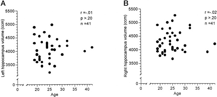
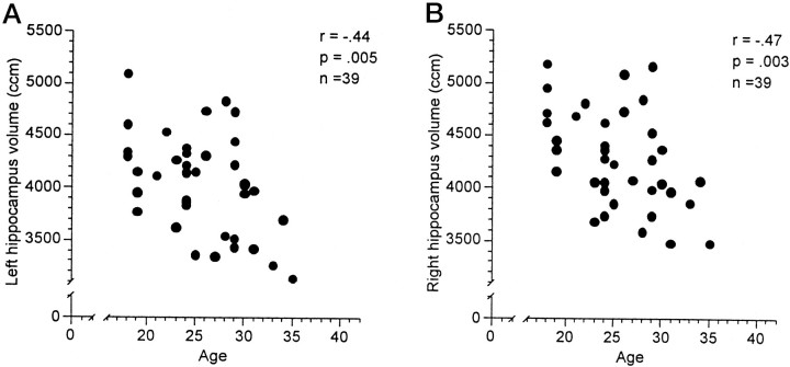
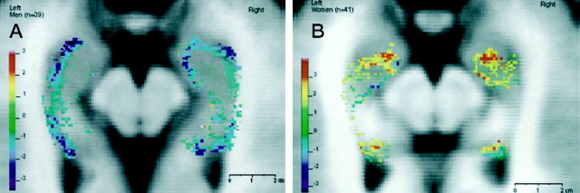
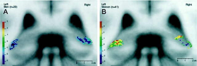
Magnetic Resonance Imaging (MRI) provides a noninvasive method for investigating brain morphology. Within the medial temporal lobe, special attention has been paid to the hippocampus (HC) and amygdala (AG) because of their role in memory, depression, emotion, and learning. Volume changes in these areas have been observed in conjunction with certain disease states, e.g. Alzheimer's disease, post-traumatic stress disorder, and depression. Aging has also been shown to result in gray matter volume loss of the overall brain, including the HC. With regard to gender specificity, results suggest a larger shrinkage for men of brain gray matter, with controversial observations being made for the HC. With recently refined MRI acquisition and segmentation protocols, the HC and AG of 80 subjects in early adulthood (39 men and 41 women, age 18-42 years) were investigated. Whereas the volume of the AG appeared to be independent of age and gender, a significant negative correlation with age for both left and right HC was found in men (r = -0.47 and -0.44, respectively) but not in women (r = 0.01 and 0.02, respectively). The volume decline in men appeared to be linear, starting at the beginning of the third life decade and approximating 1.5% per annum. Using voxel-based regressional analysis, it was shown that changes with age occurred mostly in the head and tail of the HC. This finding underscores the need to include sociodemographic variables in functional and anatomical MRI designs.
Figures

Similar articles
-
Hippocampal shape analysis using medial surfaces.Neuroimage. 2005 May 1;25(4):1077-89. doi: 10.1016/j.neuroimage.2004.12.051. Neuroimage. 2005. PMID: 15850726
-
Is there a correlation between hippocampus and amygdala volume and olfactory function in healthy subjects?Neuroimage. 2012 Jan 16;59(2):1052-7. doi: 10.1016/j.neuroimage.2011.09.024. Epub 2011 Sep 24. Neuroimage. 2012. PMID: 21967725
-
Group differences in anterior hippocampal volume and in the retrieval of spatial and temporal context memory in healthy young versus older adults.Neuropsychologia. 2010 Dec;48(14):4020-30. doi: 10.1016/j.neuropsychologia.2010.10.010. Epub 2010 Oct 12. Neuropsychologia. 2010. PMID: 20946907
-
Stress regulation in the central nervous system: evidence from structural and functional neuroimaging studies in human populations - 2008 Curt Richter Award Winner.Psychoneuroendocrinology. 2010 Jan;35(1):179-91. doi: 10.1016/j.psyneuen.2009.02.016. Psychoneuroendocrinology. 2010. PMID: 19362426 Review.
-
Regional deficits in brain volume in schizophrenia: a meta-analysis of voxel-based morphometry studies.Am J Psychiatry. 2005 Dec;162(12):2233-45. doi: 10.1176/appi.ajp.162.12.2233. Am J Psychiatry. 2005. PMID: 16330585 Review.
Cited by
-
Nonlocal regularization for active appearance model: Application to medial temporal lobe segmentation.Hum Brain Mapp. 2014 Feb;35(2):377-95. doi: 10.1002/hbm.22183. Epub 2012 Sep 15. Hum Brain Mapp. 2014. PMID: 22987811 Free PMC article.
-
Challenges and opportunities of diagnostic markers of Alzheimer's disease based on structural magnetic resonance imaging.Brain Behav. 2023 Mar;13(3):e2925. doi: 10.1002/brb3.2925. Epub 2023 Feb 16. Brain Behav. 2023. PMID: 36795041 Free PMC article. Review.
-
Deformation field morphometry reveals age-related structural differences between the brains of adults up to 51 years.J Neurosci. 2008 Jan 23;28(4):828-42. doi: 10.1523/JNEUROSCI.3732-07.2008. J Neurosci. 2008. PMID: 18216191 Free PMC article.
-
Association of depressive symptoms with hippocampal volume in 1936 adults.Neuropsychopharmacology. 2014 Feb;39(3):770-9. doi: 10.1038/npp.2013.271. Epub 2013 Oct 4. Neuropsychopharmacology. 2014. PMID: 24220026 Free PMC article.
-
Brain Volume Metric Analysis Is Correlated with Aging Changes and Sex Differences in Thai Older Adults.Dement Geriatr Cogn Dis Extra. 2025 Jan 27;15(1):47-57. doi: 10.1159/000543774. eCollection 2025 Jan-Dec. Dement Geriatr Cogn Dis Extra. 2025. PMID: 40093354 Free PMC article.
References
-
- Ashtari M, Greenwald BS, Kramer-Ginsberg E, Hu J, Wu H, Patel M, Aupperle P, Pollack S. Hippocampal/amygdala volumes in geriatric depression. Psychol Med. 1999;29:629–638. - PubMed
-
- Baenziger O, Martin E, Steinlin M, Good M, Largo R, Burger R, Fanconi S, Duc G, Buchli R, Rumpel H. Early pattern recognition in severe perinatal asphyxia: a prospective MRI study. Neuroradiology. 1993;35:437–442. - PubMed
-
- Bartzokis G, Altshuler LL, Greider T, Curran J, Keen B, Dixon WJ. Reliability of medial temporal lobe volume measurements using reformatted 3D images. Psychiatry Res. 1998;82:11–24. - PubMed
-
- Christiansen P, Larsson HBW, Thomsen C. Age dependent white matter lesions and brain volume changes in healthy volunteers. Acta Radiol. 1994;35:117–122. - PubMed
Publication types
MeSH terms
LinkOut - more resources
Full Text Sources
Medical