

Expression of neuronal connexin36 in AII amacrine cells of the mammalian retina
- PMID: 11150340
- PMCID: PMC6762459
- DOI: 10.1523/JNEUROSCI.21-01-00230.2001
Expression of neuronal connexin36 in AII amacrine cells of the mammalian retina
Abstract
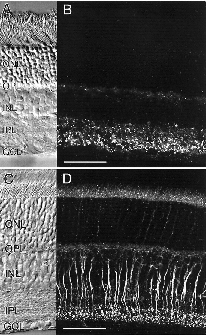
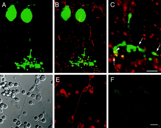
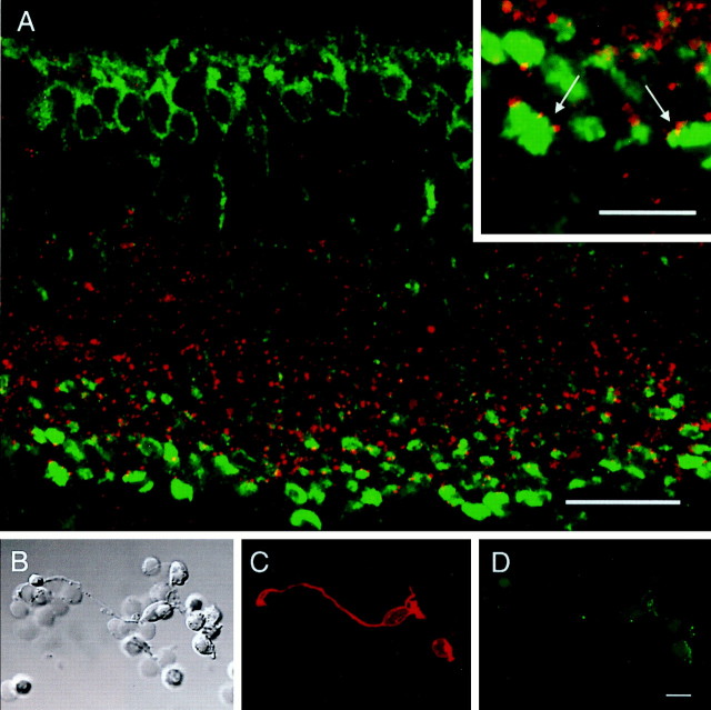
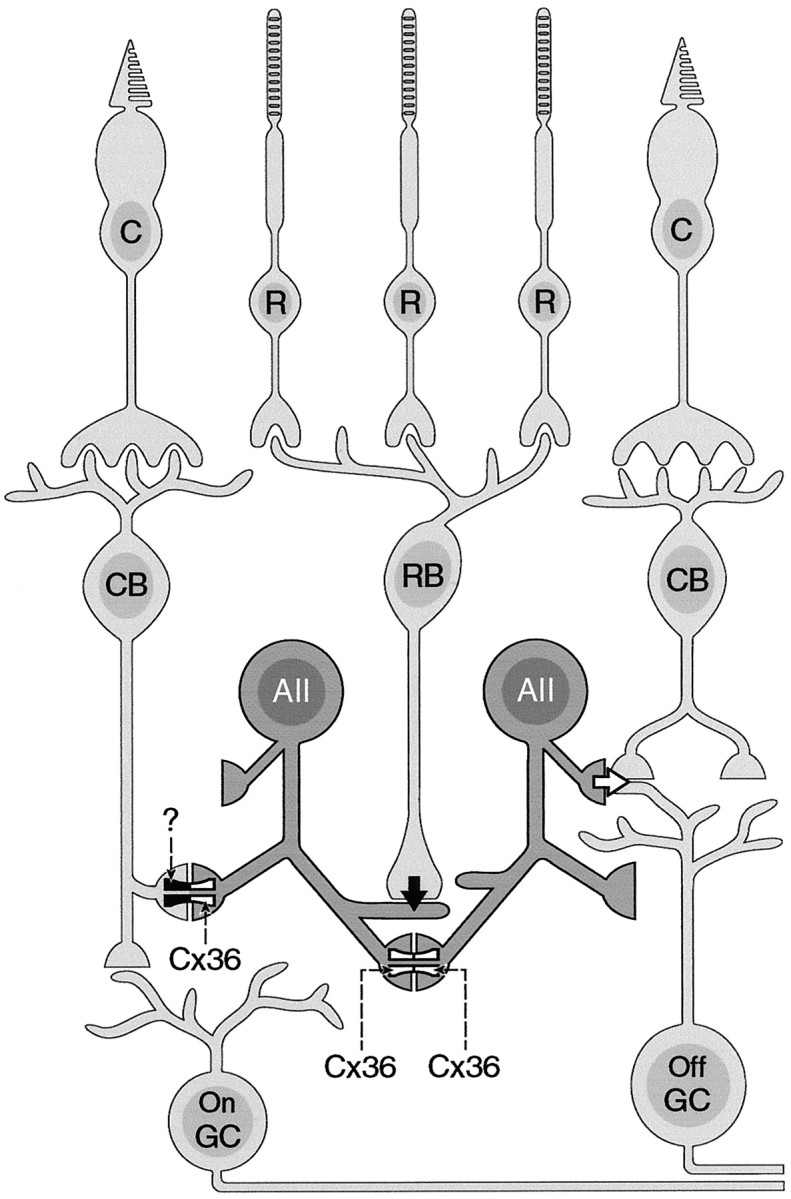
We have studied the expression pattern of neuronal connexin36 (Cx36) in the mouse and rat retina. In vertical sections of both retinas, a polyclonal antibody directed against Cx36 produced punctate labeling in the inner plexiform layer (IPL). Intense immunoreactivity was localized to the entire OFF sublamina of the IPL, and much weaker staining could be observed in the ON sublamina. Double-labeling experiments in the rat retina with antibodies directed against parvalbumin indicate that Cx36 is expressed on dendrites of AII amacrine cells. Cx36-like immunoreactivity in sublamina a of the IPL did not overlap with lobular appendages or cell bodies of AII amacrine cells. In a mouse retinal slice preparation, AII amacrine and ON cone bipolar cells were intracellularly injected with Neurobiotin and counterstained with antibody against Cx36. Punctate labeling appeared to be in register with dendritic arborization of AII amacrines and cone bipolar cells in the ON sublamina of the IPL. Whereas AII amacrine cells isolated from the rat retina clearly displayed Cx36-like immunoreactivity, isolated ON cone bipolar cells were negative for Cx36. Axon terminals of rod bipolar cells were decorated with Cx36-positive contacts but did not express Cx36 themselves. These results indicate that Cx36 is expressed by AII amacrine cells in homologous and heterologous gap junctions made with AII amacrines and cone bipolar cells, respectively. The heterologous gap junctions appear to be heterotypic, because ON cone bipolar cells do not express Cx36.
Figures


Similar articles
-
The immunocytochemical localization of connexin 36 at rod and cone gap junctions in the guinea pig retina.Eur J Neurosci. 2003 Dec;18(11):2925-34. doi: 10.1046/j.1460-9568.2003.03049.x. Eur J Neurosci. 2003. PMID: 14656288
-
Localization of heterotypic gap junctions composed of connexin45 and connexin36 in the rod pathway of the mouse retina.Eur J Neurosci. 2006 Sep;24(6):1675-86. doi: 10.1111/j.1460-9568.2006.05052.x. Eur J Neurosci. 2006. PMID: 17004931
-
Connexin-36 distribution and layer-specific topography in the cat retina.Brain Struct Funct. 2019 Jul;224(6):2183-2197. doi: 10.1007/s00429-019-01876-y. Epub 2019 Jun 6. Brain Struct Funct. 2019. PMID: 31172263 Free PMC article.
-
Multiple neuronal connexins in the mammalian retina.Cell Commun Adhes. 2003 Jul-Dec;10(4-6):425-30. doi: 10.1080/cac.10.4-6.425.430. Cell Commun Adhes. 2003. PMID: 14681052 Review.
-
Somatostatin and somatostatin subtype 2A expression in the mammalian retina.Microsc Res Tech. 2000 Jul 15;50(2):103-11. doi: 10.1002/1097-0029(20000715)50:2<103::AID-JEMT2>3.0.CO;2-X. Microsc Res Tech. 2000. PMID: 10891874 Review.
Cited by
-
Increased Connexin36 Phosphorylation in AII Amacrine Cell Coupling of the Mouse Myopic Retina.Front Cell Neurosci. 2020 Jun 1;14:124. doi: 10.3389/fncel.2020.00124. eCollection 2020. Front Cell Neurosci. 2020. PMID: 32547367 Free PMC article.
-
An Update on Connexin Gap Junction and Hemichannels in Diabetic Retinopathy.Int J Mol Sci. 2021 Mar 21;22(6):3194. doi: 10.3390/ijms22063194. Int J Mol Sci. 2021. PMID: 33801118 Free PMC article. Review.
-
Electrical synapses in retinal ON cone bipolar cells: subtype-specific expression of connexins.Proc Natl Acad Sci U S A. 2005 Sep 13;102(37):13313-8. doi: 10.1073/pnas.0505067102. Epub 2005 Sep 6. Proc Natl Acad Sci U S A. 2005. PMID: 16150718 Free PMC article.
-
Dendrodendritic electrical synapses between mammalian retinal ganglion cells.J Neurosci. 2004 Nov 17;24(46):10553-67. doi: 10.1523/JNEUROSCI.3319-04.2004. J Neurosci. 2004. PMID: 15548670 Free PMC article.
-
Gap Junctions Contribute to Differential Light Adaptation across Direction-Selective Retinal Ganglion Cells.Neuron. 2018 Oct 10;100(1):216-228.e6. doi: 10.1016/j.neuron.2018.08.021. Epub 2018 Sep 13. Neuron. 2018. PMID: 30220512 Free PMC article.
References
-
- Al-Ubaidi MR, White TW, Ripps H, Poras I, Avner P, Gomès D, Bruzzone R. Functional properties, developmental regulation, and chromosomal localization of murine connexin36, a gap-junctional protein expressed preferentially in retina and brain. J Neurosci Res. 2000;59:813–826. - PubMed
-
- Beyer EC, Paul DL, Goodenough DA. Connexin family of gap junction proteins. J Membr Biol. 1990;116:187–194. - PubMed
-
- Bruzzone R, White TW, Goodenough DA. The cellular internet: on-line with connexins. BioEssays. 1996;18:709–718. - PubMed
-
- Chun MH, Han SH, Chung JW, Wässle H. Electron microscopic analysis of the rod pathway of the rat retina. J Comp Neurol. 1993;332:421–432. - PubMed
-
- Condorelli DF, Parenti R, Spinella F, Trovato Salinaro A, Belluardo N, Cardile V, Cicirata F. Cloning of a new gap junction gene (Cx36) highly expressed in mammalian brain neurons. Eur J Neurosci. 1998;10:1202–1208. - PubMed
Publication types
MeSH terms
Substances
LinkOut - more resources
Full Text Sources
Other Literature Sources
Miscellaneous