Regulation by the medial amygdala of copulation and medial preoptic dopamine release
- PMID: 11150352
- PMCID: PMC6762439
- DOI: 10.1523/JNEUROSCI.21-01-00349.2001
Regulation by the medial amygdala of copulation and medial preoptic dopamine release
Abstract
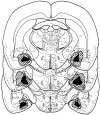
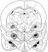
The medial preoptic area (MPOA) is a critical integrative site for male copulatory behavior in most vertebrate species. Extracellular dopamine (DA) is increased in the MPOA of male rats immediately before and during copulation. DA agonists microinjected into the MPOA of male rats facilitate and DA antagonists inhibit sexual behavior. A major source of input to the MPOA is the medial amygdala (MeA), which processes and relays olfactory information to the MPOA. We now report that microinjections of a DA agonist into the MPOA of animals with excitotoxic lesions of the amygdala restored copulatory ability that was lost after the lesions. Moreover, radio-frequency lesions of the MeA impaired copulation and blocked the increases in extracellular DA seen in animals with sham lesions during exposure to a receptive female and during copulation. Thus, both copulatory ability and the MPOA DA response, during exposure to a receptive female and during copulation, are facilitated by input from the MeA to the MPOA.
Figures


Similar articles
-
Testosterone restoration of copulatory behavior correlates with medial preoptic dopamine release in castrated male rats.Horm Behav. 2001 May;39(3):216-24. doi: 10.1006/hbeh.2001.1648. Horm Behav. 2001. PMID: 11300712
-
Getting his act together: roles of glutamate, nitric oxide, and dopamine in the medial preoptic area.Brain Res. 2006 Dec 18;1126(1):66-75. doi: 10.1016/j.brainres.2006.08.031. Epub 2006 Sep 11. Brain Res. 2006. PMID: 16963001 Review.
-
Stimulation of the medial amygdala enhances medial preoptic dopamine release: implications for male rat sexual behavior.Brain Res. 2001 Nov 2;917(2):225-9. doi: 10.1016/s0006-8993(01)03031-1. Brain Res. 2001. PMID: 11640908
-
Nitric oxide promotes medial preoptic dopamine release during male rat copulation.Neuroreport. 1996 Dec 20;8(1):31-4. doi: 10.1097/00001756-199612200-00007. Neuroreport. 1996. PMID: 9051747
-
Dopamine, the medial preoptic area, and male sexual behavior.Physiol Behav. 2005 Oct 15;86(3):356-68. doi: 10.1016/j.physbeh.2005.08.006. Epub 2005 Aug 30. Physiol Behav. 2005. PMID: 16135375 Review.
Cited by
-
Estrogen regulation of cell proliferation and distribution of estrogen receptor-alpha in the brains of adult female prairie and meadow voles.J Comp Neurol. 2005 Aug 22;489(2):166-79. doi: 10.1002/cne.20638. J Comp Neurol. 2005. PMID: 15984004 Free PMC article.
-
Copulation induces expression of the immediate early gene Arc in mating-relevant brain regions of the male rat.Behav Brain Res. 2019 Oct 17;372:112006. doi: 10.1016/j.bbr.2019.112006. Epub 2019 Jun 3. Behav Brain Res. 2019. PMID: 31170433 Free PMC article.
-
Sex, drugs and gluttony: how the brain controls motivated behaviors.Physiol Behav. 2011 Jul 25;104(1):173-7. doi: 10.1016/j.physbeh.2011.04.057. Epub 2011 May 5. Physiol Behav. 2011. PMID: 21554895 Free PMC article. Review.
-
Preoptic glutamate facilitates male sexual behavior.J Neurosci. 2006 Feb 8;26(6):1699-703. doi: 10.1523/JNEUROSCI.4176-05.2006. J Neurosci. 2006. PMID: 16467517 Free PMC article.
-
Oxytocin, Erectile Function and Sexual Behavior: Last Discoveries and Possible Advances.Int J Mol Sci. 2021 Sep 26;22(19):10376. doi: 10.3390/ijms221910376. Int J Mol Sci. 2021. PMID: 34638719 Free PMC article. Review.
References
-
- Baum MJ, Everitt BJ. Increased expression of c-fos in the medial preoptic area after mating in male rats: role of afferent inputs from the medial amygdala and midbrain central tegmental field. Neuroscience. 1992;50:627–646. - PubMed
-
- Bitran D, Hull EM. Pharmacological analysis of male rat sexual behavior. Neurosci Biobehav Rev. 1987;11:365–389. - PubMed
-
- Coolen LM, Peters HJ, Veening JG. Fos immunoreactivity in the rat brain following consummatory elements of sexual behavior: a sex comparison. Brain Res. 1996;738:67–82. - PubMed
-
- Coolen LM, Peters HJ, Veening JG. Anatomical interrelationships of the medial preoptic area and other brain regions activated following male sexual behavior: a combined fos and tract-tracing study. J Comp Neurol. 1998;397:421–435. - PubMed
-
- de Jonge FH, Louwerse AL, Ooms MP, Evers P, Endert E, Van de Poll NE. Lesions of the SDN-POA inhibit sexual behavior of male Wistar rats. Brain Res Bull. 1989;23:483–492. - PubMed
Publication types
MeSH terms
Substances
Grants and funding
LinkOut - more resources
Full Text Sources
