. 2001 Feb;75(3):1252-64.
doi: 10.1128/JVI.75.3.1252-1264.2001.

Characterization of cell lines carrying self-replicating hepatitis C virus RNAs

Affiliations

Characterization of cell lines carrying self-replicating hepatitis C virus RNAs

T Pietschmann et al. J Virol. 2001 Feb.

Abstract

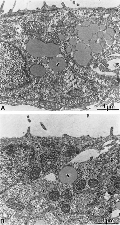
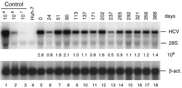
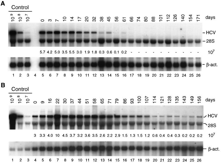
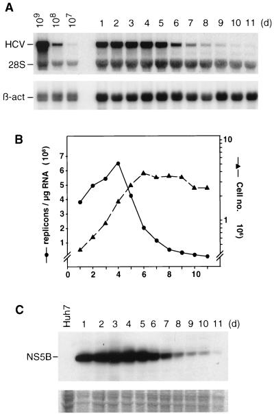
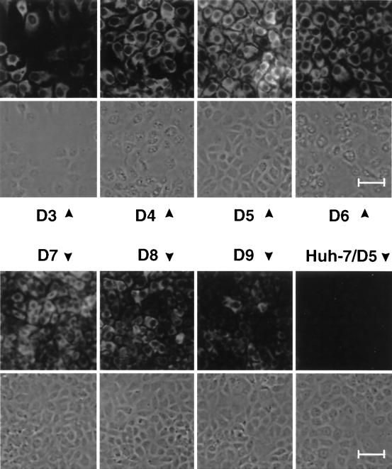
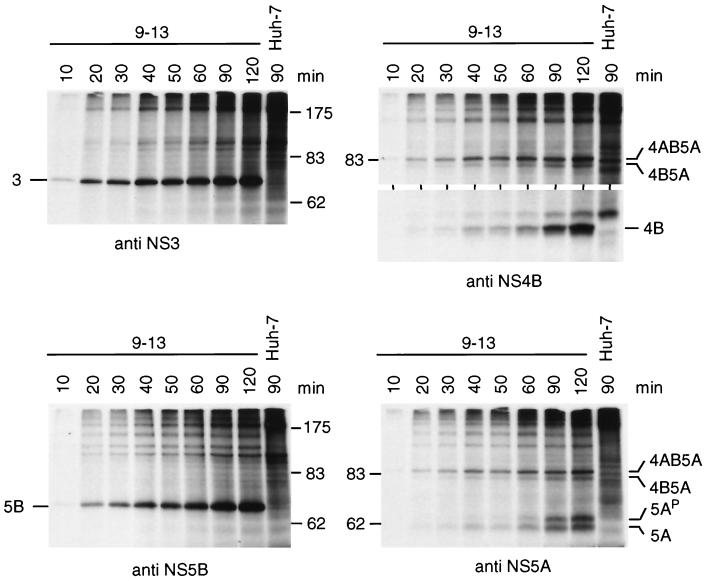
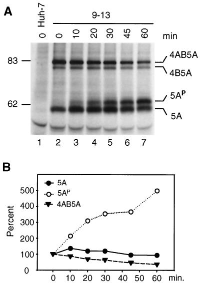
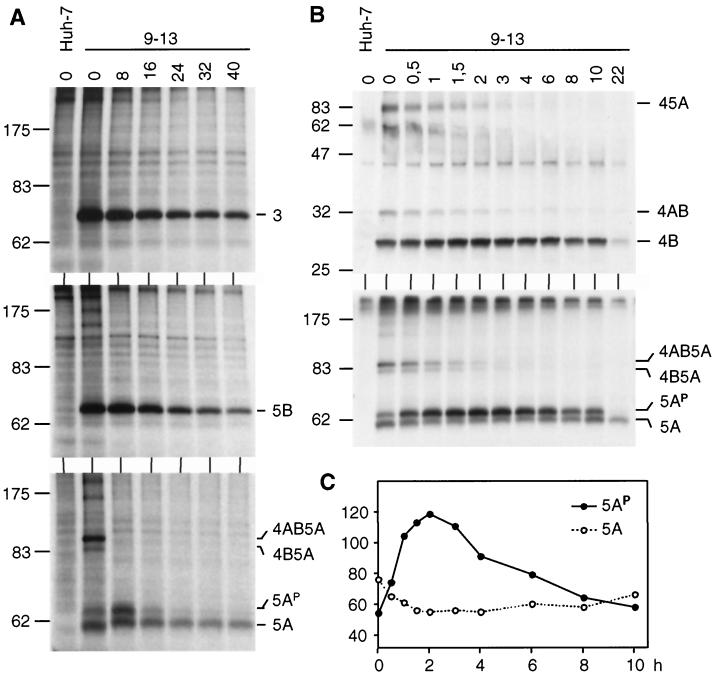
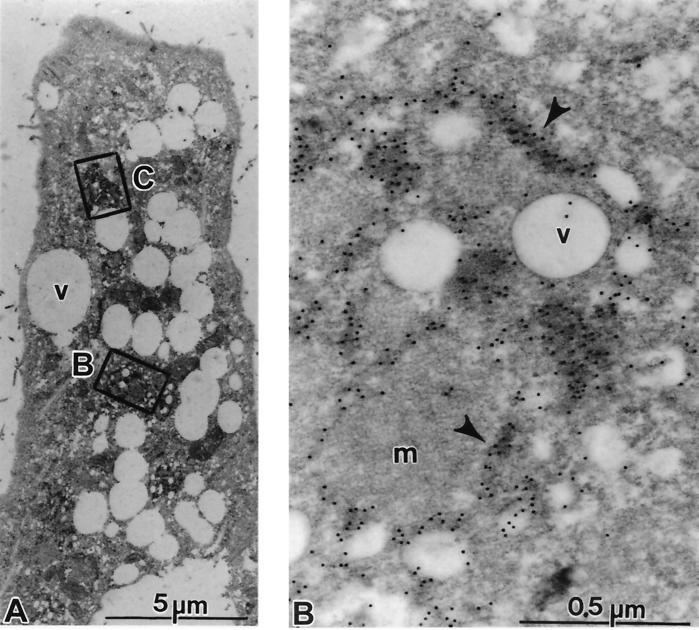
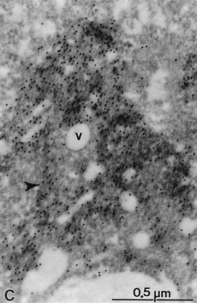
Subgenomic selectable RNAs of the hepatitis C virus (HCV) have recently been shown to self-replicate to high levels in the human hepatoma cell line Huh-7 (V. Lohmann, F. Körner, J. O. Koch, U. Herian, L. Theilmann, and R. Bartenschlager, Science 285:110-113, 1999). Taking advantage of this cell culture system that allows analyses of the interplay between HCV replication and the host cell, in this study we characterized two replicon-harboring cell lines that have been cultivated for more than 1 year. During this time, we observed no signs of cytopathogenicity such as reduction of growth rates or ultrastructural changes. High levels of HCV RNAs were preserved in cells passaged under continuous selection. When selective pressure was omitted replicon levels dropped, but depending on culture conditions the RNAs persisted for more than 10 months. A tight coupling of the amounts of HCV RNA and proteins to host cell growth was observed. Highest levels were found in exponentially growing cells, followed by a sharp decline in resting cells, suggesting that cellular factors required for RNA replication and/or translation vary in abundance and become limiting in resting cells. Studies of polyprotein processing revealed rapid cleavages at the NS3/4A and NS5A/B sites resulting in a rather stable NS4AB5A precursor that was processed slowly into individual products. Half-lives (t(1/2)s) of mature proteins ranged from 10 to 16 h, with the exception of the hyperphosphorylated form of NS5A, which was less stable (t(1/2), approximately 7 h). Results of immunoelectron microscopy revealed an association of the majority of viral proteins with membranes of the endoplasmic reticulum, suggesting that this is the site of RNA replication. In summary, replicon-bearing cells are a good model for viral persistence, and they allow the study of various aspects of the HCV life cycle.


Figures

FIG. 1
FIG. 1
Lack of ultrastructural changes in cells with HCV replicons. Analysis of ultrathin sections obtained from cultures of Huh-7 control cells (A) or replicon cell line 5-15 (B). Both cultures display a similar cellular architecture with a variable number of vesicles (v) of various sizes. The vesiculation was dependent on the cell density of the cultures and not on the expression of HCV proteins. No specific morphological alterations could be detected in the replicon-bearing cells.
FIG. 2
FIG. 2
Stability of HCV replicon in cell line 9-13 passaged under continuous G418 selection. Cells were regularly passaged three times a week in the presence of G418 (1 mg/ml) and total RNA was isolated from cells at given time points. After denaturing agarosegel electrophoresis, HCV- and β-actin-specific RNAs were detected by Northern bloting using 32P-labeled riboprobes complementary to the HCV IRES and neo or a β-actin-specific antisense RNA, respectively, and quantified by phosphorimaging. The number of replicon RNA molecules was determined by comparison with the serial dilution of in vitro transcripts (lanes 1 to 3). β-Actin RNA served as a control to correct for the amount of total RNA loaded in each lane of the gel (∼2 μg). The result obtained with total RNA from the parental Huh-7 cells is shown in lane 4. Numbers between both panels refer to the number of replicon RNA molecules (108) contained in 1 μg of total RNA of the respective sample. Note that the control RNA is a mixture of given numbers of in vitro transcripts and 2 μg of total RNA from the parental Huh-7 cells that was used as carrier. The positions of HCV RNA, 28S rRNA, and β-actin mRNA (β-act.) are given to the right. Analogous results were obtained with cell line 5-15 (not shown).
FIG. 3
FIG. 3
Stability of HCV replicons in cells passaged in the absence of selection. (A) Cells were passaged three times a week in a way that two-fifths of the cells were harvested for RNA preparation and three-fifths of the cells were seeded in a new culture flask. (B) Cells were passaged three times a week at a dilution of 1:5; i.e., four-fifths were harvested and one-fifth was seeded in a new culture flask. Total RNA was prepared from harvested cells and 4 μg of each preparation was analyzed for the amount of HCV RNA by Northern blotting as described in the legend to Fig. 2.
FIG. 4
FIG. 4
Fluctuations of HCV RNA replication levels during cell passage. Cells of cell line 9-13 that had been regularly passaged under continuous selection were seeded in multiple culture dishes and harvested at regular intervals between 1 and 11 days postseeding. Harvested cells were counted, and each half of these cells were used to prepare total RNA or a total lysate for protein analysis. (A) Total RNA was analyzed by Northern blotting as described in the legend to Fig. 2. (B) Quantification of HCV RNA and the number of cells at the time of harvest. The amounts of replicon RNA (left y axis) were determined by phosphorimaging using the Northern blot shown in panel A. The numbers of cells are given in a log scale (right y axis). (C) Proteins contained in 8 × 10⁵ cells were loaded in each lane of the gel, and NS5B was detected by Western bloting To demonstrate comparable amounts of proteins in each lane, a gel was loaded in parallel and stained after electrophoresis with Coomassie brilliant blue. A portion of this gel is shown in the lower panel. The result obtained with the parental Huh-7 cells is shown in the left lane. d, day.
FIG. 5
FIG. 5
Fluctuations of NS5B expression in a growing culture of cell line 9-13. Cells were seeded on coverslips and harvested at given times postseeding (days [D]). NS5B was detected by indirect immunofluorescence. A representative section of each sample is shown both by phase-contrast microscopy and immunofluorescence. All pictures were taken at a magnification of × 180. The smaller size of the older cells is due to confluency (compare, e.g., day 4 with day 7). Bar, 50 μm.
FIG. 6
FIG. 6
Processing kinetics of the NS3-5B polyprotein as determined by pulse labeling. 9-13 cells were seeded 60 h prior to metabolic radiolabeling of proteins with 100 μCi of labeling mix per ml for the time periods indicated above the lanes. Cells were lysed, and HCV-specific proteins were isolated by immunoprecipitation using antisera monospecific for NS3, NS4B, NS5A, or NS5B. HCV proteins and the positions of protein molecular weight standards (in kilodaltons) are specified in each panel. Results obtained with the parental Huh-7 cells radiolabeled for 90 min are shown in the right lane of each panel. Only the upper and lower portion of the gel is shown in case of the immunoprecipitations with the NS4B-specific antiserum.
FIG. 7
FIG. 7
Kinetics of generation of NS5A proteins. (A) 9-13 cells were seeded 60 h prior to radiolabeling with [35S]methionine-cysteine (200 μCi/ml) for 10 min followed by incubation with nonradioactive medium for the times given above the lanes. Cells were lysed, and NS5A proteins were analyzed by immunoprecipitation, SDS-PAGE, and autoradiography. (B) Quantification of NS5A-specific bands by phosphorimaging. Values obtained at time point zero were set at 100%.
FIG. 8
FIG. 8
Half-lives of HCV proteins. (A) Cells of cell line 9-13 were pulse-labeled for 30 min with [35S]methionine-cysteine, followed by incubation with nonradioactive medium for various times as given above each lane (hours). Cells were lysed, and HCV proteins were analyzed by immunoprecipitation, SDS-PAGE, and autoradiography. (B) Pulse-chase experiments were performed as in panel A, but cells were lysed at shorter intervals during the chase period. HCV-specific bands are identified to the right of each panel; the positions of molecular weight standards (in kilodaltons) are given to the left. (C) The fully processed NS5A and its hyperphosphorylated form shown in panel B were quantified by phosphorimaging, and the values (given in photo-stimulated light units) are plotted against the chase time.
FIG. 9
FIG. 9
Subcellular localization of HCV proteins. Cells of cell line 5-15 were processed 4 days postseeding for immunoelectron microscopy without osmium fixation as described in Materials and Methods. The sections were probed with a mixture of polycional antisera directed against NS3, NS4B, NS5A, and NS5B, followed by 12-nm-diameter colloidal gold particles conjugated to anti-rabbit antibodies. (A) The cell in low magnification overview displays a strong vesiculation and a couple of gold-labeled areas (boxed). (B and C) Enlargements of the areas indicated by rectangles in panel A. (B) Accumulations of gold label in elongated structures representing cisternae of the ER (arrowheads). Note the scarcity of gold particles on vesicles (v), on mitochondria (m), or outside the labeled cisternae. (C) Gold-labeled cisternae are easily identified (arrowhead). Additional antibody binding could be seen on numerous submembranous structures around small vesicles (v).
FIG. 9
FIG. 9
Subcellular localization of HCV proteins. Cells of cell line 5-15 were processed 4 days postseeding for immunoelectron microscopy without osmium fixation as described in Materials and Methods. The sections were probed with a mixture of polycional antisera directed against NS3, NS4B, NS5A, and NS5B, followed by 12-nm-diameter colloidal gold particles conjugated to anti-rabbit antibodies. (A) The cell in low magnification overview displays a strong vesiculation and a couple of gold-labeled areas (boxed). (B and C) Enlargements of the areas indicated by rectangles in panel A. (B) Accumulations of gold label in elongated structures representing cisternae of the ER (arrowheads). Note the scarcity of gold particles on vesicles (v), on mitochondria (m), or outside the labeled cisternae. (C) Gold-labeled cisternae are easily identified (arrowhead). Additional antibody binding could be seen on numerous submembranous structures around small vesicles (v).

References

    1. Asabe S I, Tanji Y, Satoh S, Kaneko T, Kimura K, Shimotohno K. The N-terminal region of hepatitis C virus-encoded NS5A is important for NS4A-dependent phosphorylation. J Virol. 1997;71:790–796. - PMC - PubMed
    1. Ausubel F M, Brent R, Kingston R E, Moore D D, Seidman J G, Smith J A, Struhl K, editors. Current protocols in molecular biology. New York, N.Y: John Wiley and Sons; 1987.
    1. Barba G, Harper F, Harada T, Kohara M, Goulinet S, Matsuura Y, Eder G, Schaff Z, Chapman M J, Miyamura T, Brechot C. Hepatitis C virus core protein shows a cytoplasmic localization and associates to cellular lipid storage droplets. Proc Natl Acad Sci USA. 1997;94:1200–1205. - PMC - PubMed
    1. Barik S, Banerjee A K. Phosphorylation by cellular casein kinase II is essential for transcriptional activity of vesicular stomatitis virus phosphoprotein P. Proc Natl Acad Sci USA. 1992;89:6570–6574. - PMC - PubMed
    1. Barik S, Banerjee A K. Sequential phosphorylation of the phosphoprotein of vesicular stomatitis virus by cellular and viral protein kinases is essential for transcription activation. J Virol. 1992;66:1109–1118. - PMC - PubMed

Publication types