. 2001 Feb;75(3):1378-86.
doi: 10.1128/JVI.75.3.1378-1386.2001.

Activation of Kaposi's sarcoma-associated herpesvirus (human herpesvirus 8) lytic replication by human cytomegalovirus

Affiliations

Activation of Kaposi's sarcoma-associated herpesvirus (human herpesvirus 8) lytic replication by human cytomegalovirus

J Vieira et al. J Virol. 2001 Feb.

Abstract

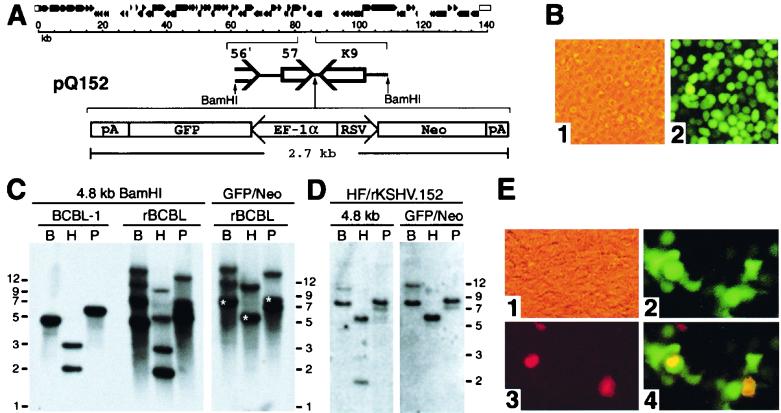
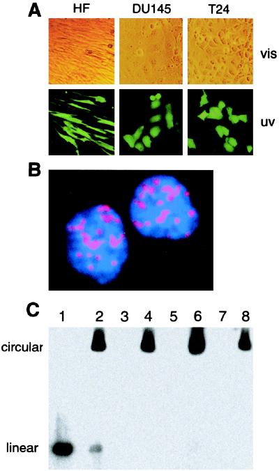
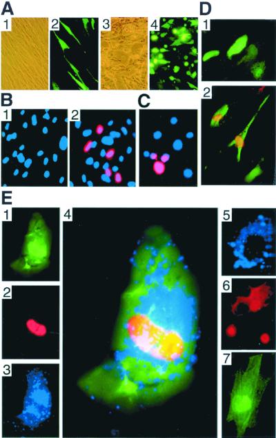
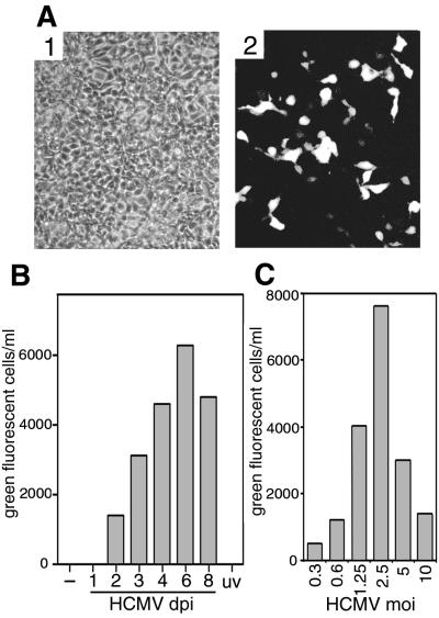
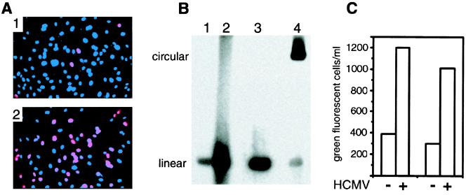
The majority of Kaposi's sarcoma-associated herpesvirus (KSHV)-infected cells identified in vivo contain latent KSHV, with lytic replication in only a few percent of cells, as is the case for the cells of Kaposi's sarcoma (KS) lesions. Factors that influence KSHV latent or lytic replication are not well defined. Because persons with KS are often immunosuppressed and susceptible to many infectious agents, including human cytomegalovirus (HCMV), we have investigated the potential for HCMV to influence the replication of KSHV. Important to this work was the construction of a recombinant KSHV, rKSHV.152, expressing the green fluorescent protein (GFP) and neo (conferring resistance to G418). The expression of GFP was a marker of KSHV infection in cells of both epithelial and endothelial origin. The rKSHV.152 virus was used to establish cells, including human fibroblasts (HF), containing only latent KSHV, as demonstrated by latency-associated nuclear antigen expression and Gardella gel analysis. HCMV infection of KSHV latently infected HF activated KSHV lytic replication with the production of infectious KSHV. Dual-color immunofluorescence detected both the KSHV lytic open reading frame 59 protein and the HCMV glycoprotein B in coinfected cells, and UV-inactivated HCMV did not activate the production of infectious KSHV-GFP. In addition, HCMV coinfection increased the production of KSHV from endothelial cells and activated lytic cycle gene expression in keratinocytes. These data demonstrate that HCMV can activate KSHV lytic replication and suggest that HCMV could influence KSHV pathogenesis.


Figures

FIG. 1
FIG. 1
Recombinant KSHV. (A) Top, schematic diagram of the KSHV genome (46). Bottom, components of pQ152, which was used to construct recombinant virus. An expanded segment of the KSHV genome shows the 4.8-kb BamHI fragment containing ORFs 57 and K9. This fragment was used for the insertion of the GFP/Neo cassette between the polyadenylation sites for ORFs 57 and K9. (B) Photomicrographs of BCBL-1 cells that were transfected with pQ152 and grown with G418 selection to generate recombinant rKSHV.152 virus 5 weeks postelectroporation (×100). Panel 1, phase contrast; panel 2, fluorescence. (C) Hybridization analysis, following gel electrophoresis, of DNA isolated from BCBL-1, and BCBL-1 with rKSHV.152, digested with BamHI, HindIII, or PstI. Left panel: analysis with the 4.8-kb BamHI fragment labeled with 32P as a probe. Right panel: analysis of BCBL-1 with rKSHV.152 with the GFP/Neo construct labeled with 32P used as a probe. B, BamHI; H, HindIII; P, PstI. Fragment sizes predicted from the BC-1 sequence (46) are as follows: BamHI, 4,774 bp; HindIII, 2,030 and 3,001 bp; and PstI, 5,907 bp. The predicted bands from a correct recombination event with the addition of the 2.7-kb GFP/Neo cassette are marked by an (∗). (D) Hybridization analysis of DNA isolated from HF infected with rKSHV.152 following digestion with BamHI, HindIII, or PstI and gel electrophoresis. Left panel: autoradiogram of HF/rKSHV.152 DNA hybridized with the 32P-labeled KSHV BamHI 4.8-kb fragment. Right panel: autoradiogram of HF/rKSHV.152 DNA hybridized with the 32P-labeled GFP/Neo as a probe. B, BamHI; H, HindIII; P, PstI. Fragment sizes expected for a correct recombination event would be 7.5 kb for BamHI, 5.7 kb for HindIII, and 8.6 kb for PstI, based on the published sequence of BC-1(46). (E) 293 cells inoculated with virus isolated from BCGL-1 cells containing rKSHV.152. Infected 293 cells were examined for GFP expression and the expression of ORF 59 protein using the MAb 11D1 visualized with Alexa 594 (red). Shown are photomicrographs of infected 293 cells 2 dpi (×100). Panel 1, phase contrast; panel 2, fluorescence with filters for GFP; panel 3, fluorescence with filters for Alexa 594 (red); panel 4, merged image from the green and red filters.
FIG. 2
FIG. 2
Analysis of ORF73/LANA and viral DNA in long-term rKSHV.152-infected cells. (A) Cultures of T24, DU145, and HF infected with rKSHV.152, selected with G418, and photographed with phase contrast (vis) and fluorescence (uv) (×100). (B) IFA detection of KSHV ORF 73 protein nuclear expression in HF with a rabbit polyclonal antibody to ORF 73 protein and visualized with a biotinylated anti-rabbit antibody reacted with strepavidin Alexa 594 (red). Cells were counterstained with DAPI, resulting in the blue nuclei (×2,000). (C) Gardella gel analysis of viral DNA present in long-term rKSHV.152-infected cultures. Lane 1, KSHV virion isolated from the media of TPA-induced BCBL-1 cells; lane 2, BCBL-1 cells; lane 3, HF; lane 4, HF/rKSHV.152; lane 5, T24; lane 6, T24/rKSHV.152; lane 7, DU145; lane 8, DU145/rKSHV.152.
FIG. 3
FIG. 3
Detection of viral proteins. (A) HF infected with rKSHV.152 and HF coinfected with rKSHV.152 and HCMV, photographed with phase contrast or fluorescence. Shown for HF/rKSHV.152 are phase contrast (panel 1) and fluorescence (panel 2) (×100). Shown for HF/rKSHV.152 infected with HCMV are phase contrast (panel 3) and fluorescence (panel 4). (B) Expression of the KSHV lytic ORF 59 protein induced by HCMV. HF infected with rKSHV.152, minus and plus HCMV infection, were analyzed for the presence of the lytic cycle 59 protein with MAb 11D1 visualized with Alexa 594 (red) and stained with DAPI (blue) (×200). Panel 1, HF with rKSHV.152; panel 2, HF with rKSHV.152 infected with HCMV. (C) Identification of ORF 59 expression in HF coinfected with rKSHV.152 and HSV. HF latently infected with rKSHV.152 were infected with HSV-1 and 2 days later were examined for ORF59 expression with MAb 11D1 visualized with Alexa 594 (red) and stained with DAPI (blue). (D) Visualization of GFP and nuclear ORF 59 protein with MAb 11D1 and Alexa 594 (red) in keratinocytes infected with rKSHV.152 (×200) (panel 1) or rKSHV.152 and HCMV (panel 2). (E) Identification of the KSHV ORF 59 protein and HCMV gB in fibroblasts infected by rKSHV.152 and HCMV. Photomicrographs show the detection of GFP expression (panel 1), KSHV ORF 59 protein with MAb 11D1 visualized with Alexa 594 (red) (panel 2), and HCMV gB with MAb 7-17 detected with Cy5 (blue) (panel 3). Panel 4, merged image showing ORF 59, gB, and GFP in coinfected cells (×600); panel 5, HF infected with HCMV reacted with antibodies 7-17 (blue) and 11D1 (red), demonstrating that 11D1 did not react with an HCMV-infected cell; panel 6, BCBL-1 cells induced with TPA for 2 days and reacted with MAb 11D1 (red) and MAb 7-17 (blue), showing that 7-17 does not react with KSHV proteins; panel 7, HF infected with rKSHV.152 reacted with MAb 11D1 and MAb 7-17.
FIG. 4
FIG. 4
Induction of KSHV lytic replication by HCMV. (A) Gardella gel analysis of KSHV from HF and HF coinfected with HCMV. Lane 1, KSHV isolated virions; lane 2, BCBL-1 cells; lane 3, BCBL-1 cells induced with TPA for 2 days; lane 4, HF containing rKSHV.152; lane 5, HF with rKSHV.152 and infected with HCMV 2 dpi; lane 6, HF infected with HCMV. The gel was probed with the 4.8-kb BamHI fragment (Fig. 1) labeled with 32P. The positions of circular and linear viral DNA are indicated, as determined by the positions of DNA from BCBL-1 cells and the DNA from purified virions, respectively. (B) Analysis of cell-free KSHV DNA. Viral DNA in cell-free media from HF infected with rKSHV.152 and from HF coinfected with rKSHV.152 and HCMV was analyzed for viral DNA that was resistant to DNase, with and without prior treatment with proteinase K and/or NP-40, as indicated (+, with; −, without). Viral DNA was detected by PCR for the ORF 26 region, and the product was identified with a 32P-labeled specific probe by liquid hybridization prior to gel electrophoresis (30).
FIG. 5
FIG. 5
Infectious rKSHV.152 from cultures coinfected with HCMV. (A) 293 cells infected by virus harvested from HF coinfected by rKSHV.152 and HCMV. HF/rKSHV.152 cultures coinfected with HCMV for 3 days were sonicated, the cell debris was pelleted, and the supernatant was used to infect 293 cells, which were photographed 2 dpi (×100). Panel 1, phase contrast; panel 2, fluorescence. (B) Kinetics of KSHV virus induction by HCMV. Infectious rKSHV.152 from HF carrying rKSHV.152 was determined from cultures not infected with HCMV (−), from cultures infected with HCMV and harvested at the times indicated (dpi), or from cultures infected with UV-inactivated HCMV harvested 3 dpi (uv). The cells and media were harvested and sonicated, and the cellular debris was pelleted. Sample supernatants were used to inoculate 293 cells, and the number of GFP-positive cells was determined 1 dpi. A representative experiment of four separate experiments is shown. (C) Influence of HCMV MOI on production of KSHV. HF with rKSHV.152 were infected with HCMV at the indicated MOI. Four days post-HCMV infection cultures were harvested and sonicated, cellular debris was pelleted, the supernatant was used to inoculate 293 cells, and the number of GFP-positive cells was determined. A representative experiment of three separate experiments is shown.
FIG. 6
FIG. 6
HCMV activation of KSHV in HUVEC. (A) Visualization of the nuclear expression of the lytic cycle ORF 59 protein with MAb 11D1 in HUVEC infected with rKSHV.152, minus and plus HCMV. Panel 1, HUVEC infected with rKSHV.152 3 dpi reacted with MAb 11D1 and Alexa 594 (red) and stained with DAPI (blue); panel 2, HUVEC coinfected with rKSHV.152 and HCMV reacted with MAb 11D1 and Alexa 594 (red) and stained with DAPI (blue). (B) Gardella gel analysis of KSHV DNA isolated from HUVEC infected with rKSHV.152, minus and plus HCMV. Lane 1, HUVEC infected with rKSHV.152; lane 2, HUVEC coinfected with rKSHV.152 and HCMV; lane 3, KSHV virion; lane 4, BCBL-1 cells. The positions of linear and circular KSHV genomes are indicated. (C) Presentation of two experiments showing the number of GFP-positive 293 cells resulting from infection with virus harvested from HUVEC cultures infected with rKSHV.152 and coinfected with rKSHV.152 and HCMV.

Similar articles

Cited by

References

    1. Adams V, Kempf W, Hassam S, Briner J, Schmid M, Moos R, Pfaltz M. Detection of several types of human papilloma viruses in AIDS-associated Kaposi's sarcoma. J Med Virol. 1995;46:189–193. - PubMed
    1. Ambinder R F, Robertson K D, Tao Q. DNA methylation and the Epstein-Barr virus. Semin Cancer Biol. 1999;9:369–375. - PubMed
    1. Beral V, Newton R. Overview of the epidemiology of immunodeficiency-associated cancers. J Natl Cancer Inst Monogr. 1998;23:1–6. - PubMed
    1. Beral V, Peterman T A, Berkelman R L, Jaffe H W. Kaposi's sarcoma among persons with AIDS: a sexually transmitted infection? Lancet. 1990;335:123–128. - PubMed
    1. Blasig C, Zietz C, Haar B, Neipel F, Esser S, Brockmeyer N H, Tschachler E, Colombini S, Ensoli B, Sturzl M. Monocytes in Kaposi's sarcoma lesions are productively infected by human herpesvirus 8. J Virol. 1997;71:7963–7968. - PMC - PubMed

Publication types