. 2001 Feb;75(3):1387-400.
doi: 10.1128/JVI.75.3.1387-1400.2001.

Adenovirus serotype 7 retention in a late endosomal compartment prior to cytosol escape is modulated by fiber protein

Affiliations

Adenovirus serotype 7 retention in a late endosomal compartment prior to cytosol escape is modulated by fiber protein

N Miyazawa et al. J Virol. 2001 Feb.

Abstract

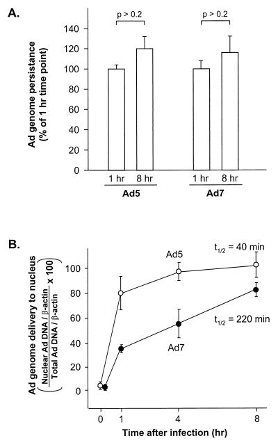
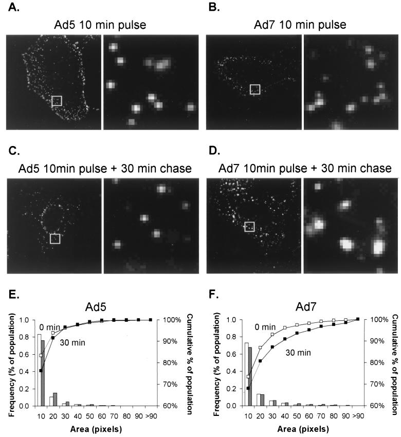
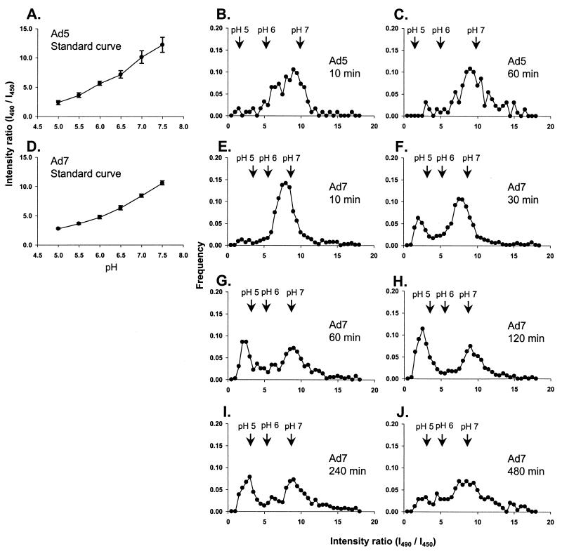
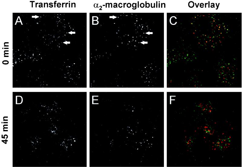
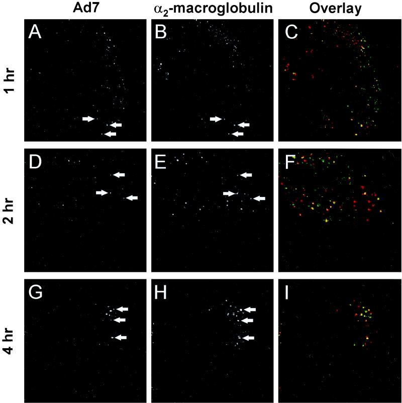
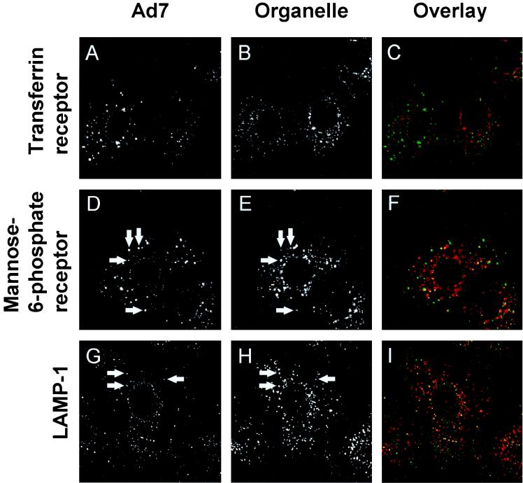
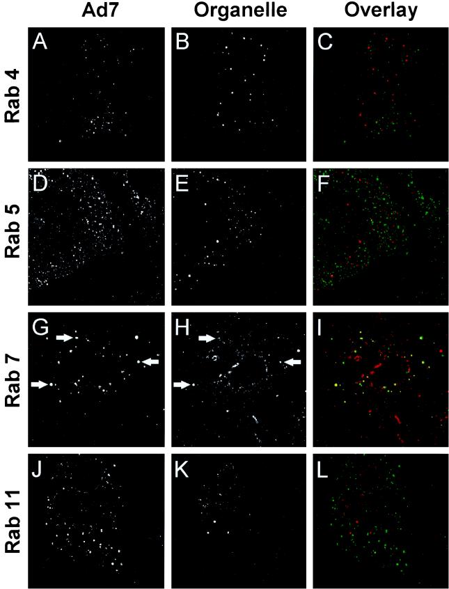
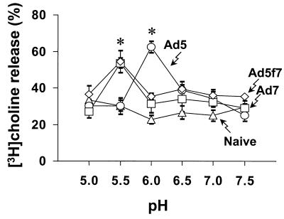
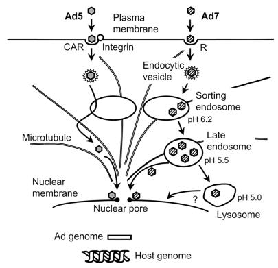
The intracellular trafficking of adenovirus (Ad) subgroup B (e.g., Ad7) differs from that of subgroup C (e.g., Ad5) in that Ad5 rapidly escapes from endocytic compartments following infection whereas Ad7 accumulates in organelles. To assess the hypothesis that Ad7 is targeted to the lysosomal pathway, Ad7 and Ad5 were conjugated with fluorophores and their trafficking in A549 epithelial cells was analyzed by fluorescence microscopy. Within 1 h after infection, Ad7, but not Ad5, accumulated in the cytoplasm of A549 cells. The pH in the environment of Ad5 was nearly neutral (pH 7), while Ad7 occupied acidic compartments (pH 5) over the first 2 h with a gradual shift toward neutrality by 8 h. Ad7 partially colocalized with alpha(2)-macroglobulin and late endosomal and lysosomal marker proteins, including Rab7, mannose-6-phosphate receptor, and LAMP-1. The pH optimum for membrane lysis by Ad7, as well as a chimeric Ad5 capsid that expressed the Ad7 fiber (Ad5fiber7), was pH 5.5, while that for lysis by Ad5 was pH 6.0. Thus, the native trafficking pathway for Ad7 involves residence in late endosomes and lysosomes, with information encoded in the Ad7 fiber acting as a pH-dependent trigger for membrane lysis and escape to the cytosol.


Figures

FIG. 1
FIG. 1
Quantitative evaluation of Ad5 and Ad7 genome persistence in cells and delivery of the genome to the nucleus at various times after a 10-min infection. A549 cells were infected with Ad5 or Ad7 (103 particles/cell) at 37°C for 10 min. The cells were washed and incubated for 0 to 8 h at 37°C. They were then harvested, and DNA was extracted from the total-cell lysate or from isolated nuclei. Ad DNA was quantified by a fluorogenic PCR assay using probes for Ad5 or Ad7 and compared to an internal standard (cellular β-actin gene). (A). Ad DNA persistence in A549 cells. The amounts of Ad5 and Ad7 DNA in A549 cells were compared at 1 and 8 h after infection. Data are presented as a percentage of the DNA content at 1 h (B). Percentage of the Ad genome delivered to the nucleus. The data are presented as the ratio of nuclear Ad genome copies (normalized to nuclear β-actin gene copies) to total-cell lysate Ad genome copies (normalized to lysate β-actin gene copies). Shown are the analyses for Ad5 and Ad7. The data are presented as the mean ± standard error of three experiments.
FIG. 2
FIG. 2
Localized intracellular accumulation of Ad7 compared to Ad5. A549 cells were incubated with Cy3-Ad (1011 particles/ml) for 10 min at 37°C, washed, and immediately fixed (10-min pulse) or incubated for 30 min at 37°C prior to fixation (10-min pulse plus 30-min chase). (A to D) Images of Cy3-Ad5 and Ad7. For each pair of panels, the right panels are ×8 magnifications of the box in the left panel; the ×8 magnification provides an overview of the regions and extent of localized accumulation of the fluorophore-labeled virus. (E) Histogram of the frequency of Cy3-Ad5 as a function of the pixel number. Open bars and open squares (□) represent a 10-min pulse, whereas shaded bars and solid squares (■) represent a 10-min pulse followed by a 30-min chase (F) Histogram of the frequency of Cy3-Ad7 as a function of pixel number. The cumulative percentage curve of Ad7 (■) was shifted toward the right at 30 min, indicating an increase in the mean pixel area of the fluorescent spots as infection progressed. The dimensions of each box analyzed are 3.5 by 3.5 μm.
FIG. 3
FIG. 3
Comparison of the pH of compartments containing Ad5 or Ad7. A549 cells were infected with carboxyfluorescein-Ad (1011 particles/ml) for 10 min at 37°C and incubated at 37°C for various times. pH measurements were performed in living cells by recording carboxyfluorescein emission intensity following excitation at either 490 or 450 nm. (A). Standard curve of carboxyfluorescein-Ad5 using cells equilibrated with 50 mM methylamine buffers (pH 5.0 to 7.5). The fluorescence intensity ratio (I490/I450) is related to the pH of the standard medium. Bars show the 95% confidence interval. (B) Carboxyfluorescein-Ad5, 10-min pulse (infection) and 10-min chase (incubation). (C) Carboxyfluorescein-Ad5, 10-min pulse and 60-min chase. (D) Standard curve of carboxyfluorescein-Ad7. (E to J) Carboxyfluorescein-Ad7, 10-min pulse with various chase times as indicated (10 to 480 min).
FIG. 4
FIG. 4
Endocytic trafficking in A549 cells. The cells were labeled with Cy3-α2M (10 μg/ml) and FITC-Tf (10 μg/ml) for 15 min, washed, and incubated for 0 or 45 min. The left column shows FITC-Tf; the center column shows Cy3-α2M; and the right column shows a color overlay of the signals (FITC-Tf, green; Cy3-α2M, red; colocalization, yellow). (A) FITC-Tf, without incubation. (B) Cy3-α2M, without incubation. (C) Overlay of panels A and B (D) FITC-Tf, 45-min incubation. (E) Cy3-α2M, 45-min incubation. (F) Overlay of panels D and E. Arrows indicate examples of colocalization of Cy3-α2M and FITC-Tf. Field width, 50 μm.
FIG. 5
FIG. 5
Colocalization of Ad7 with α2M. A549 cells were infected with carboxyfluorescein-Ad7 (1011 particles/ml) for 10 min at 37°C, loaded with Cy3-α2M (10 μg/ml) for 15 min, and then incubated for an additional 45 or 105 min at 37°C. The left column shows carboxyfluorescein-Ad7, the center column shows Cy3-α2M, and the right column shows a color overlap of the two signals (carboxyfluorescein-Ad7, green; Cy3-α2M, red; colocalization yellow). (A) Carboxyfluorescein-Ad7, 1 h after infection. (B) Cy3-α2M, 1 h after Ad7 infection, (C) Overlay of panels A and B. (D) Carboxyfluorescein-Ad7, 2 h after infection. (E) Cy3-α2M, 2 h after Ad7 infection. (F) Overlay of panels D and E. (G) Carboxyfluorescein-Ad7, 4 h after infection (H) Cy3-α2M, 4 h after Ad7 infection. (I) Overlay of panels G and H. Arrows indicate examples of colocalization of Ad7 and Cy3-α2M. Field width, 50 μm.
FIG. 6
FIG. 6
Colocalization of Ad7 with endosomal proteins. A549 cells were infected with carboxyfluorescein-Ad7 (1011 particles/ml) for 10 min at 37°C, incubated for 60 min at 37°C, and then fixed and permeabilized. The cells were incubated at 23°C for 1 h with primary antibodies against Tf receptor, mannose-6-phospate receptor, or LAMP-1 and localized using Texas red-conjugated secondary antibodies. The left column shows carboxyfluorescein-Ad, the center column shows organelle-specific protein staining, and the right column shows overlap (Ad7, green; specific protein, red; overlap), yellow). Very little colocalization of Ad7 with Tf. receptor was observed. However, Ad7 was partially colocalized with mannose-6-phosphate (late endosomes) and LAMP-1 (late endosomes and lysosomes). Arrows indicate examples of colocalization. Field width, 40 μm.
FIG. 7
FIG. 7
Colocalization of Ad7 with small GTP binding proteins found in late endosomes and lysosomes. A549 cells were infected with carboxyfluorescein-Ad7 (1011 particles/ml) for 10 min at 37°C, incubated for 60 min at 37°C, and then fixed and permeabilized. The cells were incubated at 23°C for 1 h with primary antibodies against Rab proteins, using Texas red-conjugated secondary antibodies. The left column shows carboxyfluorescein-Ad, the center column shows organelle-specific protein staining, and the right column shows overlap (Ad7, green; specific protein, red; colocalization, yellow). Very little colocalization of Ad7 with Rab 4, Rab 5, or Rab 11 was observed. However, Ad7 was partially colocalized with Rab 7 (late endosomes and lysosomes). Arrows indicate examples of colocalization. Field width, 40 μm.
FIG. 8
FIG. 8
Quantitative comparison of the motility of Ad5 and Ad7 at different time points. A549 cells were infected with Cy3-Ad5 or Cy3-Ad7 (1011 particles/ml) for 10 min, at 37°C, washed out, and incubated at 37°C for 30 or 240 min. Digital segments of the fluorescent image fields were analyzed with respect to the number of linear translocations (>2 μm) observed. Data are normalized with respect to the number of viruses visible in the field and the duration of observation. The motility of Ad7 at 240 min after infection was increased significantly compared with that 30 min after infection, whereas the motility of Ad5 decreased over the same interval.
FIG. 9
FIG. 9
Effect of pH on Ad-dependent membrane lysis. KB cells (2 × 10⁴ cells) were incubated for 1 h in medium containing [3H] choline, washed, chilled to 4°C, and then treated with 1011 particles of Ad5, Ad7, or the chimeric virus Ad5fiber7 for 1 h. The medium was replaced with permeability buffer at various pH, containing sodium azide to prevent endocytosis of the viruses. Cells with bound viruses were then warmed to 37°C for 1 h to permit lysis. The percent [3H]choline release was determined by measuring 3H released into the medium compared with the counts remaining in cells. Data from Ad5, Ad7, Ad5fiber7 (Ad5f7), and naive control are shown as mean values ± standard error. The activity of Ad5-induced membrane lysis was significantly greater than that of Ad7- or Ad5fiber7-induced lysis at pH 6.0; the activity of Ad5-induced membrane lysis was significantly less than that of Ad7- or Ad5fiber7-induced lysis at pH 5.5 (P < 0.05 for each comparison).
FIG. 10
FIG. 10
Summary of intracellular trafficking of subgroup B and subgroup C Ad. Subgroup C Ad (e.g., Ad5) is internalized via interaction with a high affinity receptor (coxsackie-virus-Ad receptor [CAR]) and a secondary receptor integrin and then enters the cell via receptor-mediated endocytosis. When the endocytic compartment containing Ad5 fuses with a sorting endosome (pH 6.2), Ad5 breaks out the early endosome, escapes to the cytosol, and translocates to the nucleus along microtubules within 1 h. Subgroup B Ad (e.g., Ad7) binds to cells via interaction with an unidentified receptor (R) and is internalized via receptor-mediated endocytosis. The endocytic compartment containing Ad7 fuses with sorting endosomes, but unlike Ad5, Ad7 does not disrupt the sorting-endosome membrane, remaining inside that organelle as it matures to become a late endosome (pH 5.5) and finally a lysosome (pH 5.0). Ad7 escapes from late endosomes and/or lysosomes and translocates to the nucleus. Following equally rapid internalization (t½ = 2 to 3 min) (33), Ad5 reaches the nucleus rapidly (t½ = 40 min) while Ad7 trafficking progresses more slowly (t½ = 220 min).

Similar articles

Cited by

References

    1. Abrahamsen K, Kong H L, Mastrangeli A, Brough D, Lizonova A, Crystal R G, Falck-Pedersen E. Construction of an adenovirus type 7a Ela vector. J Virol. 1997;71:8946–8951. - PMC - PubMed
    1. Bai M, Harfe B, Freimuth P. Mutations that alter an Arg-Gly-Asp (RGD) sequence in the adenovirus type 2 penton base protein abolish its cell-rounding activity and delay virus reproduction in flat cells. J Virol. 1993;67:5198–5205. - PMC - PubMed
    1. Bergelson J M, Cunningham J A, Droguett G, Kurt-Jones E A, Krithivas A, Hong J S, Horwitz M S, Crowell R L, Finberg R W. Isolation of a common receptor for coxsackie B viruses and adenoviruses 2 and 5. Science. 1997;275:1320–1323. - PubMed
    1. Blumenthal R, Seth P, Willingham M C, Pastan I. pH-dependent lysis of liposomes by adenovirus. Biochemistry. 1986;25:2231–2237. - PubMed
    1. Chardonnet Y, Dales S. Early events in the interaction of adenoviruses with HeLa cells. I. Penetration of type 5 and intracellular release of the DNA genome. Virology. 1970a;40:462–477. - PubMed

Publication types

LinkOut - more resources