. 2001 Feb;75(3):1437-49.
doi: 10.1128/JVI.75.3.1437-1449.2001.

Mutations in hepatitis C virus RNAs conferring cell culture adaptation

Affiliations

Mutations in hepatitis C virus RNAs conferring cell culture adaptation

V Lohmann et al. J Virol. 2001 Feb.

Abstract

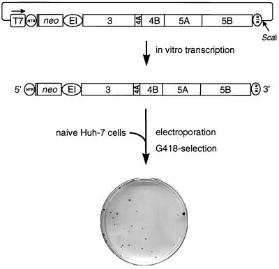
As an initial approach to studying the molecular replication mechanisms of hepatitis C virus (HCV), a major causative agent of acute and chronic liver disease, we have recently developed selectable self-replicating RNAs. These replicons lacked the region encoding the structural proteins and instead carried the gene encoding the neomycin phosphotransferase. Although the replication levels of these RNAs within selected cells were high, the number of G418-resistant colonies was reproducibly low. In a search for the reason, we performed a detailed analysis of replicating HCV RNAs and identified several adaptive mutations enhancing the efficiency of colony formation by several orders of magnitude. Adaptive mutations were found in nearly every nonstructural protein but not in the 5' or 3' nontranslated regions. The most drastic effect was found with a single-amino-acid substitution in NS5B, increasing the number of colonies approximately 500-fold. This mutation was conserved with RNAs isolated from one cell line, in contrast to other amino acid substitutions enhancing the efficiency of colony formation to a much lesser extent. Interestingly, some combinations of these nonconserved mutations with the highly adaptive one reduced the efficiency of colony formation drastically, suggesting that some adaptive mutations are not compatible.


Figures

FIG. 1
FIG. 1
Experimental approach used to establish cell lines carrying self-replicating HCV RNAs. neo, neomycin phosphotransferase gene; EI, EMCV IRES; T7, promoter of the bacteriophage T7 RNA polymerase. Since for optimal HCV IRES activity ∼36 nucleotides of the core open reading frame are required, 12 amino acid residues of the core protein are fused to the amino terminus of the neomycin phosphotransferase. For details, see the text.
FIG. 2
FIG. 2
Sequence analysis of HCV polyproteins recloned from cell line 9-13 and result of functional testing of different HCV fragments. The parental replicon construct is shown at the top, with numbers below referring to nucleotide positions of the HCV genome (for details, see the legend to Fig. 1). HCV polyproteins from eight independent clones are shown as open bars. The positions of nucleotide and amino acid sequence deviations from the original consensus sequence are indicated by vertical lines and labeled as follows: black, amino acid substitution; gray, silent nucleotide change; ↓, nonsense mutation; formula image
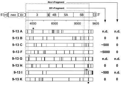
, frameshift mutation. The single conserved amino acid substitution in NS5B is marked with a diamond. The positions of the recognition sequences for the restriction enzymes SfiI and NcoI used for subcloning into the parental construct are indicated with dotted lines. The names of the corresponding constructs are given at the left, and the CFU/μg in vitro transcript obtained with each of the subcloned NcoI or SfiI fragments after selection with G418 (500 μg/ml) is given on the right. n.d., not determined.
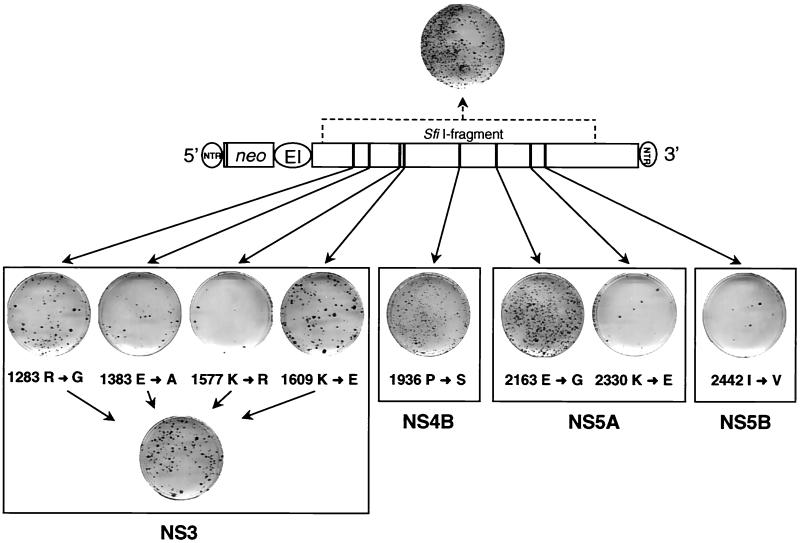
FIG. 3
FIG. 3
Single-amino-acid substitution in NS5A increases ECF almost 100-fold. The basic construct with the amino acid substitutions found in the SfiI fragment of clone 9-13F is shown in the center. Results obtained with single-amino-acid substitutions are given below. Colonies shown on each cell culture dish were obtained after transfection of Huh-7 cells with 500 ng of each mutant or construct 9-13F, shown at the top. The individual amino acid substitutions are written below each stained culture dish, with the arrow pointing to the substituting residue. Numbers refer to the amino acid position of the HCV polyprotein. A summary of these results is given in Table 3.
FIG. 4
FIG. 4
Identification of adaptive mutations in the 3′-proximal region of the HCV replicon. The basic construct is shown at the top, with the restriction sites used for subcloning indicated above. Three fragments corresponding to the 5′ NTR up to the 3′ end of neo, the EMCV IRES up to the amino-terminal region of NS3, and the carboxy-terminal half of NS5B up to the end of the 3′ NTR were amplified by RT-PCR from cell line 9-13 and inserted into the parental replicon construct. From 16 to 18 clones from each fragment (indicated with the pictograms at the top of the table) were isolated, and 300 ng of the corresponding in vitro transcripts was used for transfection into naive Huh-7 cells. The CFU/μg of in vitro transcript generated from each replicon construct is given at the left, and the number of clones in each category is shown on the right. For the 3′ fragment, five clones were obtained with >10,000 CFU/μg of RNA. Note that for the nonadapted parental replicon, the CFU/μg of RNA is 1 to 100 (indicated by shading).
FIG. 5
FIG. 5
Sequence analysis of replicons carrying various 3′-terminal fragments that were isolated from cell line 9-13. The region subcloned into the parental construct and composed of the 3′-terminal region of NS5B (beginning at nucleotide position 8499) and the tripartite 3′ NTR is given on the top. The amino acid substitution in NS5B found in each subclone is indicated with a black vertical line and labeled with a diamond; the positions of all other amino acid changes are depicted with gray lines. Nucleotide substitutions in the 3′ NTR are indicated with gaps and labeled with a star. The CFU/μg of in vitro transcript obtained with each clone is given on the right. The tripartite structure of the 3′ NTR composed of the variable region (VR), the poly (U/UC)n tract, and the highly conserved X tail is indicated. Numbers to the left refer to the individual clone. Note that all cloned fragments carried some minor rearrangements in the poly (U/UC)n tract.
FIG. 6
FIG. 6
Evidence that replicon 9-13F/NcoI is not cytopathogenic. Replicon rep5B/Gly2884 (10 ng) was transfected into naive Huh-7 cells either alone or together with 10, 100, or 1,000 ng of the potentially cytopathogenic replicon rep9-13F/NcoI. As a control, cotransfections were performed with rep5B/Gly2884 and given amounts of an inactive rep9-13F/NcoI mutant carrying a deletion of 10 amino acid residues spanning the active site of the NS5B RNA polymerase. About 4 weeks posttransfection, cells were fixed and stained with Coomassie brilliant blue. Colonies obtained after transfection of rep5B/Gly2884 and selection with G418 are shown on the top; results obtained in cotransfections are given below. Representative plates are shown.
FIG. 7
FIG. 7
Evidence for the incompatibility of adaptive mutations. Replicons were generated carrying both the highly adaptive NS5B mutation at position 2884 and either all four mutations in NS3 or one of the given mutations in NS4B, NS5A, or NS5B found in the SfiI fragment of clone 9-13F. About 4 weeks after transfection into naive Huh-7 cells, they were fixed and stained with Coomassie brilliant blue. The result obtained after transfection of the parental replicon rep5B/Gly2884 is shown on the top, and results obtained with the combination mutants are shown below. Note that in several independent experiments, no or only one colony was obtained with the combination of the highly adaptive NS5B and NS5A mutations (2884 R→G and 2163 E→G). A summary of these results is given in Table 3.
FIG. 8
FIG. 8
Location of adaptive mutations in the three-dimensional structures of the NS3 helicase (A) and the NS5B RdRp (B). The three domains of the NS3 helicase are color coded according to Kim et al. (32). The two residues altered in the adapted replicons are located on the surface of the molecule, and they are given in ball-and-stick representation. The three-dimensional structure of the NS5B RdRp as viewed from the front is shown in panel B, and individual domains (thumb, palm, and fingers) are marked with different colors. The palm subdomain located at the base is closed on either side by the fingers and thumb subdomains and at the back by the loops. The arginine residue replaced by glycine with the adapted replicon is given in ball-and-stick representation.

References

    1. Asabe S I, Tanji Y, Satoh S, Kaneko T, Kimura K, Shimotohno K. The N-terminal region of hepatitis C virus-encoded NS5A is important for NS4A-dependent phosphorylation. J Virol. 1997;71:790–796. - PMC - PubMed
    1. Ausubel F M, Brent R, Kingston R E, Moore D D, Seidman J G, Smith J A, Struhl K, editors. Current protocols in molecular biology. New York, N.Y: John Wiley and Sons; 1987.
    1. Bartenschlager R, Ahlborn-Laake L, Mous J, Jacobsen H. Kinetic and structural analyses of hepatitis C virus polyprotein processing. J Virol. 1994;68:5045–5055. - PMC - PubMed
    1. Bartenschlager R, Lohmann V. Replication of hepatitis C virus. J Gen Virol. 2000;81:1631–1648. - PubMed
    1. Beard M R, Abell G, Honda M, Carroll A, Gartland M, Clarke B, Suzuki K, Lanford R, Sangar D V, Lemon S M. An infectious molecular clone of a Japanese genotype 1b hepatitis C virus. Hepatology. 1999;30:316–324. - PubMed

Publication types