. 2001 Feb;75(3):1450-8.
doi: 10.1128/JVI.75.3.1450-1458.2001.

Genetic evidence of an essential role for cytomegalovirus small capsid protein in viral growth

Affiliations

Genetic evidence of an essential role for cytomegalovirus small capsid protein in viral growth

E M Borst et al. J Virol. 2001 Feb.

Abstract

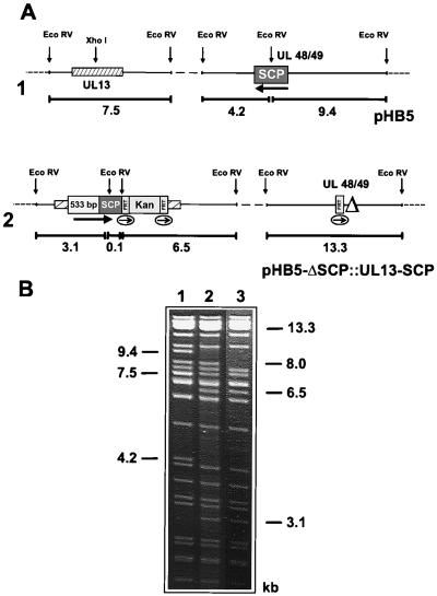
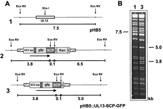
Many steps in the replication cycle of cytomegalovirus (CMV), like cell entry, capsid assembly, and egress of newly synthesized virions, have not been completely analyzed yet. In order to facilitate these studies, we decided to construct a recombinant CMV that incorporates the green fluorescent protein (GFP) into the nucleocapsid. A comparable herpes simplex virus type 1 (HSV-1) mutant has recently been generated by fusion of the GFP open reading frame (ORF) with the HSV-1 ORF encoding small capsid protein (SCP) VP26 (P. Desai and S. Person, J. Virol. 72:7563-7568, 1998). Recombinant CMV genomes expressing a fusion protein consisting of GFP and the SCP were constructed by the recently established bacterial artificial chromosome mutagenesis procedure. In transfected cells, the SCP-GFP fusion protein localized to distinct foci in the nucleus that may represent sites for capsid assembly (assemblons). However, no viable progeny was reconstituted from these mutant CMV genomes. CMV genomes with deletion of the SCP ORF also did not give rise to infectious virus. Rescue of the mutation by insertion of the SCP gene at an ectopic position in an SCP knockout genome indicates that, in contrast to the HSV-1 SCP, the CMV SCP is essential for viral growth. Expression of the SCP-GFP fusion protein together with the authentic SCP blocked the CMV infection cycle, suggesting that the SCP-GFP fusion protein exerts a dominant-negative effect on the assembly of new virions. The results of this study are discussed with regard to recently published data about the structure of the CMV virion and its differences from the HSV-1 virion.


Figures

FIG. 1
FIG. 1
Alignment of the amino acid sequences of the HCMV (upper line) and MCMV (lower line) SCPs. Identical (vertical lines) and conserved (colons) residues and the sites of GFP insertion into the SCPs are indicated. Amino acid positions are on the right. Dots indicate gaps that were introduced into the MCMV SCP sequences to achieve optimal alignment.
FIG. 2
FIG. 2
Construction of HCMV BAC plasmids encoding an SCP-GFP fusion protein (A) and structural analysis of the mutated BAC plasmids (B). (A) Structure of the genomic region of the HCMV BAC plasmids containing the SCP ORF (diagram 1) or the SCP-GFP ORF (diagrams 2 and 3). The arrow indicates the orientation of the SCP ORF. The kanamycin resistance marker (Kan) was excised from BAC plasmid pHB5::SCP-GFP-Kan (diagram 2) by Flp recombinase-mediated site-specific recombination, resulting in BAC plasmid pHB5::SCP-GFP (diagram 3). Fragment sizes are indicated in kilobase pairs below each diagram. (B) DNA of the HCMV BAC plasmids was digested with EcoRV, separated on a 0.7% agarose gel, and stained with ethidium bromide. The sizes of the EcoRV fragments characteristic of parental BAC plasmid pHB5 (lane 1) and mutant BAC plasmids pHB5::SCP-GFP-Kan (lane 2) and pHB5::SCP-GFP (lane 3) are indicated.
FIG. 3
FIG. 3
Subnuclear localization of the SCP-GFP fusion protein. HCMV BAC plasmid pHB5::SCP-GFP was transfected into RPE cells, and expression of the SCP-GFP protein was analyzed 2 days later by confocal laser scanning microscopy.
FIG. 4
FIG. 4
Deletion of the ORF encoding the SCP. (A) Structures of the HCMV BAC plasmids with the EcoRV fragments containing the SCP ORF (diagram 1), after replacement of the SCP ORF with a kanamycin resistance marker (Kan) (diagram 2), and after excision of the kanamycin resistance marker (diagram 3). Fragment sizes in kilobase pairs are shown below the diagrams. (B) Structural analysis of HCMV BAC plasmids pHB5 and pHB5-ΔSCP. DNA of the BAC plasmids was subjected to EcoRV digestion, followed by gel electrophoresis. Relevant DNA bands are indicated.
FIG. 5
FIG. 5
Rescue of the SCP knockout mutation by ectopic insertion of the gene for SCP. (A) Structures of the genomic regions containing the UL13 and UL48/49 ORFs of BAC plasmid pHB5 (diagram 1) and of BAC plasmid pHB5-ΔSCP::UL13-SCP after deletion of the UL48/49 ORF and ectopic insertion of the gene for SCP (diagram 2). The black arrows indicate the orientation of the SCP ORF at its original (diagram 1) and ectopic (diagram 2) positions, and the thin arrows display the orientation of the FRT sites. Fragment sizes are indicated below the diagrams in kilobase pairs. (B) DNA of HCMV BAC plasmids pHB5 (lane 1) and pHB5-ΔSCP::UL13-SCP (lane 2) and of the corresponding virus reconstituted from BAC plasmid pHB5-ΔSCP::UL13-SCP (lane 3) was digested with EcoRV and analyzed by gel electrophoresis. The sizes of the DNA bands characteristic of pHB5 (lane 1), pHB5-ΔSCP::UL13-SCP (lane 2), and the viral DNA (lane 3) are indicated.
FIG. 6
FIG. 6
Insertion of the SCP-GFP gene within the UL13 ORF. (A) Structures of the region of the HCMV BAC plasmids containing the UL13 ORF prior to (diagram 1) and after (diagram 2) insertion of the SCP-GFP gene. The kanamycin resistance marker was excised by Flp recombinase, resulting in BAC plasmid pHB5::UL13-SCP-GFP (diagram 3). The arrows indicate the orientation of the ectopic SCP-GFP gene. Fragment sizes in kilobase pairs are indicated below the diagrams. (B) Ethidium bromide-stained agarose gel showing the EcoRV-digested DNA of BAC plasmids pHB5 (lane 1) and pHB5::UL13-SCP-GFP (lane 3). Relevant DNA fragments are marked. Note that the 7.5-kb band of pHB5 and the 5.0-kb band of pHB5::UL13-SCP-GFP are double bands.

References

    1. Adler H, Messerle M, Wagner M, Koszinowski U H. Cloning and mutagenesis of the murine gammaherpesvirus 68 genome as an infectious bacterial artificial chromosome. J Virol. 2000;74:6964–6974. - PMC - PubMed
    1. Baldick C J, Jr, Shenk T. Proteins associated with purified human cytomegalovirus particles. J Virol. 1996;70:6097–6105. - PMC - PubMed
    1. Booy F P, Trus B L, Newcomb W W, Brown J C, Conway J F, Steven A C. Finding a needle in a haystack: detection of a small protein (the 12-kDa VP26) in a large complex (the 200-MDa capsid of herpes simplex virus) Proc Natl Acad Sci USA. 1994;91:5652–5656. - PMC - PubMed
    1. Borst E M, Hahn G, Koszinowski U H, Messerle M. Cloning of the human cytomegalovirus (HCMV) genome as an infectious bacterial artificial chromosome in Escherichia coli: a new approach for construction of HCMV mutants. J Virol. 1999;73:8320–8329. - PMC - PubMed
    1. Borst E M, Messerle M. Development of a cytomegalovirus vector for somatic gene therapy. Bone Marrow Transplant. 2000;25(Suppl. 2):S80–S82. - PubMed

Publication types

LinkOut - more resources