. 2001 Feb;75(3):1533-9.
doi: 10.1128/JVI.75.3.1533-1539.2001.

Rapid CD4(+) T-cell loss induced by human immunodeficiency virus type 1(NC) in uninfected and previously infected chimpanzees

Affiliations

Rapid CD4(+) T-cell loss induced by human immunodeficiency virus type 1(NC) in uninfected and previously infected chimpanzees

F J Novembre et al. J Virol. 2001 Feb.

Erratum in

  • J Virol 2001 Jun;75(11):5441

Abstract

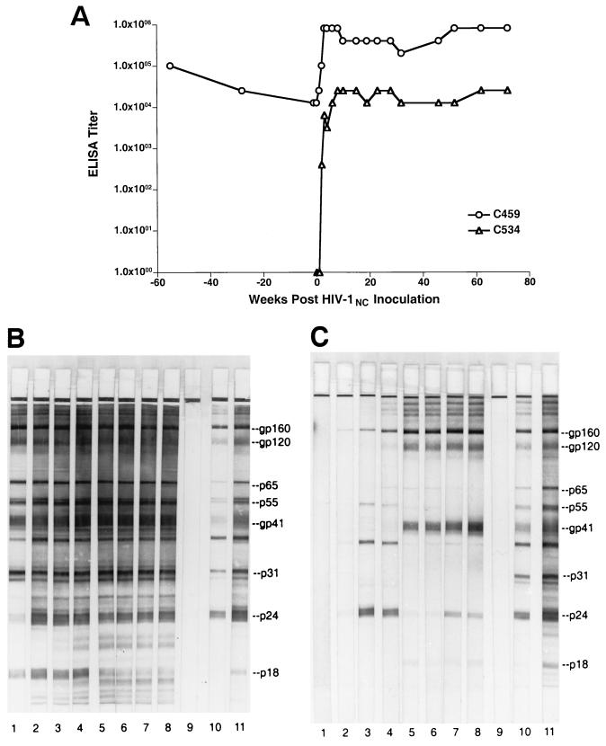
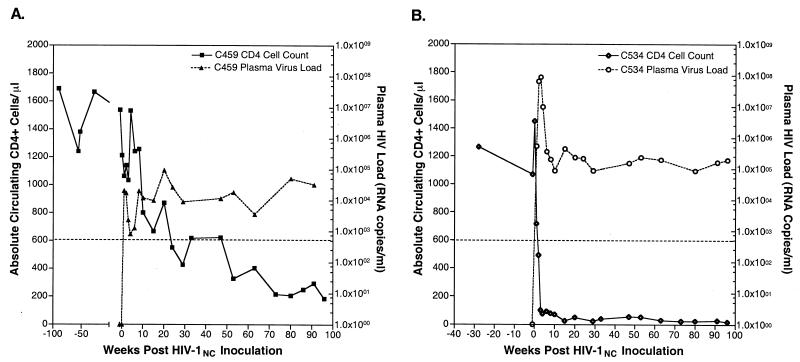
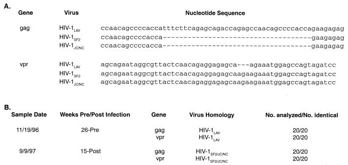
To investigate the pathogenicity of a virus originating in a chimpanzee with AIDS (C499), two chimpanzees were inoculated with a plasma-derived isolate termed human immunodeficiency virus type 1(NC) (HIV-1(NC)). A previously uninfected chimpanzee, C534, experienced rapid peripheral CD4(+) T-cell loss to fewer than 26 cells/microl by 14 weeks after infection. CD4(+) T-cell depletion was associated with high plasma HIV-1 loads but a low virus burden in the peripheral lymph node. The second chimpanzee, C459, infected 13 years previously with HIV-1(LAV), experienced a more protracted course of peripheral CD4(+) T-cell loss after HIV-1(NC) inoculation, resulting in fewer than 200 cells/microl by 96 weeks postinoculation. The quantities of viral RNA in the plasma and peripheral lymph node from C459 were below the lower limits of detection prior to inoculation with HIV-1(NC) but were significantly and persistently increased after superinfection, with HIV-1(NC) representing the predominant viral genotype. These results show that viruses derived from C499 are more pathogenic for chimpanzees than any other HIV-1 isolates described to date.


Figures

FIG. 1
FIG. 1
HIV-specific antibody responses following HIV-1NC infection in chimpanzees. (A) Plasma samples obtained from C459 and C534 at the indicated times postinfection were used to quantify HIV-specific antibody responses using a commercially available whole-virus ELISA. (B and C) Qualitative analyses of viral antibody were performed using commercially available Western blot strips. In panel B, plasma samples were from animal C459; in panel C, plasma samples from animal C534. Lane 1, day of inoculation; lane 2, 2 weeks postinoculation; lane 3, 8 weeks postinoculation; lane 4, 15 weeks postinoculation; lane 5, 32 weeks postinoculation; lane 6, 53 weeks postinoculation; lane 7, 63 weeks postinoculation; lane 8, 80 weeks postinoculation; lane 9, plasma from an uninfected chimpanzee; lane 10, weak positive control serum; lane 11, strong positive control serum.
FIG. 2
FIG. 2
Peripheral CD4+ T-cell levels and plasma HIV loads in HIV-1NC-infected chimpanzees. At the indicated times pre- and post-HIV-1NC inoculation, blood was obtained from chimpanzees C459 (A) and C534 (B) for use in determination of circulating levels of CD4+ CD2+ T cells by flow cytometry and for use in the quantitation of plasma viral RNA levels. The dotted line represents the cutoff level for the quantitation of plasma HIV levels (800 copies/ml).
FIG. 3
FIG. 3
Distribution of productively infected cells in lymph nodes from HIV-1NC-infected chimpanzees. In situ hybridization for HIV-1 RNA is shown for chimpanzees C459 (A and B) and C534 (C and D), using the chromagen nitroblue tetrazolium-BCIP (5-bromo-4-chloro-3-indolylphosphate) and nuclear fast red counterstain. (A) Productively infected cells are not apparent in sections of lymph ode from HIV-1 infected chimpanzee C459 prior to inoculation with HIV-1NC (×100). (B) Numerous infected cells (dark blue stain) are seen in the paracortex of a lymph node collected from C459 3 weeks after superinfection with HIV-1NC (×100). (C) Lymph node from C543 (uninfected chimpanzee) prior to infection with HIV-1NC (×100). (D) HIV-1-infected cell (arrow) shown in the paracortex of a lymph node from C534 3 weeks after infection with HIV-1NC. Arrowheads delineate the margins of a germinal center (×200). Note the absence of secondary follicles in lymph nodes from HIV-infected, nonprogressor C459 (A) and HIV-negative C534 (C).
FIG. 4
FIG. 4
Nucleotide sequence analysis of HIV-1 genes in chimpanzee C459. (A) Sequence alignments of portions of gag and vpr genes of HIV-1LAV, HIV-1SF2, and HIV-1JC/NC, illustrating major differences. (B) PBMC from chimpanzee C459 obtained pre- and post-HIV-1NC infection (sample dates 11/19/96 and 9/9/97, respectively) were used to prepare genomic DNA for use as template in PCR experiments. Amplified DNA was cloned into the pGEM-T vector. Twenty clones obtained from each sample were analyzed by DNA sequencing. Results show that, prior to HIV-1NC infection, sequences were most similar to HIV-1LAV (the virus used to inoculate animal C459 in 1984) but that, after HIV-1NC inoculation, sequences were most similar to HIV-1SF2 and HIV-1JC/NC (26).

References

    1. Alter H J, Eichberg J W, Masur H, Saxinger W C, Gallo R, Macher A M, Lane H C, Fauci A S. Transmission of HTLV-III infection from human plasma to chimpanzees: an animal model for AIDS. Science. 1984;226:549–552. - PubMed
    1. Benveniste R E, Arthur L O, Tsai C C, Sowder R, Copeland T D, Henderson L E, Oroszlan S. Isolation of a lentivirus from a macaque with lymphoma: comparison with HTLV-III/LAV and other lentiviruses. J Virol. 1986;60:483–490. - PMC - PubMed
    1. Bogers W M, Koornstra W H, Dubbes R H, ten Haaft P J, Verstrepen B E, Jhagjhoorsingh S S, Haaksma A G, Niphuis H, Laman J D, Norley S, Schuitemaker H, Goudsmit J, Hunsmann G, Heeney J L, Wigzell H. Characteristics of primary infection of a European human immunodeficiency virus type 1 clade B isolate in chimpanzees. J Gen Virol. 1998;79:2895–2903. - PubMed
    1. Castro B A, Walker C M, Eichberg J W, Levy J A. Suppression of human immunodeficiency virus replication by CD8+ cells from infected and uninfected chimpanzees. Cell Immunol. 1991;132:246–255. - PubMed
    1. Connor R I, Montefiori D C, Binley J M, Moore J P, Bonhoeffer S, Gettie A, Fenamore E A, Sheridan K E, Ho D D, Dailey P J, Marx P A. Temporal analyses of virus replication, immune responses, and efficacy in rhesus macaques immunized with a live, attenuated simian immunodeficiency virus vaccine. J Virol. 1998;72:7501–7509. - PMC - PubMed

Publication types

MeSH terms