. 2001 Feb;21(3):794-810.
doi: 10.1128/MCB.21.3.794-810.2001.

Multifaceted regulation of cell cycle progression by estrogen: regulation of Cdk inhibitors and Cdc25A independent of cyclin D1-Cdk4 function

Affiliations

Multifaceted regulation of cell cycle progression by estrogen: regulation of Cdk inhibitors and Cdc25A independent of cyclin D1-Cdk4 function

J S Foster et al. Mol Cell Biol. 2001 Feb.

Abstract

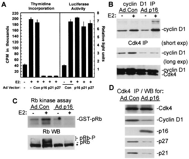
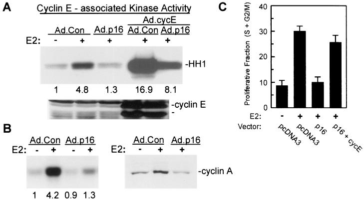
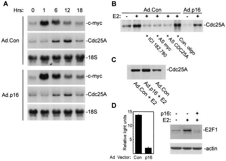
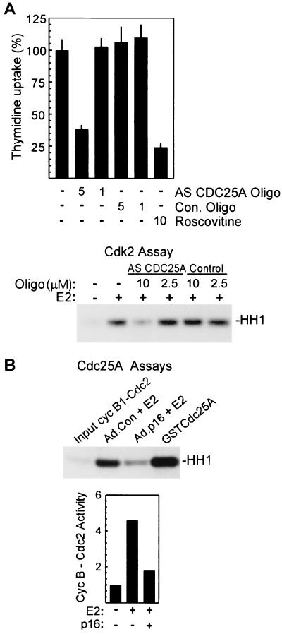
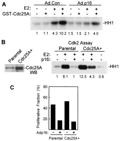
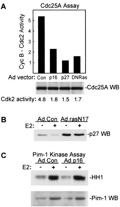
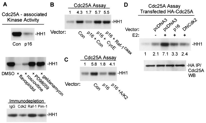
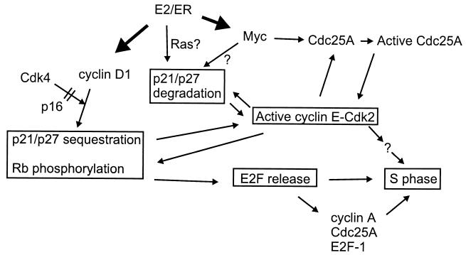
Estrogens induce proliferation of estrogen receptor (ER)-positive MCF-7 breast cancer cells by stimulating G(1)/S transition associated with increased cyclin D1 expression, activation of cyclin-dependent kinases (Cdks), and phosphorylation of the retinoblastoma protein (pRb). We have utilized blockade of cyclin D1-Cdk4 complex formation through adenovirus-mediated expression of p16(INK4a) to demonstrate that estrogen regulates Cdk inhibitor expression and expression of the Cdk-activating phosphatase Cdc25A independent of cyclin D1-Cdk4 function and cell cycle progression. Expression of p16(INK4a) inhibited G(1)/S transition induced in MCF-7 cells by 17-beta-estradiol (E(2)) with associated inhibition of both Cdk4- and Cdk2-associated kinase activities. Inhibition of Cdk2 activity was associated with delayed removal of Cdk-inhibitory activity in early G(1) and decreased cyclin A expression. Cdk-inhibitory activity and expression of both p21(Cip1) and p27(Kip1) was decreased, however, in both control and p16(INK4a)-expressing cells 20 h after estrogen treatment. Expression of Cdc25A mRNA and protein was induced by E(2) in control and p16(INK4a)-expressing MCF-7 cells; however, functional activity of Cdc25A was inhibited in cells expressing p16(INK4a). Inhibition of Cdc25A activity in p16(INK4a)-expressing cells was associated with depressed Cdk2 activity and was reversed in vivo and in vitro by active Cdk2. Transfection of MCF-7 cells with a dominant-negative Cdk2 construct inhibited the E(2)-dependent activation of ectopic Cdc25A. Supporting a role for Cdc25A in estrogen action, antisense CDC25A oligonucleotides inhibited estrogen-induced Cdk2 activation and DNA synthesis. In addition, inactive cyclin E-Cdk2 complexes from p16(INK4a)-expressing, estrogen-treated cells were activated in vitro by treatment with recombinant Cdc25A and in vivo in cells overexpressing Cdc25A. The results demonstrate that functional association of cyclin D1-Cdk4 complexes is required for Cdk2 activation in MCF-7 cells and that Cdk2 activity is, in turn, required for the in vivo activation of Cdc25A. These studies establish Cdc25A as a growth-promoting target of estrogen action and further indicate that estrogens independently regulate multiple components of the cell cycle machinery, including expression of p21(Cip1) and p27(Kip1).


Figures

FIG. 1
FIG. 1
Blockade of estrogen-induced G1/S transition in MCF-7 cells by p16INK4a is associated with inhibition of cyclin D1-Cdk4 complex formation and pRb kinase activity. (A, left panel) Cdk inhibitor effects on E2-induced DNA synthesis. MCF-7 cells were infected with adenoviral vectors for p16INK4a, p21Cip1, p27Kip1, or with control adenovirus (MOI = 50), growth arrested, and treated with 10 nM E2 as indicated. Thymidine incorporation was measured from 20 to 32 h after treatment with E2 and is given as the mean ± standard deviation from four replicates. (A, right panel) Analysis of estrogen-induced transcriptional activity in MCF-7/MVLN cells. MCF-7/MVLN cells were infected with the indicated adenoviral vectors (MOI = 50), growth arrested, and treated with 10 nM E2 for 24 h. Results from the luciferase assay are given as relative light units (mean ± standard deviation from three replicates) based on an arbitrary designation. (B) Analysis of de novo-synthesized cyclin D1 and Cdk4. MCF-7 cells were infected with Ad.Con or Ad.p16 (MOI = 50) followed by growth arrest and treatment with 10 nM E2 as indicated. Cultures were labeled for 2 h with Tran35S label, 4 h after estrogen treatment. Immunoprecipitations (IP) were performed with antibodies to cyclin D1 (upper panel) and Cdk4 (lower panel). Two different exposures are given for Cdk4 precipitates. (C) Assay of Cdk4-associated kinase activity. MCF-7 cells were infected with Ad.p16 and Ad.Con (MOI = 50), growth arrested, and treated for 6 h with 10 nM E2 as indicated. Lysates were assayed for Cdk4-associated pRb kinase activity in an immunocomplex assay with GST-pRb as the substrate. (C, lower panel) Western blotting analysis of total pRb phosphorylation: MCF-7 cultures were infected as indicated and treated for 20 h with E2. Western blot analysis of pRb was performed with whole-cell lysates. Slowly migrating, phosphorylated pRb forms are indicated (pRb-P). Nonspecific bands on Western blots are indicated by an asterisk. (D) Analysis of p21Cip1-p27Kip1 association with Cdk4. MCF-7 cells were infected as indicated, followed by growth arrest and a 10-h treatment with 10 nM E2. Proteins in Cdk4 immunoprecipitates were analyzed by Western blotting (WB) analysis for p16INK4a, cyclin D1, p21Cip1, p27Kip1, and Cdk4.
FIG. 2
FIG. 2
Expression of p16INK4a prevents Cdk2 activation in E2-treated MCF-7 cells. (A) For assay of E2-induced cyclin E-Cdk2 activity, MCF-7 cells were infected with Ad.Con or Ad.p16 (MOI = 50). Additional cultures were coinfected with Ad.cycE as indicated (all MOIs = 50). Cultures were growth arrested and treated with 10 nM E2 for 12 h, and cyclin E-associated histone kinase activity was determined with equal amounts of lysate. The histone band (HH1) is indicated. Relative kinase activities based on densitometry are indicated under the appropriate lanes. Western blot analysis of cyclin E expression in the same lysates is given in the lower panel with identical arrangement of lanes. (B, left panel) Effects of p16INK4a expression on Cdk2-associated kinase activity in MCF-7 cells. MCF-7 cells were infected with the indicated adenoviral vectors, growth arrested, and treated for 20 h with E2. Histone kinase activity was determined with Cdk2 immunoprecipitates. Numbers represent relative kinase activities. (B, right panel) Cyclin A expression in control and p16INK4a-expressing MCF-7 cells was determined by Western blotting of whole-cell lysates from MCF-7 cells infected as indicated and treated for 20 h with E2. (C) Reversal of p16INK4a-mediated G1 arrest by cyclin E. MCF-7/tTA cells were transfected with pEGFPN1 along with control vector (pcDNA3), pBPSTRI-p16 (p16), or pMTcyclin E (cycE). The cultures were growth arrested and treated for 24 h with E2, and the proliferative fraction (S+G2/M) was determined by DNA content analysis of the transfected population as given in Materials and Methods. The results are given as the mean ± standard deviation from three independent experiments.
FIG. 3
FIG. 3
Estradiol downregulates Cdk-inhibitory activity and decreases expression of p21Cip1 and p27Kip1 proteins in MCF-7 cells arrested in G1 by p16INK4a expression. (A) Effects of p16INK4a expression on downregulation of Cdk-inhibitory activity in MCF-7 cells. Cdk-inhibitory activity in Ad.Con- and Ad.p16-infected MCF-7 cells was assayed after treatment of growth-arrested cells with 10 nM E2 for the indicated times. Whole-cell lysates were mixed with Cdk2 immunoprecipitates followed by assay of histone kinase activity. Results are presented in graphic form based on densitometric measurements. (B, left panels) Expression of p21Cip1 and p27Kip1 proteins in MCF-7 cells. Cultures were infected with Ad.p16 and Ad.Con (MOI = 50), growth arrested, and treated with 10 nM E2 for 20 h. Lysates were assayed by Western blotting (WB) for expression of p27Kip1, p21Cip1, p16INK4a, and actin as indicated. (B, right panels) Levels of Cdk2, p21Cip1, and p27Kip1 proteins in cyclin E immunoprecipitates (IP) from cultures treated in the same fashion were analyzed by Western blotting. (C) Proteasomal regulation of Cdk inhibitor levels. Ad.p16-infected MCF-7 cells were growth arrested and treated with 10 nM E2 and the specific proteasome inhibitor MG132 (10 μM) as indicated. Lysates prepared after 20 h were analyzed by Western blotting for p21Cip1, p27Kip1, and actin (upper three panels). In the lower panel, cyclin E immunoprecipitates from cultures treated in the same fashion were analyzed for p27Kip1 and p21Cip1 content by Western blotting. (D) Estrogen regulation of ectopic p27Kip1 expression. MCF-7 cells were infected with Ad.p27, growth arrested, and treated for 20 h with E2. Expression of p27Kip1 was determined by Western blotting of whole-cell lysates. Lysates of uninfected, growth-arrested MCF-7 cells (NV) were included for comparison. (E) Western blot analysis of p27Kip1 (T187A) expression in MCF-7 cells. MCF-7 cells were transfected with the HA-p27-T187A vector, growth arrested, and treated with E2 for 20 h. Expression of the p27Kip1 mutant was determined by Western blot analysis of lysates with anti-HA antibodies.
FIG. 4
FIG. 4
Estradiol induces expression of Cdc25A mRNA and protein in MCF-7 cells. (A) MCF-7 cells were infected with Ad.p16 or Ad.Con (MOI = 50), growth arrested, and treated for the indicated times with 10 nM E2. Total RNA was analyzed for expression of c-myc and Cdc25A mRNA by Northern blotting. Membranes were reprobed for 18S rRNA as a loading control. (B) Cdc25A protein synthesis. Ad.p16- and Ad.Con-infected MCF-7 cultures were growth arrested and treated with 10 nM E2 or 10 nM E2 plus 500 nM ICI 182,780 as indicated. Cultures were labeled with Tran35S-label from 10 to 12 h after treatment. Control (Con.), antisense CDC25A, or c-myc antisense (AS) oligonucleotides (oligo) (10 μM) were added to the culture medium 18 h before treatment and remained in the culture until labeling. Equal amounts of protein lysates were immunoprecipitated with monoclonal anti-Cdc25A (Ab-3) antibodies and analyzed by SDS-PAGE. (C) Western blot analysis of Cdc25A expression. MCF-7 cultures were infected with Ad.Con or Ad.p16, growth arrested, and treated for 20 h with E2 as indicated. Cdc25A was immunoprecipitated with monoclonal anti-Cdc25A (Ab-3) and analyzed by Western blotting with anti-Cdc25A (N15). (D, left panel) E2F-dependent transcriptional activity was measured in lysates of Ad.Con- and Ad.p16-infected cells 30 h after treatment with E2 as given in Materials and Methods. Values are given as relative light units (mean ± standard deviation from three replicates). Values given represent estrogen-induced transcription, with activity in untreated cells subtracted. (D, right side panels) Western blot analysis of E2F-1 and actin expression in whole-cell lysates of MCF-7 cells treated as described above in panel C.
FIG. 5
FIG. 5
Cdc25A is required for estrogen-induced Cdk2 activation and DNA synthesis in MCF-7 cells and is inactive in cells expressing p16INK4a. (A, upper panel) Inhibition of S-phase entry by antisense (AS) CDC25A oligonucleotides (Oligo). DNA synthesis in E2-treated MCF-7 cells was assayed as given above in Fig. 1. Antisense oligonucleotides were added to the culture medium at the indicated concentrations 18 h before estrogen treatment and remained in the culture throughout the assay. Values represent percent thymidine incorporation relative to that in estrogen-treated control (Con.) cultures and are given as the mean ± standard deviation from four replicates. (A, lower panel) Inhibition of Cdk2 activation by antisense CDC25A. MCF-7 cells were treated with antisense CDC25A or control oligonucleotides as described above, and lysates prepared 12 h after E2 treatment were assayed for cyclin E-associated histone kinase activity. (B) Assay of endogenous Cdc25A activity. Cdc25A immunoprecipitates were prepared from MCF-7 cultures treated as indicated, and Cdc25A was eluted from the beads as described in Materials and Methods. The function of eluted Cdc25A was assayed as activation of cyclin B1-Cdc2 complexes measured in a standard histone kinase assay. In lane 4, maximal activation is demonstrated by treatment of complexes with purified recombinant GST-Cdc25A. Results from a separate experiment are presented graphically in the lower portion of panel B, with activity in lysates of untreated, Ad.Con-infected cells taken as 1.
FIG. 6
FIG. 6
Cyclin E-Cdk2 complexes from p16INK4a-expressing MCF-7 cells are activated in vitro and in vivo by Cdc25A. (A) Activation of cyclin E-Cdk2 complexes by Cdc25A in vitro. MCF-7 cells were infected with Ad.Con or Ad.p16, growth arrested, and treated for 20 h with E2 as indicated. Cyclin E immunoprecipitates were incubated with GST-Cdc25A and assayed for histone kinase activity. Relative kinase activities from densitometric measurements are given beneath the figure. (B) Effect of in vivo overexpression of Cdc25A on Cdk2 activity in MCF-7 cells. (Left panel) Cdc25A expression was assayed in parental MCF-7 cells and in cells transduced with retroviral vector for Cdc25A. Western blot analysis of Cdc25A immunoprecipitates from proliferating parental and Cdc25A-overexpressing cells is shown. (Right panel) Cdk2-associated kinase activity was assayed in lysates of parental and Cdc25A-overexpressing MCF-7 cells infected with Ad.Con or Ad.p16. Cultures were growth arrested and treated for 20 h with 10 nM E2 as indicated. Relative activity based on densitometry is provided beneath each lane. (C) Flow cytometric analysis of estrogen-induced S-phase entry in parental and Cdc25A-overexpressing MCF-7 cells is given. Parental and Cdc25A-overexpressing MCF-7 cells were infected with Ad.Con or Ad.p16 as indicated and growth arrested, and the proliferative fraction (S+G2/M) was determined 24 h after E2 treatment by flow cytometric analysis of DNA content.
FIG. 7
FIG. 7
Cdc25A activation in vivo is inhibited by p27Kip1 and by dominant-negative Ras. (A, top panel) Effects of transduction with Ad.p16, Ad.p27, and Ad.RasN17 on generation of Cdc25A activity in MCF-7 cells. MCF-7 cells were infected with Ad.Con, Ad.p16, Ad.RasN17, or Ad.p27 as indicated, and Cdc25A activity was assayed following growth arrest and a 20-h estrogen treatment. Results are presented in graph form with input cyclin B1-Cdc2 activity taken as a value of 1. (A, lower panel) Western blotting (WB) analysis of Cdc25A expression in MCF-7 cells. Cdc25A was immunoprecipitated from 600 μg of the same lysates assayed above. Relative Cdk2 activities assayed in the same lysates are given below the panel based on densitometric analysis. (B) Expression of p27Kip1 was assayed by Western blotting of lysates from MCF-7 cells infected with Ad.Con and Ad.rasN17 vectors following growth arrest and 20-h treatment with estrogen as indicated. (C) Pim-1-associated histone kinase activity (upper panel) was determined as given in Materials and Methods with lysates of control and Ad.p16-infected MCF-7 cells 20 h after E2 treatment. Western blot analysis of Pim-1 protein expression in the same lysates is given in the lower panel.
FIG. 8
FIG. 8
Cdc25A is activated in vivo by Cdk2. (A) Analysis of Cdc25A-associated kinase activity. (A, top panel) Control-infected and Ad.p16-infected MCF-7 cells were growth arrested and treated for 12 h with E2. Lysates were precleared and incubated with GST-Cdc25A-coated beads, and histone kinase activity associating with Cdc25A was measured as given in Materials and Methods. (A, middle panel) The in vitro assay of Cdc25A-associated kinase activity was carried out with the addition of flavopiridol, roscovitine, PD9059, or geldanamycin (all 2.5 μM). DMSO was used as a solvent control. Lysates were from estrogen-treated MCF-7 cells. (A, bottom panel) Immunodepletion analysis of Cdc25A-associated kinase activity is shown. Lysates of estrogen-treated MCF-7 cells were subjected to immunodepletion with the indicated specific antibodies or control goat IgG before incubation with Cdc25A beads and assay. (B) Reversal of Cdc25A inhibition in vivo. MCF-7 cells were infected with Ad.Con and Ad.p16 vectors along with Ad.Con, Ad.cycE, or Ad.Raf-1caax as indicated. Cdc25A activity was assayed following growth arrest and a 20-h treatment with E2. (C) Reactivation of Cdc25A in vitro. Cdc25A immunoprecipitates from lysates of estrogen-treated Ad.p16-infected MCF-7 cells prepared as described above were incubated in vitro with soluble, active cyclin A-Cdk2 complexes as described in Materials and Methods and assayed for activation of cyclin B1-Cdc2 complexes. Activity in Cdc25A immunoprecipitates from Ad.Con-infected cells is given for comparison. In panels C and D, relative activity based on densitometry is given above the respective lanes. (D) Activation and inhibition of ectopic Cdc25A activity. MCF-7/tTA cells were transfected with pBI-HACdc25A along with control plasmid (pcDNA3), pBPSTRI-p16 (p16), or dominant-negative Cdk2 vector (DNCdk2). Following growth arrest and E2 treatment (20 h), Cdc25A activity was assayed in anti-HA immunoprecipitates as given in Materials and Methods. Relative activity is given under each lane. In the lower panel, equal expression of HA-Cdc25A was verified by anti-Cdc25A immunoblot analysis of anti-HA immunoprecipitates prepared from the same lysates.
FIG. 9
FIG. 9
Schematic model of estrogen-mediated promotion of S-phase entry. Transcriptional activation of Myc and cyclin D1 expression in early G1 (dark arrows) facilitates cyclin E-Cdk2 activation in mid-to-late G1- and S-phase entry. Expression of cyclin D1 and complex formation with Cdk4 leads to sequestration of p21Cip1 and p27Kip1 Cdk inhibitors and initiates phosphorylation and inactivation of pocket proteins, including pRb. Conversely, expression of p16INK4a prevents cyclin D1-Cdk4 association, delays removal of Cdk-inhibitory activity, and effectively inhibits pocket protein phosphorylation and release of E2F transcription factors. Estrogen downregulates expression of both p21Cip1 and p27Kip1 independent of cyclin D1-Cdk4 function and at least in part through the proteasome. It is not yet clear to what extent this is related to estrogen-induced Myc expression, Ras activation, or induction of as-yet-unidentified mediators. Myc further participates in cyclin E-Cdk2 activation by eliciting Cdc25A expression. As suggested in earlier studies, full activation of both Cdc25A and Cdk2 hinges upon interaction and mutual activation between these two regulators. Ultimately, active cyclin E-Cdk2 likely elicits S-phase entry both through contribution to pocket protein phosphorylation and E2F release and through phosphorylation of additional, unknown mediators of S-phase entry. Upon inactivation of pocket proteins, derepression at E2F-dependent promoters and consequent induction of cyclin A, Cdc25A, and E2F-1 provides further reinforcement for G1/S transition and progression through the S phase.

References

    1. Altucci L, Addeo R, Cicatiello L, Dauvois S, Parker M G, Truss M, Beato M, Sica V, Bresciani F, Weisz A. 17beta-Estradiol induces cyclin D1 gene transcription, p36D1–p34cdk4 complex activation and p105Rb phosphorylation during mitogenic stimulation of G(1)-arrested human breast cancer cells. Oncogene. 1996;12:2315–2324. - PubMed
    1. Altucci L, Addeo R, Cicatiello L, Germano D, Pacilio C, Battista T, Cancemi M, Petrizzi V B, Bresciani F, Weisz A. Estrogen induces early and timed activation of cyclin-dependent kinases 4, 5, and 6 and increases cyclin messenger ribonucleic acid expression in rat uterus. Endocrinology. 1997;138:978–984. - PubMed
    1. Blomberg I, Hoffmann I. Ectopic expression of Cdc25A accelerates the G1/S transition and leads to premature activation of cyclin E- and cyclin A-dependent kinases. Mol Cell Biol. 1999;19:6183–6194. - PMC - PubMed
    1. Brenner A J, Stampfer M R, Aldaz C M. Increased p16 expression with first senescence arrest in human mammary epithelial cells and extended growth capacity with p16 inactivation. Oncogene. 1998;17:199–205. - PubMed
    1. Cangi M G, Cukor B, Suong P, Signoretti S, Moreira G, Jr, Ranashinge M, Cady B, Pagano M, Loda M. Role of the Cdc25A phosphatase in human breast cancer. J Clin Investig. 2000;106:753–761. - PMC - PubMed

Publication types

MeSH terms

Substances