. 2001 Feb;21(3):902-15.
doi: 10.1128/MCB.21.3.902-915.2001.

Gas6 induces growth, beta-catenin stabilization, and T-cell factor transcriptional activation in contact-inhibited C57 mammary cells

Affiliations

Gas6 induces growth, beta-catenin stabilization, and T-cell factor transcriptional activation in contact-inhibited C57 mammary cells

S Goruppi et al. Mol Cell Biol. 2001 Feb.

Abstract

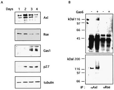
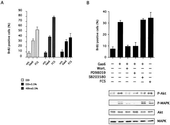
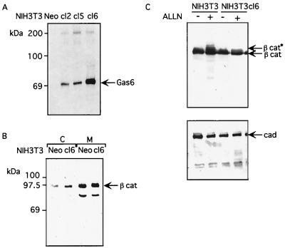
Gas6 is a growth factor related to protein S that was identified as the ligand for the Axl receptor tyrosine kinase (RTK) family. In this study, we show that Gas6 induces a growth response in a cultured mammalian mammary cell line, C57MG. The presence of Gas6 in the medium induces growth after confluence and similarly causes cell cycle reentry of density-inhibited C57MG cells. We show that Axl RTK but not Rse is efficiently activated by Gas6 in density-inhibited C57MG cells. We have analyzed the signaling required for the Gas6 proliferative effect and found a requirement for PI3K-, S6K-, and Ras-activated pathways. We also demonstrate that Gas6 activates Akt and concomitantly inhibits GSK3 activity in a wortmannin-dependent manner. Interestingly, Gas6 induces up-regulation of cytosolic beta-catenin, while membrane-associated beta-catenin remains unaffected. Stabilization of beta-catenin in C57MG cells is correlated with activation of a T-cell factor (TCF)-responsive transcriptional element. We thus provide evidence that Gas6 is mitogenic and induces beta-catenin proto-oncogene stabilization and subsequent TCF/Lef transcriptional activation in a mammary system. These results suggest that Gas6-Axl interaction, through stabilization of beta-catenin, may have a role in mammary development and/or be involved in the progression of mammary tumors.


Figures

FIG. 1
FIG. 1
Gas6 is a growth factor for C57MG cells. (A) Analysis of Gas6 growth-promoting effect on different cell lines. Cells representing REF, rat SMC, IMR90, SA0S, MCF7, NIH 3T3, and C57MG were seeded at 104 cells per cm2 in 6-cm petri dishes. After 24 h from plating in 10% FCS, 400 ng of recombinant Gas6/ml was added to the medium for 3 days. After this time, MTT was added to the medium to monitor the cell number for 4 h. All the experiments were performed in duplicate at least three times. The lower panel shows the morphology of Gemsa-stained Gas6-treated C57MG cells. (B) Analysis of C57MG growth rate in the presence of Gas6. C57MG cells were similarly seeded in 6-cm-diameter petri dishes and labeled with MTT on days 1, 2, 3, and 4 after seeding in the presence or absence of 400 ng of Gas6/ml in the culture medium. (C) Analysis of Gas6-induced increase of C57MG cell number. Cells were seeded as described above at half concentration, and the increase in cell number was evaluated after 1, 2, 3, and 4 days of culture in the presence or absence of 400 ng of Gas6/ml in complete medium. After trypsinization the cells were counted using a bright-light hemacytometer Neubauer chamber (Sigma). All the experiments shown were carried out in duplicate.
FIG. 2
FIG. 2
Gas6 activates Axl RTK in C57MG mammary cells. (A) Analysis of Axl and Rse RTK protein expression and regulation during the assessment of density growth arrest. C57MG cells were seeded as described for Fig. 1A, and the cellular lysates were prepared after 1, 2, 3, and 4 days by adding Laemmli loading buffer directly to the petri dish. Equal amounts of total proteins, as determined using the Bio-Rad assay, were loaded on SDS-PAGE gels and were blotted to nitrocellulose membranes. Western blots were decorated separately by using rabbit affinity-purified polyclonal antibodies for Axl, Gas1, and p27; a goat polyclonal antibody for Rse; or a monoclonal antibody for tubulin. The complexes were evidenced by incubation with a secondary peroxidase-conjugated antibody and the ECL method. (B) Gas6 activates Axl RTK. Western blot analysis with antiphosphotyrosine antibodies of Axl and Rse RTKs immunoprecipitated (IP) from density-inhibited C57MG cells. Both antibodies recognizing the C-terminal region (upper panel) and the N-terminal region of Axl and Rse RTKs were used in this analysis. The results shown here are representative of several experiments. −, absence of Gas6; +, presence of Gas6.
FIG. 3
FIG. 3
Gas6 induces proliferation of density-inhibited C57MG cells. Analysis of S-phase induction in C57MG cells undergoing density-dependent inhibition. C57MG cells were plated 104 per cm2 in a petri dish and were allowed to achieve density-dependent inhibition in the presence of serum (10% FCS) for 3 days. Unt, untreated. The ability to induce DNA synthesis was tested by adding 400 ng of Gas6/ml to the starvation medium together with 50 μM BrdU to monitor S-phase entry. After 20 h, cells were fixed with paraformaldehyde and processed for immunofluorescence with anti-BrdU monoclonal antibodies. Secondary antibody was anti-mouse and FITC conjugated. Total nuclei were stained with PI. When wortmannin (Wort) (100 nM), LY294002 (10 μM), rapamycin (20 ng/ml), SB203580 (20 μM), or PD98059 (20 μM) were used, each was added to the starvation medium (for 30, 30, 30, 60, and 60 min respectively) before addition of Gas6. No significant effect on 10% FCS-induced cell cycle reentry was found for these drugs (<5%). The percentage of BrdU incorporation was calculated as the ratio of nuclei positive for FITC (BrdU) and total nuclei (P.I.). The mean of four independent experiments carried out in duplicate together with the standard deviation of the mean is reported here. Immunofluorescence analysis was performed using an Axiovert 35 microscope (Zeiss), and images were obtained with a confocal laser scan microscope (Zeiss).
FIG. 4
FIG. 4
Gas6 addition activates Akt and inhibits GSK3 in C57MG cells. (A) Gas6 induces the activation of Akt. Western blot analysis with antibodies specific to the threonine 308 of Akt (Akt-P) was performed on total cellular lysates from density-inhibited C57MG stimulated (+) or not (−) for 20 min with 400 ng of Gas6/ml. An identical blot was decorated with anti-Akt to monitor the total amount of the kinase in the extracts (lower panel). In separate experiments, cells were treated with 100 nM wortmannin (Wort.) or 20 μM PD98059 before addition of Gas6 to the medium. (B) Gas6 induces the inactivation of GSK3. The activation state of endogenous GSK3 α and β isoforms after Gas6 addition to C57MG cells undergoing DDI was determined by Western blotting with specific antibodies recognizing only the phosphorylated, inactive form of the kinase. Cells were stimulated for 0, 10, and 20 min before Western blot analysis. An identical Western blot was stained with anti-GSK3 antibody to monitor the total amount of the kinase in the samples (lower panel). (C) Adding wortmannin prevents Gas6-dependent GSK3 regulation. Western blot analysis of GSK3 regulation was performed as described for panel B. Total cellular lysates from density-inhibited C57MG were stimulated (+) or not (−) for 20 min with 400 ng of Gas6/ml; in separate experiments, cells were treated with 100 nM wortmannin (Wort.) or 20 μM PD98059 before addition of Gas6 to the medium.
FIG. 5
FIG. 5
Gas6 does not act as a growth-potentiating factor for C57MG cells. (A) Gas6-induced proliferation is maintained in serum-starved C57MG cells. C57MG cells were plated at 104 per cm2 in 10% FCS–DMEM and were allowed to achieve DDI in the presence of serum for 3 days. Then cells were either maintained in the same medium (DDI), or the medium was changed to 0.5% FCS (DDI + 0.5%) and further incubated for 24 h. Serum-starved cells (48 h; 0.5%) were obtained by changing the medium to 0.5% FCS on the day after seeding and were incubated for 2 days. The ability to induce DNA synthesis was tested by adding to the culture medium 400 ng of Gas6/ml together with 50 μM BrdU for 20 h. An immunofluorescence assay was performed as described in Materials and Methods. Unt, untreated. (B) Analysis of Gas6-induced downstream signaling on serum-starved DDI C57MG cells. DDI + 0.5% cells were prepared and treated as described for panel A. When wortmannin (100 nM), PD98059 (20 μM), or SB203580 (20 μM) was used, each was added to the culture medium for 1 h before stimulation with Gas6 (400 ng/ml). Entry into the S phase was monitored after 20 h as described above. An analysis of Akt and MAPK activation was performed: Western blotting with antibodies specific to Thr308 of Akt (P-Akt) or raised against Thr202/Tyr204 of p44/42 MAPK (P-MAPK). C57MG cells were prepared as above, and 1 h before Gas6 stimulation, the medium was changed to serum-free DMEM. In separate experiments, cells were treated with wortmannin (100 nM), PD98059 (20 μM), or SB203580 (20 μM) before Gas6 stimulation. Parallel membranes were decorated with anti-Akt or anti-MAPK to monitor the total amount of kinase in the lysate. Cells were separately stimulated with 10% FCS and used as positive control (FCS). −, absence; +, presence.
FIG. 6
FIG. 6
Gas6 interferes with β-catenin protein stability in C57MG cells. (A) Replicate cultures of control cells (−) or C57MG cells stimulated overnight with Gas6 (+) were pulse labeled (0 h) with [35S]methionine for 30 min and chased in the absence of the label for 1 and 2 h. At each time point, cellular extracts were prepared and immunoprecipitated with antibodies specific either to β-catenin (upper panel) or Axl RTK (lower panel). The immunocomplexes were analyzed by SDS-PAGE followed by fluorography. (B) Western blot analysis of β-catenin association. Confluent C57MG cells undergoing density-dependent inhibition were stimulated (Gas6) or not (−) for 30 min with 400 ng of Gas6/ml. After this time, equivalent amounts of protein extracts were immunoprecipitated with antibodies specific to either cadherins (IP:cad), α-catenin (IP αcat), or GSK3 (IP:GSK3). Immunocomplexes were resolved by SDS–10% PAGE and were blotted to nitrocellulose membranes. Western blotting was carried out with specific antibodies to β-catenin. The position of β-catenin is indicated by arrows.
FIG. 7
FIG. 7
Gas6 induces TCF/Lef-dependent transcription in C57MG mammary cells. (A) Gas6 induces TCF-dependent transcriptional activity. Replicate petri dishes of C57MG cells were transfected with either wt (pTOPCAT) or mutant (pFOPCAT) TCF/Lef reporter constructs (5 μg) as described in Materials and Methods. A separate petri dish was transfected simultaneously with pTOPCAT and wt β-catenin (4 μg) (β-cat) as positive control. The day after in separate experiments, either 400 ng of Gas6/ml or 100 ng of PDGFbb/ml was added to the cell medium and CAT activities were measured after 2 more days. (B) Wortmannin blocks Gas6-induced TCF-dependent transcription activation. Replicate petri dishes of C57MG cells were transfected with wild-type (pTOPCAT) TCF/Lef reporter constructs (5 μg). A separate petri dish was transfected simultaneously with pTOPCAT and wild-type β-catenin (4 μg) (β-cat) as positive control. One day later in separate experiments, either 400 ng of Gas6/ml or Gas6 and 1 μM wortmannin (Wort) were added to the cell medium and CAT activities were measured after 2 more days. The thin layer-chromatography in this figure is representative of three separate experiments.
FIG. 8
FIG. 8
Stable Gas6-expressing NIH 3T3 fibroblasts have higher cytosolic β-catenin protein levels. (A) Analysis of Gas6 expression in stable cell lines. NIH 3T3 cells were cotransfected with pGDSV7gas6 and a plasmid carrying the neomycin phosphotransferase cDNA. Relative levels of Gas6 expression were assessed by immunoprecipitating the supernatant of [35S]methionine-labeled clones (cl) with specific anti-Gas6 antibodies (56). A clone transfected only with neomycin gene resistance (Neo) was used as control. The immunocomplexes were analyzed by SDS–10% PAGE followed by fluorography. (B) Gas6 induces an increase in cytosolic β-catenin protein in NIH 3T3 cells. A Western blot analysis of steady-state β-catenin protein levels in cytosolic (C) and membranous (M) protein fractions of control neomycin-expressing (Neo) and Gas6-expressing (cl6) NIH 3T3 cells was performed. Cellular extracts were prepared and fractionated, as described in Materials and Methods. Equal amounts of protein fractions were separated by SDS–10% PAGE, transferred to nitrocellulose membranes, and analyzed by Western blotting using anti-β-catenin antibody. (C) Ubiquitination of β-catenin is reduced in stable Gas6-expressing NIH 3T3 fibroblasts. Cellular lysates of control (NIH3T3) or Gas6-expressing (NIH3T3cl6) NIH 3T3 cells were treated (+) or not (−) with 25 μM ALLN for 4 h before lysis. Equal amounts of total cellular proteins were analyzed by Western blotting using anti-β-catenin antibody (upper panel) or anti-cadherin as a control (lower panel). Addition of ALLN proteosome inhibitor to cells generates higher-molecular-weight forms of β-catenin (β cat∗) but not of cadherin (cad) (1).

Similar articles

Cited by

References

    1. Aberle H, Bauer A, Stappert J, Kispert A, Kemler R. beta-catenin is a target for the ubiquitin-proteasome pathway. EMBO J. 1997;16:3797–3804. - PMC - PubMed
    1. Alessi D R, Cuenda A, Cohen P, Dudley D T, Saltiel A R. PD098059 is a specific inhibitor of the activation of mitogen-activated protein kinase kinase in vitro e in vivo. J Biol Chem. 1995;270:27489–27494. - PubMed
    1. Allen M P, Zeng C, Schneider K, Xiong X, Meintzer M K, Bellosta P, Basilico C, Varnum B, Heidenreich K A, Wierman M E. Growth arrest-specific gene 6 (Gas6)/adhesion related kinase (Ark) signaling promotes gonadotropin-releasing hormone neuronal survival via extracellular signal-regulated kinase (ERK) and Akt. Mol Endocrinol. 1999;13:191–201. - PubMed
    1. Barker N, Morin P J, Clevers H. The Yin-Yang of TCF/beta-catenin signaling. Adv Cancer Res. 2000;77:1–24. - PubMed
    1. Barnard D, Sun H, Baker L, Marshall M S. In vitro inhibition of Ras-Raf association by short peptides. Biochem Biophys Res Commun. 1998;247:176–180. - PubMed

Publication types

MeSH terms

Substances