. 2001 Jan;125(1):252-65.
doi: 10.1104/pp.125.1.252.

Nodal endoplasmic reticulum, a specialized form of endoplasmic reticulum found in gravity-sensing root tip columella cells

Collaborators, Affiliations

Nodal endoplasmic reticulum, a specialized form of endoplasmic reticulum found in gravity-sensing root tip columella cells

H Q Zheng et al. Plant Physiol. 2001 Jan.

Abstract

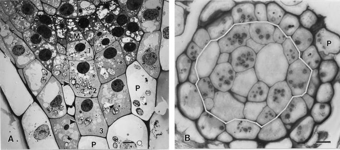
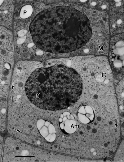
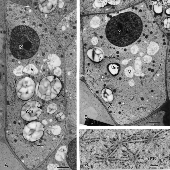
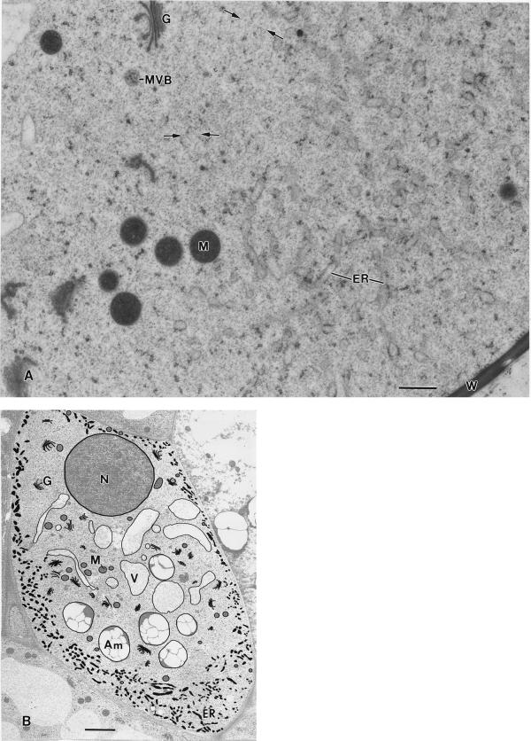
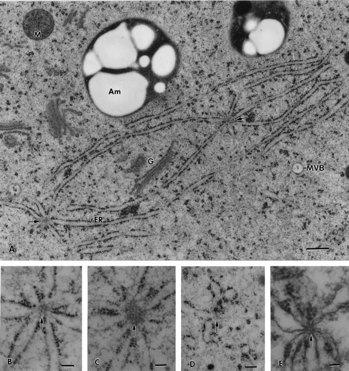
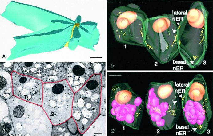
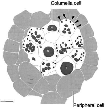
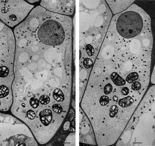
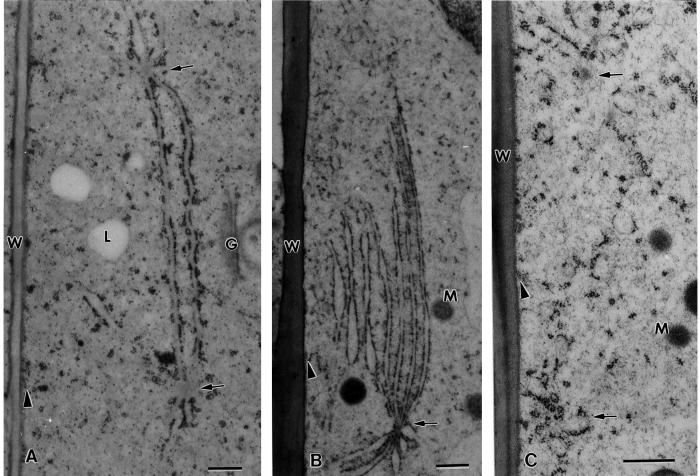
The endoplasmic reticulum (ER) of columella root cap cells has been postulated to play a role in gravity sensing. We have re-examined the ultrastructure of columella cells in tobacco (Nicotiana tabacum) root tips preserved by high-pressure freezing/freeze-substitution techniques to gain more precise information about the organization of the ER in such cells. The most notable findings are: the identification of a specialized form of ER, termed "nodal ER," which is found exclusively in columella cells; the demonstration that the bulk of the ER is organized in the form of a tubular network that is confined to a peripheral layer under the plasma membrane; and the discovery that this ER-rich peripheral region excludes Golgi stacks, vacuoles, and amyloplasts but not mitochondria. Nodal ER domains consist of an approximately 100-nm-diameter central rod composed of oblong subunits to which usually seven sheets of rough ER are attached along their margins. These domains form patches at the interface between the peripheral ER network and the ER-free central region of the cells, and they occupy defined positions within central and flanking columella cells. Over one-half of the nodal ER domains are located along the outer tangential walls of the flanking cells. Cytochalasin D and latrunculin A cause an increase in size and a decrease in numbers of nodal ER domains. We postulate that the nodal ER membranes locally modulate the gravisensing signals produced by the sedimenting amyloplasts, and that the confinement of all ER membranes to the cell periphery serves to enhance the sedimentability of the amyloplasts in the central region of columella cells.


Figures

Figure 1
Figure 1
Longitudinal (A) and cross (B) sections through 5-d-old tobacco root tips grown vertically but placed horizontally for approximately 2 min during the mounting of the samples for high pressure freezing. A, Electron micrographs depicting a meristematic (M) columella cell initial, and three stories of derived columella cells (nos. 1, 2, and 3). Most of the columella cell amyloplasts (arrowheads) appear sedimented toward the lower, distal cell wall. B, Light micrograph of a root tip cross section at the level of the second tier columella cells (cells demarcated by the white line). Due to the offset organization of the columella cells (see asterisks), the section includes the amyloplast-containing layer (arrowheads) of some cells but not others. The columella cells are surrounded by one or two layers of vacuolated peripheral (P) cells. Bar = 5 μm.
Figure 2
Figure 2
Meristematic (M) columella cell initial and a derived first story columella (C) cell. The arrows point to nodal ER domains in a columella cell. N, Nucleus; Am, amyloplast. Bar = 2 μm.
Figure 3
Figure 3
Electron micrographs of a flanking file (A) and a central file (B) columella cell of a tobacco root tip. In the flanking file cell, nodal ER membranes (small arrows) are seen along the external lateral wall and in the basal region, whereas in the central file cell the nodal ER membranes are seen only adjacent to the lateral walls. All of the nodal ER domains are positioned at the interface between the ER-rich cell cortex and the amyloplast-containing central region of the cells. Note how in the flanking cell the nodal ER regions form barriers that prevent the adjacent amyloplasts (Am) from approaching the plasma membrane. In contrast, where the tubular ER in the cell cortex is not shielded by nodal ER membranes the amyloplasts can get much closer to the plasma membrane (white arrows). C, Set of closely spaced and interconnected nodal ER domains from the basal region of a flanking file cell. rER, Rough ER; N, nucleus. A and B, bar = 2 μm; C, bar = 0.2 μm.
Figure 4
Figure 4
A, Electron micrograph of the interface region between the ER-rich cortical and the ER-devoid central region of a columella cell. The ER is organized in the form of interconnected 100- to 150-nm diameter tubules that carry small patches of polysomes. The spaces between the ER tubules are filled by a cytosolic matrix that seamlessly blends with the cytosolic matrix in the central region. The scarcity of organelles in the central region is striking, and in some places (small arrows) thin, actin-like filaments can be detected (see also Fig. 3 B). MVB, Multivesicular body. B, Micrograph of a longitudinal/slightly tangential section through a tobacco flanking file columella cell in which the major membranous organelles have been traced to highlight their distribution and particularly the distribution of the ER in the cell cortex. Note that the Golgi stacks (G), vacuoles (V), and amyloplasts (Am) are all confined to the central ER-devoid region of the cells and that only some mitochondria (M) have penetrated the tubular ER network region in the cell cortex. A, Bar = 0.5 μm; B, bar = 2 μm.
Figure 5
Figure 5
Higher magnification micrographs of nodal ER membrane domains. A, Two nodal ER domains from the basal region of a flanking file cell that are interconnected by two shared ER cisternae. All of the ER cisternae that appear attached to the nodal rods (arrows) are typical sheet-like, rough ER (rER) membranes. B through E, Effects of drugs and fixatives on the structural organization of the nodal rods. B, Control cell nodal ER domain in which the nodal rod appears composed of a small number of oblong subunits. C, Nodal ER from a root tip exposed to 40 μm cytochalasin D for 1 h to disrupt actin filaments. Note the increased diameter of the nodal rod and the oblong shape of the subunits that form the rod. D, Likely former nodal ER domain from a root tip sample treated with 1 μm propyzamide, a microtubule disrupting drug, for 1 h. The central rod of the nodal ER domain appears completely disrupted. E, Nodal ER domain as seen in a root tip preserved by chemical fixation (2% [v/v] glutaraldehyde, 1% [v/v] OsO4 with 0.8% [v/v] potassium ferricyanide). The central rod structure is greatly reduced in diameter and resembles a cross-sectioned microtubule. G, Golgi; M, mitochondrion; MVB, multivesicular body. A, Bar = 0.5 μm; B through E, bar = 0.1 μm.
Figure 6
Figure 6
A, Model of a nodal ER domain based on six 70-nm-thick serial sections. The yellow structure corresponds to the approximately 100-nm-diameter central rod element and the blue structures to the rough ER cisternae that are attached along their margins to the central rod. B, One of 198 serial 100-nm-thick serial sections used for the three-dimensional reconstruction of the three columella cells depicted in Figure 6, C and D. The red lines highlight the three reconstructed cells. Cell 1 corresponds to a very young second tier flanking file cell and contains only a limited number of nodal ER domains; cell 2 corresponds to a partially expanded, central file second tier cell, and cell 3 to a nearly fully expanded, flanking file second tier cell. C, Model of the three cells outlined in B. The cell walls are displayed in light green, the nuclei are colored orange, and the nodal rods correspond to the yellow lines. Note that most of the lateral nodal ER domains are organized into patches, most of which are found near the equatorial regions of the cells. Only in the more mature flanking cells are basal nodal ER domains seen. D, Model in which the three cells of C are shown as separate units. The cells have also been rotated to optimize the viewing of the nodal ER domain patches. The purple structures correspond to amyloplasts, which are not sedimented due to the root tip manipulations prior to freezing. A, Bar = 0.5 μm; B, bar = 3 μm; C and D, bar = 5 μm.
Figure 7
Figure 7
Reconstruction of a 0.2-μm-thick cross-section at the level of the second tier columella cells of a tobacco root cap. The columella cells are shown in white and the surrounding peripheral cells in light gray. Due to the offset type of arrangement of the columella cells, some of the cells appear cross-sectioned at the level of their nuclei (N), whereas others are cross-sectioned at the level of their amyloplasts (Am; see also Fig. 1B). The sites of nodal ER domains are indicated with stars. Note that the majority of the nodal ER domains are located along the external periclinal walls of the outer flanking file cells (arrows). Bar = 5 μm.
Figure 8
Figure 8
Effects of drugs that disrupt microfilaments (A) and microtubules (B) on the distribution and organization of nodal ER domains (arrows) in flanking file columella cells. In A, the 1-h treatment with 1 μm latrunculin A is seen to lead to much larger nodal ER domains (see also Figs. 5C and 9B) along the lateral walls, but the number of these domains decreases (data not shown). Note also the proliferation of rough ER membrane sheets (asterisk) between the nucleus and the adjacent upper cell wall. The cell in B was treated with 10 μm propyzamide for 1 h. Only small fragments of what might have been former nodal ER domains (arrows) are seen. N, Nucleus; Am, amyloplast; V, vacuole. Higher magnification views of the nodal ER domains of these samples are shown in Figure 9. Bars = 2 μm.
Figure 9
Figure 9
Higher magnification views of nodal ER domains (arrows) of a control cell (A), a cell treated with 1 μm latrunculin A for 1 h (B), and a cell treated with 10 μm propyzamide for 1 h (C). Note the greatly expanded rough ER membrane sheets associated with the nodal ER domain in the latrunculin A sample and the loss of nodal ER domains in the propyzamide-treated specimen. W, Cell wall; G, Golgi; M, mitochondrion; L, lipid body; arrowheads, cortical microtubules. Compare with Figure 8. Bar = 0.5 μm.

Similar articles

Cited by

References

    1. Baluska F, Kreibaum A, Vitha S, Parker JS, Barlow PW, Sievers A. Central root cap cells are depleted of endoplasmic microtubules and actin microfilament bundles: implications for their role as gravity-sensing statocytes. Protoplasma. 1997;196:212–223. - PubMed
    1. Barlow PW. The root cap. In: Torrey J, Clarkson DT, editors. The Development and Function of Roots. London: Academic Press; 1975. pp. 21–54.
    1. Barlow PW, Hawes CR, Horne JC. Structure of amyloplasts and endoplasmic reticulum in the root caps of Lepidium sativum and Zea mays observed after selective membrane staining and by high-voltage electron microscopy. Planta. 1984;160:363–371. - PubMed
    1. Blancaflor EB, Fasano JM, Gilroy S. Mapping the functional roles of cap cells in the response of Arabidopsis primary roots to gravity. Plant Physiol. 1998;116:213–222. - PMC - PubMed
    1. Buckley IK. Studies in fixation for electron microscopy using cultures cells. Lab Invest. 1973;29:398–410. - PubMed

Publication types

MeSH terms

LinkOut - more resources