

N-acetylcysteine attenuates TNF-alpha-induced p38 MAP kinase activation and p38 MAP kinase-mediated IL-8 production by human pulmonary vascular endothelial cells
- PMID: 11156586
- PMCID: PMC1572545
- DOI: 10.1038/sj.bjp.0703787
N-acetylcysteine attenuates TNF-alpha-induced p38 MAP kinase activation and p38 MAP kinase-mediated IL-8 production by human pulmonary vascular endothelial cells
Abstract
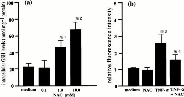
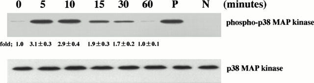
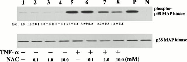
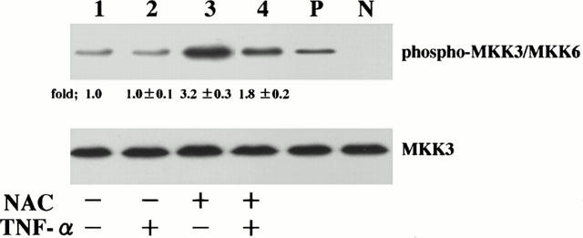
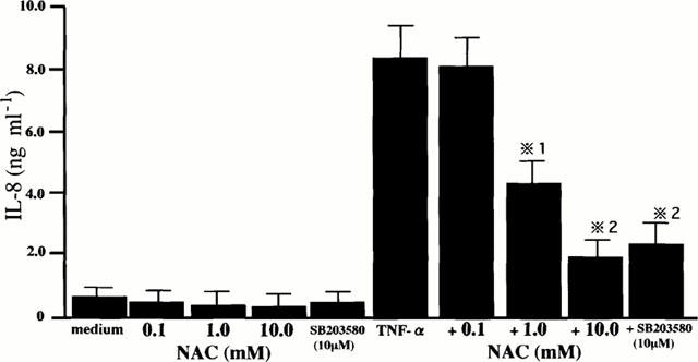
1. We have previously shown that tumour necrosis factor-alpha (TNF-alpha) activates p38 mitogen-activated protein (MAP) kinase to produce interleukin-8 (IL-8) by human pulmonary vascular endothelial cells. Reactive oxygen species (ROS) including H(2)O(2) generated by TNF-alpha can act as signalling intermediates for cytokine induction; therefore, scavenging ROS by anti-oxidants is important for the regulation of cytokine production. However, the effect of N-acetylcysteine (NAC), which acts as a precursor of glutathione (GSH) synthesis, on TNF-alpha-induced activation of p38 MAP kinase pathway and p38 MAP kinase-mediated IL-8 production by human pulmonary vascular endothelial cells has not been determined. To clarify these issues, we examined the effect of NAC on TNF-alpha-induced activation of p38 MAP kinase, MAP kinase kinase (MKK) 3 and MKK6 which are upstream regulators of p38 MAP kinase, and p38 MAP kinase-mediated IL-8 production. 2. Human pulmonary vascular endothelial cells that had been preincubated with NAC were stimulated with TNF-alpha and then the activation of p38 MAP kinase and MKK3/MKK6 in the cells and IL-8 concentrations in the culture supernatants were determined. 3. Intracellular GSH levels increased in NAC-treated cells. 4. NAC attenuated TNF-alpha-induced activation of p38 MAP kinase and MKK3/MKK6. 5. NAC attenuated p38 MAP kinase-mediated IL-8 production by TNF-alpha-stimulated cells. 6. These results indicate that the cellular reduction and oxidation (redox) regulated by intracellular GSH is critical for TNF-alpha-induced activation of p38 MAP kinase pathway and p38 MAP kinase-mediated IL-8 production by human pulmonary vascular endothelial cells, and we emphasize that anti-oxidant therapy is an important strategy for the treatment of acute lung injury.
Figures
Similar articles
-
Intracellular glutathione regulates tumour necrosis factor-alpha-induced p38 MAP kinase activation and RANTES production by human bronchial epithelial cells.Clin Exp Allergy. 2001 Jan;31(1):144-51. Clin Exp Allergy. 2001. PMID: 11167962
-
p38 Mitogen-activated protein kinase regulates IL-8 expression in human pulmonary vascular endothelial cells.Eur Respir J. 1999 Jun;13(6):1357-64. Eur Respir J. 1999. PMID: 10445612
-
Regulation by intracellular glutathione of TNF-alpha-induced p38 MAP kinase activation and RANTES production by human pulmonary vascular endothelial cells.Allergy. 2000 May;55(5):463-9. doi: 10.1034/j.1398-9995.2000.00455.x. Allergy. 2000. PMID: 10843427
-
Modulation of protein kinase activity and gene expression by reactive oxygen species and their role in vascular physiology and pathophysiology.Arterioscler Thromb Vasc Biol. 2000 Oct;20(10):2175-83. doi: 10.1161/01.atv.20.10.2175. Arterioscler Thromb Vasc Biol. 2000. PMID: 11031201 Review.
-
Molecular mechanisms of N-acetylcysteine actions.Cell Mol Life Sci. 2003 Jan;60(1):6-20. doi: 10.1007/s000180300001. Cell Mol Life Sci. 2003. PMID: 12613655 Free PMC article. Review.
Cited by
-
TNF-α-Induced cPLA2 Expression via NADPH Oxidase/Reactive Oxygen Species-Dependent NF-κB Cascade on Human Pulmonary Alveolar Epithelial Cells.Front Pharmacol. 2016 Nov 25;7:447. doi: 10.3389/fphar.2016.00447. eCollection 2016. Front Pharmacol. 2016. PMID: 27932980 Free PMC article.
-
Pulmonary endothelium in acute lung injury: from basic science to the critically ill.Intensive Care Med. 2004 Sep;30(9):1702-14. doi: 10.1007/s00134-004-2370-x. Epub 2004 Jul 16. Intensive Care Med. 2004. PMID: 15258728 Review.
-
Evaluating the Efficacy of L-N-acetylcysteine and Dexamethasone in Combination to Provide Otoprotection for Electrode Insertion Trauma.J Clin Med. 2020 Mar 6;9(3):716. doi: 10.3390/jcm9030716. J Clin Med. 2020. PMID: 32155788 Free PMC article.
-
Redox activation of DUSP4 by N-acetylcysteine protects endothelial cells from Cd²⁺-induced apoptosis.Free Radic Biol Med. 2014 Sep;74:188-199. doi: 10.1016/j.freeradbiomed.2014.06.016. Epub 2014 Jun 26. Free Radic Biol Med. 2014. PMID: 24973647 Free PMC article.
-
Advances in the Use of N-Acetylcysteine in Chronic Respiratory Diseases.Antioxidants (Basel). 2023 Sep 2;12(9):1713. doi: 10.3390/antiox12091713. Antioxidants (Basel). 2023. PMID: 37760016 Free PMC article. Review.
References
-
- ANDERSON M.E. Determination of glutathione and glutathione disulfide in biological samples. Meth. Enzymol. 1985;113:548–555. - PubMed
-
- CHABOT F., MITCHELL J.A., CUTERIDGE J.M.C., EVANS T.W. Reactive oxygen species in acute lung injury. Eur. Respir. J. 1998;11:745–757. - PubMed
-
- CLERK A., FULLER S.J., MICHAEL A., SUGDEN P.H. Stimulation of ‘stress-regulated' mitogen-activated protein kinases (stress-activated protein kinases/c-jun N-terminal kinases and p38-mitogen-activated protein kinase) in perfused rat hearts by oxidative and other stresses. J. Biol. Chem. 1998;273:7228–7234. - PubMed
-
- COTGREAVE I., MOLDEUS P., SCHUPPE I. The metabolism of N-acetylcysteine by human endothelial cells. Biochem. Pharmacol. 1991;42:13–16. - PubMed
-
- DAVIS R.J. MAPKs: new JNK expand the group. Trends Biochem. Sci. 1994;19:470–473. - PubMed
Publication types
MeSH terms
Substances
LinkOut - more resources
Full Text Sources
Miscellaneous
