. 2001 Jan;132(1):354-67.
doi: 10.1038/sj.bjp.0703810.

Knockdown of spinal metabotropic glutamate receptor 1 (mGluR(1)) alleviates pain and restores opioid efficacy after nerve injury in rats

Affiliations

Knockdown of spinal metabotropic glutamate receptor 1 (mGluR(1)) alleviates pain and restores opioid efficacy after nerve injury in rats

M E Fundytus et al. Br J Pharmacol. 2001 Jan.

Abstract

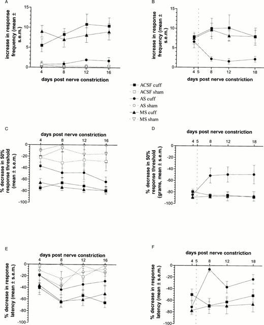
1. Nerve injury often produces long-lasting spontaneous pain, hyperalgesia and allodynia that are refractory to treatment, being only partially relieved by clinical analgesics, and often insensitive to morphine. With the aim of assessing its therapeutic potential, we examined the effect of antisense oligonucleotide knockdown of spinal metabotropic glutamate receptor 1 (mGluR(1)) in neuropathic rats. 2. We chronically infused rats intrathecally with either vehicle, or 50 microg day(-1) antisense or missense oligonucleotides beginning either 3 days prior to or 5 days after nerve injury. Cold, heat and mechanical sensitivity was assessed prior to any treatment and again every few days after nerve injury. 3. Here we show that knockdown of mGluR(1) significantly reduces cold hyperalgesia, heat hyperalgesia and mechanical allodynia in the ipsilateral (injured) hindpaw of neuropathic rats. 4. Moreover, we show that morphine analgesia is reduced in neuropathic rats, but not in sham-operated rats, and that knockdown of mGluR(1) restores the analgesic efficacy of morphine. 5. We also show that neuropathic rats are more sensitive to the excitatory effects of intrathecally injected N-methyl-D-aspartate (NMDA), and have elevated protein kinase C (PKC) activity in the spinal cord dorsal horn, two effects that are reversed by knockdown of mGluR(1). 6. These results suggest that activity at mGluR(1) contributes to neuropathic pain through interactions with spinal NMDA receptors and PKC, and that knockdown of mGluR(1) may be a useful therapy for neuropathic pain in humans, both to alleviate pain directly, and as an adjunct to opioid analgesic treatment.


Figures

Figure 1
Figure 1
Western blot analysis. Representative Western blots (from a single animal per group) and histogram summaries (n=3 per group) from Western blot analysis of lumbar spinal cords and the thalamus/periaqueductal region of brains taken from ACSF-, AS- and MS-treated rats after 7 days of oligonucleotide infusion, and 4 days after nerve constriction (induced by implantation of a cuff around one sciatic nerve) or sham-surgery. Depicted in the histograms is the mean of the binding density scores for each group (±s.e.m.). (A) Mean binding density of anti-rat mGluR1 IgG in lumbar spinal cord. AS treatment induced a 57% decrease of mGluR1 protein compared to vehicle treatment in neuropathic rats, and a 38% decrease in mGluR1 protein compared to vehicle treatment in sham-operated rats. MS treatment induced a 1% decrease in mGluR1 protein compared to vehicle treatment in neuropathic rats, and a 44% increase in mGluR1 protein compared to vehicle treatment in sham-operated rats. (B) Mean binding density of anti-rat mGluR1 IgG in thalamus/periaqueductal region of brain. AS treatment induced a 25% decrease in neuropathic rats, and an 18% decrease in mGluR1 protein in sham-operated rats. MS treatment induced a 4% increase in neuropathic rats, and a 10% decrease in sham-operated rats, compared to vehicle-treatment. (C) Mean binding density of anti-rat mGluR5 IgG in lumbar spinal cord. AS treatment induced a slight decrease in mGluR5 protein in neuropathic rats (8%), and a more pronounced decrease in sham-operated rats (26%). MS treatment induced an increase (54%) in neuropathic rats, and a decrease (19%) in sham-operated rats in mGluR5 protein compared to vehicle treatment.
Figure 2
Figure 2
DHPG-induced spontaneous nociceptive behaviours. Mean time spent exhibiting nociceptive behaviour after i.t. injection of 50 nmol DHPG in ACSF – (n=8), AS- (n=5) and MS-treated (n=5) rats. *Significantly different from ACSF-treated (P<0.05); significantly different from MS-treated (P<0.05).
Figure 3
Figure 3
Nerve-injury induced hyperalgesia and allodynia. Change in response to cold, heat and mechanical stimulation on days 4, 8, 12 and 16 days (pre-treatment group) or 4, 8, 12 and 18 days (post-treatment group) after nerve constriction in neuropathic and sham-operated rats treated intrathecally with either ACSF (cuffed, n=12 pre-treatment, n=6 post-treatment; sham, n=4 pre-treatment), AS (cuffed, n=9 pre-treatment, n=5 post-treatment; sham, n=3 pre-treatment) or MS (cuffed, n=7 pre-treatment, n=6 post-treatment; sham, n=4 pre-treatment). (A) Cold water test in pre-treatment group: Mean increase in number of responses (lifting of hindpaw) when rats stood in water at 1°C. There was a significant interaction between neuropathic condition and i.t. treatment (F(2,33)=7.91, P<0.01). Post-hoc tests showed that AS-treated neuropathic rats had a significantly lower response frequency across test days compared to ACSF- or MS-treated neuropathic rats. (B) Cold water test in post-treatment group: Mean increase in number of responses after nerve injury, both prior to i.t. infusion (day 4), and after i.t. infusion (days 8, 12 and 18). All nerve injured rats showed a large increase in response frequency in the ipsilateral hindpaw, compared to baseline, on day 4 after nerve injury, prior to i.t. oligonucleotide infusion. The dotted line at day 5 indicates when the i.t. infusion of oligonucleotides began. There was a significant drug×day interaction (F(6,42)=2.30, P=0.05). Post-hoc Fisher's LSD t-tests showed that after i.t. infusion, AS-treated neuropathic rats showed a significantly lower increase in response frequency on days 8, 12 and 18 after nerve injury, compared to ACSF- and MS-treated rats regardless of test day. (C) von Frey hair test in pre-treatment group: Mean per cent decrease in 50% response threshold in grams. There were significant effects of neuropathic condition (F(1,33)=98.44, P<0.01), i.t. treatment (F(2,33)=9.23, P<0.01), and test day (F(3,99)=3.48, P<0.05). Post-hoc tests showed that neuropathic AS-treated rats had a significantly lower decrease in 50% response threshold than either ACSF- or MS-treated rats. (D) von Frey hair test in post-treatment group: Mean per cent decrease in 50% response threshold in grams. On day 4 after nerve injury, prior to i.t. drug infusion, all neuropathic rats showed a large decrease in 50% response threshold. The dotted line at day 5 indicates when the i.t. infusion of oligonucleotides began. There was a significant drug×day interaction (F(6,42)=3.35, P<0.05), and post-hoc tests showed that after i.t. infusion, neuropathic AS-treated rats had a significantly lower decrease in 50% response threshold than either ACSF- or MS-treated rats. (E) Radiant heat plantar test in pre-treatment group: Mean per cent decrease in response latency. There were significant effects of interaction between neuropathic condition and i.t. treatment (F(2,33)=3.26, P=0.05), and test day (F(3,99)=6.57, P<0.01). Post-hoc tests Fisher's LSD t-tests showed that AS-treated neuropathic rats had a significantly lower decrease in response latency than ACSF- or MS-treated neuropathic rats. (F) Radiant heat plantar test in post-treatment group: Mean per cent decrease in response latency. All nerve injured rats showed a large decrease in response latency, compared to baseline, on day 4 after nerve injury, prior to i.t. drug infusion. The dotted line at day 5 indicates when the i.t. infusion of oligonucleotides began. There was a significant drug×day interaction (F(6.42)=6.22, P<0.05), and post-hoc tests showed that after i.t. infusion, AS-treated neuropathic rats had a significantly lower decrease in response latency from days 8 to 18 after nerve injury.
Figure 4
Figure 4
Morphine dose-response curve. Morphine dose-response curve in neuropathic and sham-operated rats 4 days after nerve injury for ACSF- (n=15 each for cuffed and sham-operated), AS- (n=16, cuffed) and MS-treated (n=17, cuffed) rats. Prior to morphine injection, tail flick latencies were measured (BL2 as described in Methods). Following morphine injection, tail flick latencies were measured every 15 min from 15 – 60 min post-morphine. Latency scores were converted to percent maximum possible effect scores [%MPE=((latency −BL2)/(cutoff −BL2))*100]. From the %MPE scores, we calculated the area under the curve (AUC) from 15 – 60 min post morphine injection (maximum AUC=300) to determine the analgesic effect. The mean AUC for each treatment group at each morphine dose is illustrated in this figure. ACSF- and MS-treated neuropathic rats showed little analgesia after morphine was injected intrathecally (ED50=63.71 μg (21.38 μg-1950 mg) and 705.28 μg (95% C.I. not calculable) respectively). In contrast, AS-treated neuropathic rats showed a robust analgesic effect, and were not different from sham-operated rats (ED50=8.13 μg (5.25 – 12.02 μg) and 9.72 μg (4.68 – 20.89 μg) respectively).
Figure 5
Figure 5
[3H]-PDBu binding. [3H]-PDBu binding autoradiography in lumbar spinal cord of neuropathic and sham-operated rats. (A) Histogram summary showing the density of [3H]-PDBu binding (nCi g−1) in ACSF-, AS- and MS-treated neuropathic and sham-operated rats (n=3 rats per treatment combination, with five slides from each rat, for a total of 15 slides per group). Planned comparisons showed that ACSF-treated sham-operated rats had a lower binding density than ACSF-treated neuropathic rats; AS-treated neuropathic rats had a lower binding density than either ACSF- or MS-treated neuropathic rats. *Significantly different from ACSF-treated neuropathic rats; # significantly different from MS-treated neuropathic rats. (B) Computer-generated image (MCID) of a single representative slide from each group showing sample binding of [3H]-PDBu in ACSF-, AS- and MS-treated neuropathic and sham-operated rats.
Figure 6
Figure 6
NMDA-induced spontaneous nociceptive behaviours. Mean time spent exhibiting nociceptive behaviour in neuropathic (n=4 – 6 per dose) and sham-operated (n=4 – 6 per dose) rats given increasing doses of i.t. NMDA; and in neuropathic rats treated with either AS (n=11) or MS (n=10), and given 2.5 nmol of NMDA. *Significantly different from ACSF-treated (P<0.05); significantly different from MS-treated (P<0.05); #significantly different from sham-operated (P<0.05).

Similar articles

Cited by

References

    1. ABE T., SUGIHARA H., NAWA H., SHIGEMOTO R., MIZUNO N., NAKANISHI S. Molecular characterization of a novel metabotropic glutamate receptor mGluR5 coupled to inositol phosphate/Ca2+ signal transduction. J. Biol. Chem. 1992;267:13361–13368. - PubMed
    1. AKHTAR S., AGRAWAL S. In vivo studies with antisense oligonucleotides. Trends Pharmacol. Sci. 1997;18:12–18. - PubMed
    1. AL-GHOUL W.M., LI VOLSI G., WEINBERG R.J., RUSTIONI A. Glutamate immunocytochemistry in the dorsal horn after injury or stimulation of the sciatic nerve of rats. Brain Res. Bull. 1993;30:453–459. - PubMed
    1. ALVAREZ F.I., VILLALBA R.M., CARR P.A., GRANDES P., SOMOHANO P.M. Differential distribution of metabotropic glutamate receptors 1a, 1b, and 5 in rat spinal cord. J. Comp. Neurol. 2000;422:464–487. - PubMed
    1. BERTHELE A., BOXALL S.J., URBAN A., ANNESER J.M., ZIEGLGANSBERGER W., URBAN L., TOLLE T.R. Distribution and developmental changes in metabotropic glutamate receptor messenger RNA expression in the rat lumbar spinal cord. Brain. Res. Dev. Brain. Res. 1999;112:39–53. - PubMed

Publication types

MeSH terms

Grants and funding