

Long-term follow-up of asymptomatic patients with major artery occlusion: rate of symptomatic change and evaluation of cerebral hemodynamics
- PMID: 11156763
- PMCID: PMC7973943
Long-term follow-up of asymptomatic patients with major artery occlusion: rate of symptomatic change and evaluation of cerebral hemodynamics
Abstract
Background and purpose: The natural history of asymptomatic major cerebral artery occlusive disease is unclear. Rate of symptomatic change, appearance of new lesions on MR images, and cerebral hemodynamics were analyzed for patients with asymptomatic major cerebral artery occlusion.
Methods: This prospective study included asymptomatic patients who had occlusive disease between 1992 and 1995. MR imaging and MR angiography were used to detect internal carotid artery (ICA) or middle cerebral artery (MCA) occlusion in 3965 neurologically asymptomatic patients and for follow-up of affected patients for 67 to 105 months (mean, 79 months). Regional cerebral blood flow and cerebrovascular reserve capacity were examined by xenon-enhanced CT at rest and after the administration of acetazolamide, respectively.
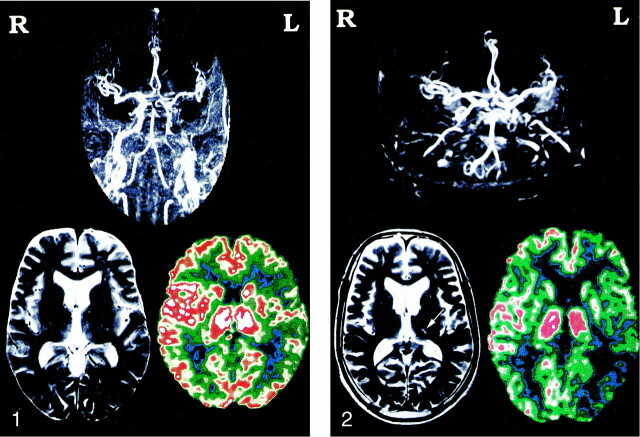
Results: Eighteen patients had MCA occlusion and 17 had ICA occlusion. During the follow-up period, five patients became symptomatic (four with MCA occlusion and one with ICA occlusion), with no significant difference (P = .332) in the rate of symptomatic change. Among these five patients, new infarction occurred on the ipsilateral side in three patients, contralateral side in one, and bilateral sides in one. New stenotic or occlusive changes occurred in three patients. The patients were divided into groups: group A, without new lesions on MR images (n = 23), and group B, with new lesions (n = 12). There was no significant difference in regional cerebral blood flow value between groups A and B in the whole hemisphere, anterior cerebral artery territory, or MCA territory. There was a significant difference in cerebrovascular reserve capacity between groups A and B between the affected side (P = .00051 and P = .00068, respectively) and the contralateral side (P = .00101 and P = .00115, respectively) for the whole hemisphere and MCA territory, and the difference was more severe on the affected side in both regions.
Conclusion: These pilot data suggest that asymptomatic MCA occlusion has a worse prognosis than does ICA occlusion. Silent events are common bilaterally. This may be because of hemodynamic factors or perhaps MCA occlusion is a marker for a more progressive type of atherosclerosis. A prospective study involving assessment of hemodynamics and baseline stroke risk factors in patients with MCA occlusion is indicated.
Figures
Comment in
-
Hemodynamic impairment and stroke risk: prove it.AJNR Am J Neuroradiol. 2001 Feb;22(2):233-4. AJNR Am J Neuroradiol. 2001. PMID: 11156758 Free PMC article. No abstract available.
References
-
- Kobayashi S, Okada K, Yamashita K. Incidence of silent lacunar lesion in normal adults and its relation to cerebral blood flow and risk factors. Stroke 1991;22:1379-1383 - PubMed
-
- Matsubayashi K, Shimada K, Kawamoto A, Ozawa T. Incidental brain lesions on magnetic resonance imaging and neurobehavioral functions in the apparently healthy elderly. Stroke 1992;23:175-180 - PubMed
-
- Nakagawa T, Hashi K. The incidence and treatment of asymptomatic, unruptured cerebral aneurysms. J Neurosurg 1994;80:217-223 - PubMed
-
- Hougaku H, Matsumoto M, Handa N, et al. Asymptomatic carotid lesions and silent cerebral infarction. Stroke 1994;25:566-570 - PubMed
-
- Shinkawa A, Ueda K, Kiyohara Y, et al. Silent cerebral infarction in a community-based autopsy series in Japan: The Hisayama Study. Stroke 1995;26:380-385 - PubMed
MeSH terms
Substances
LinkOut - more resources
Full Text Sources
Miscellaneous