

Relationship between vascular enhancement, cerebral hemodynamics, and MR angiography in cases of acute stroke
- PMID: 11156765
- PMCID: PMC7973947
Relationship between vascular enhancement, cerebral hemodynamics, and MR angiography in cases of acute stroke
Abstract
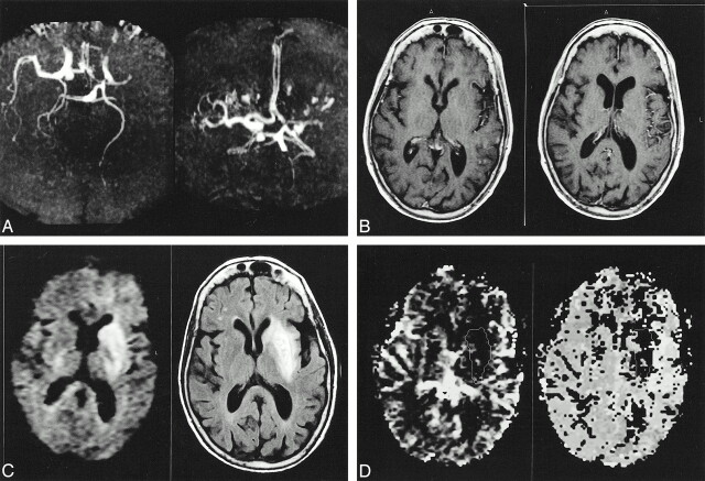
Background and purpose: The use of MR angiography and contrast-enhanced T1-weighted MR imaging in cases of acute cerebral ischemia may be helpful in the evaluation of middle cerebral artery (MCA) occlusion and leptomeningeal collaterals, respectively. The aim of our work was to investigate the relationship between MCA occlusion, T1-weighted vascular contrast enhancement, hemodynamic alterations, and tissue damage in cases of acute ischemic stroke.
Methods: We studied the MCA territory in 15 patients with acute ischemic stroke within 8 hr of symptom onset. The first MR imaging study (<8 hr after onset) comprised diffusion-weighted imaging, MR angiography, perfusion-weighted imaging, and contrast-enhanced T1-weighted MR imaging sequences. Follow-up MR imaging, performed 1 week later, consisted of MR angiography and T2-weighted fluid-attenuated inversion recovery MR imaging.
Results: Early MR angiography showed MCA stem occlusion in nine of 15 patients. Patients with MCA occlusion had significantly larger areas of abnormality on early diffusion-weighted images, significantly larger areas of altered hemodynamics, larger final lesion volumes, and poorer clinical outcome. Among the nine patients with MCA stem occlusion, vascular enhancement was marked in seven and absent in two who had complete MCA infarcts and poor clinical outcome. Among patients with MCA patency, vascular enhancement was marked in only one, mild in four, and absent in one. Patients with marked vascular enhancement had significantly larger regions of altered hemodynamics and significantly higher asymmetries in both regional cerebral blood volume and mean transit time because of increased values in the affected hemisphere.
Conclusion: Among patients with stroke with MCA occlusion, marked vascular enhancement and increased blood volume indicate efficient leptomeningeal collaterals and compensatory hemodynamic mechanisms.
Figures
References
-
- Warach S, Gaa J, Siewert B, Wielopolski P, Edelman RR. Acute human stroke studied by whole brain echo planar diffusion-weighted magnetic resonance imaging. Ann Neurol 1995;37:231-241 - PubMed
-
- Lutsep H, Albers G, DeCrespigny A, Kamat G, Marks M, Mosely M. Clinical utility of diffusion-weighted magnetic resonance imaging in the assessment of ischemic stroke. Ann Neurol 1997;41:574-580 - PubMed
-
- Röther J, Gückel F, Neff W, Schwartz A, Hennerici M. Assessment of regional cerebral blood volume in acute human stroke by use of single-slice dynamic susceptibility contrast-enhanced magnetic resonance imaging. Stroke 1996;27:1088-1093 - PubMed
-
- Sorensen A, Copen W, Ostergaard L, et al. Hyperacute stroke: simultaneous measurement of relative cerebral blood volume, relative blood flow and mean tissue transit time. Radiology 1999;210:519-527 - PubMed
MeSH terms
Substances
LinkOut - more resources
Full Text Sources
Medical